6C87
 
 | |
6CU3
 | |
6CKT
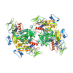
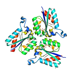
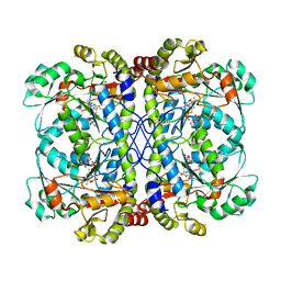
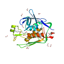
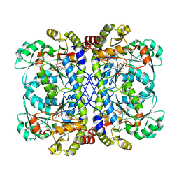
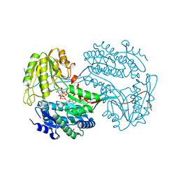
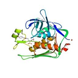
 | | Crystal structure of 2,3,4,5-tetrahydropyridine-2,6-dicarboxylate N-succinyltransferase from Legionella pneumophila Philadelphia 1 | | Descriptor: | 1,2-ETHANEDIOL, 2,3,4,5-tetrahydropyridine-2,6-dicarboxylate N-succinyltransferase | | Authors: | Seattle Structural Genomics Center for Infectious Disease (SSGCID) | | Deposit date: | 2018-02-28 | | Release date: | 2018-03-21 | | Last modified: | 2023-10-04 | | Method: | X-RAY DIFFRACTION (1.8 Å) | | Cite: | Crystal structure of 2,3,4,5-tetrahydropyridine-2,6-dicarboxylate N-succinyltransferase from Legionella pneumophila Philadelphia 1
to be published
|
|
6CK0
 | |
6CK7
 | |
6CKP
 | |
6CUM
 | |
6CW5
 | |
6CXM
 | |
6CV6
 | |
6CJA
 | |
6CAX
 | |
6CJB
 | |
6CU5
 | |
6CXY
 | |
6CUQ
 | |
6DJK
 | |
6DBB
 | |
6D6J
 | |
6D9N
 | |
6D8W
 | |
6DUI
 | |
6D46
 | |
6DEG
 | |
6D47
 | |


