Loading
PDBj
MenuPDBj@FacebookPDBj@TwitterPDBj@YouTubewwPDB FoundationwwPDB
RCSB PDBPDBeBMRBAdv. SearchSearch help
Search by PDB author
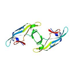
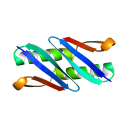
1Q10
DownloadVisualize
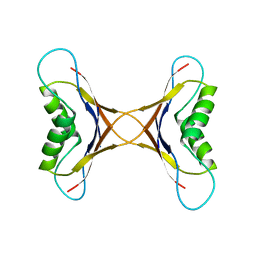
BU of 1q10 by Molmil
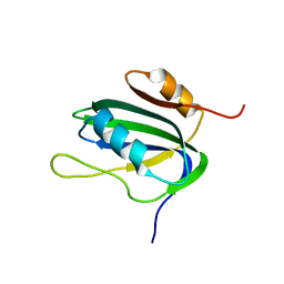
Ensemble of 40 Structures of the Dimeric Mutant of the B1 Domain of Streptococcal Protein G
Descriptor: Immunoglobulin G binding protein G
Authors:Byeon, I.J, Louis, J.M, Gronenborn, A.M.
Deposit date:2003-07-18
Release date:2003-10-14
Last modified:2021-10-27
Method:SOLUTION NMR
Cite:A Protein Contortionist: Core Mutations of GB1 that Induce Dimerization and Domain Swapping
J.Mol.Biol., 333, 2003
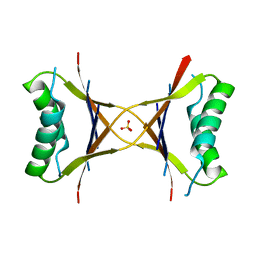
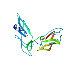
1MPE
DownloadVisualize
BU of 1mpe by Molmil
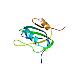
Ensemble of 20 structures of the tetrameric mutant of the B1 domain of streptococcal protein G
Descriptor: Immunoglobulin G binding protein G
Authors:Frank, M.K, Dyda, F, Dobrodumov, A, Gronenborn, A.M.
Deposit date:2002-09-12
Release date:2002-10-30
Last modified:2021-10-27
Method:SOLUTION NMR
Cite:Core mutations switch monomeric protein GB1 into an intertwined tetramer.
Nat.Struct.Biol., 9, 2002
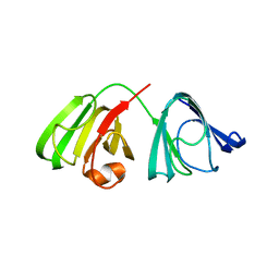
1MVK
DownloadVisualize
BU of 1mvk by Molmil
X-ray structure of the tetrameric mutant of the B1 domain of streptococcal protein G
Descriptor: Immunoglobulin G binding protein G, SULFATE ION
Authors:Frank, M.K, Dyda, F, Dobrodumov, A, Gronenborn, A.M.
Deposit date:2002-09-25
Release date:2002-10-30
Last modified:2024-02-14
Method:X-RAY DIFFRACTION (2.5 Å)
Cite:Core mutations switch monomeric protein GB1 into an intertwined tetramer.
Nat.Struct.Biol., 9, 2002
4J4E
DownloadVisualize
BU of 4j4e by Molmil
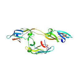
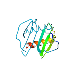
Structure of P51G Cyanovirin-N swapped trimer in the P212121 space group
Descriptor: Cyanovirin-N
Authors:Koharudin, L.M.I, Liu, L, Gronenborn, A.M.
Deposit date:2013-02-06
Release date:2013-04-03
Last modified:2023-09-20
Method:X-RAY DIFFRACTION (2.4 Å)
Cite:Different 3D domain-swapped oligomeric cyanovirin-N structures suggest trapped folding intermediates.
Proc.Natl.Acad.Sci.USA, 110, 2013
4J4G
DownloadVisualize
BU of 4j4g by Molmil
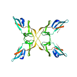
Structure of P51G Cyanovirin-N swapped tetramer in the C2 space group
Descriptor: Cyanovirin-N
Authors:Koharudin, L.M.I, Liu, L, Gronenborn, A.M.
Deposit date:2013-02-06
Release date:2013-04-03
Last modified:2023-09-20
Method:X-RAY DIFFRACTION (1.92 Å)
Cite:Different 3D domain-swapped oligomeric cyanovirin-N structures suggest trapped folding intermediates.
Proc.Natl.Acad.Sci.USA, 110, 2013
4J4F
DownloadVisualize
BU of 4j4f by Molmil
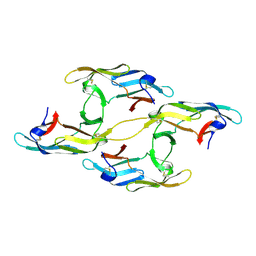
Structure of P51G Cyanovirin-N swapped tetramer in the P212121 space group
Descriptor: Cyanovirin-N
Authors:Koharudin, L.M.I, Liu, L, Gronenborn, A.M.
Deposit date:2013-02-06
Release date:2013-04-03
Last modified:2023-09-20
Method:X-RAY DIFFRACTION (1.9 Å)
Cite:Different 3D domain-swapped oligomeric cyanovirin-N structures suggest trapped folding intermediates.
Proc.Natl.Acad.Sci.USA, 110, 2013
4J4D
DownloadVisualize
BU of 4j4d by Molmil
Structure of P51G Cyanovirin-N swapped dimer in the P21212 space group
Descriptor: Cyanovirin-N
Authors:Koharudin, L.M.I, Liu, L, Gronenborn, A.M.
Deposit date:2013-02-06
Release date:2013-04-03
Last modified:2023-09-20
Method:X-RAY DIFFRACTION (2 Å)
Cite:Different 3D domain-swapped oligomeric cyanovirin-N structures suggest trapped folding intermediates.
Proc.Natl.Acad.Sci.USA, 110, 2013
4J4C
DownloadVisualize
BU of 4j4c by Molmil
Structure of P51G Cyanovirin-N swapped dimer in the P3221 space group
Descriptor: Cyanovirin-N
Authors:Koharudin, L.M.I, Liu, L, Gronenborn, A.M.
Deposit date:2013-02-06
Release date:2013-04-03
Last modified:2023-09-20
Method:X-RAY DIFFRACTION (1.9 Å)
Cite:Different 3D domain-swapped oligomeric cyanovirin-N structures suggest trapped folding intermediates.
Proc.Natl.Acad.Sci.USA, 110, 2013
4JGF
DownloadVisualize
BU of 4jgf by Molmil
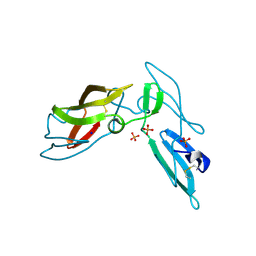
Crystal Structure of the Cataract-Causing P23T gamma D-Crystallin Mutant
Descriptor: Gamma-crystallin D
Authors:Ji, F.L, Koharudin, L.M, Jung, J.W, Gronenborn, A.M.
Deposit date:2013-03-01
Release date:2013-05-22
Last modified:2024-02-28
Method:X-RAY DIFFRACTION (2.5 Å)
Cite:Crystal structure of the cataract-causing P23T gamma D-crystallin mutant.
Proteins, 81, 2013
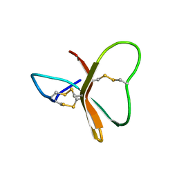
2BDS
DownloadVisualize
BU of 2bds by Molmil
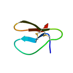
DETERMINATION OF THE THREE-DIMENSIONAL SOLUTION STRUCTURE OF THE ANTIHYPERTENSIVE AND ANTIVIRAL PROTEIN BDS-I FROM THE SEA ANEMONE ANEMONIA SULCATA. A STUDY USING NUCLEAR MAGNETIC RESONANCE AND HYBRID DISTANCE GEOMETRY-DYNAMICAL SIMULATED ANNEALING
Descriptor: BDS-I
Authors:Clore, G.M, Driscoll, P.C, Gronenborn, A.M.
Deposit date:1988-11-14
Release date:1989-04-19
Last modified:2017-11-29
Method:SOLUTION NMR
Cite:Determination of the three-dimensional solution structure of the antihypertensive and antiviral protein BDS-I from the sea anemone Anemonia sulcata: a study using nuclear magnetic resonance and hybrid distance geometry-dynamical simulated annealing.
Biochemistry, 28, 1989
1L5I
DownloadVisualize
BU of 1l5i by Molmil
30-CONFORMER NMR ENSEMBLE OF THE N-TERMINAL, DNA-BINDING DOMAIN OF THE REPLICATION INITIATION PROTEIN FROM A GEMINIVIRUS (TOMATO YELLOW LEAF CURL VIRUS-SARDINIA)
Descriptor: Rep protein
Authors:Campos-Olivas, R, Louis, J.M, Clerot, D, Gronenborn, B, Gronenborn, A.M.
Deposit date:2002-03-07
Release date:2002-09-18
Last modified:2022-02-23
Method:SOLUTION NMR
Cite:The structure of a replication initiator unites diverse aspects of nucleic acid metabolism
Proc.Natl.Acad.Sci.USA, 99, 2002
1L2M
DownloadVisualize
BU of 1l2m by Molmil
Minimized Average Structure of the N-terminal, DNA-binding domain of the replication initiation protein from a geminivirus (Tomato yellow leaf curl virus-Sardinia)
Descriptor: Rep protein
Authors:Campos-Olivas, R, Louis, J.M, Clerot, D, Gronenborn, B, Gronenborn, A.M.
Deposit date:2002-02-22
Release date:2002-09-18
Last modified:2022-02-23
Method:SOLUTION NMR
Cite:The structure of a replication initiator unites diverse aspects of nucleic acid metabolism
Proc.Natl.Acad.Sci.USA, 99, 2002
2GAT
DownloadVisualize
BU of 2gat by Molmil
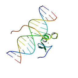
SOLUTION STRUCTURE OF THE C-TERMINAL DOMAIN OF CHICKEN GATA-1 BOUND TO DNA, NMR, REGULARIZED MEAN STRUCTURE
Descriptor: DNA (5'-D(*AP*AP*TP*GP*TP*TP*TP*AP*TP*CP*TP*GP*CP*AP*AP*C)-3'), DNA (5'-D(*GP*TP*TP*GP*CP*AP*GP*AP*TP*AP*AP*AP*CP*AP*TP*T)-3'), ERYTHROID TRANSCRIPTION FACTOR GATA-1, ...
Authors:Clore, G.M, Tjandra, N, Starich, M, Omichinski, J.G, Gronenborn, A.M.
Deposit date:1997-11-07
Release date:1998-01-28
Last modified:2022-03-09
Method:SOLUTION NMR
Cite:Use of dipolar 1H-15N and 1H-13C couplings in the structure determination of magnetically oriented macromolecules in solution.
Nat.Struct.Biol., 4, 1997
3IL8
DownloadVisualize
BU of 3il8 by Molmil
CRYSTAL STRUCTURE OF INTERLEUKIN 8: SYMBIOSIS OF NMR AND CRYSTALLOGRAPHY
Descriptor: INTERLEUKIN-8
Authors:Baldwin, E.T, Weber, I.T, St Charles, R, Xuan, J.-C, Appella, E, Yamada, M, Matsushima, K, Edwards, B.F.P, Clore, G.M, Gronenborn, A.M, Wlodawer, A.
Deposit date:1990-12-07
Release date:1992-10-15
Last modified:2012-02-29
Method:X-RAY DIFFRACTION (2 Å)
Cite:Crystal structure of interleukin 8: symbiosis of NMR and crystallography.
Proc.Natl.Acad.Sci.USA, 88, 1991
3J34
DownloadVisualize
BU of 3j34 by Molmil
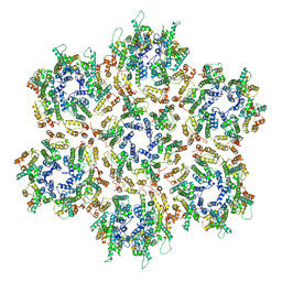
Structure of HIV-1 Capsid Protein by Cryo-EM
Descriptor: capsid protein
Authors:Zhao, G, Perilla, J.R, Yufenyuy, E, Meng, X, Chen, B, Ning, J, Ahn, J, Gronenborn, A.M, Schulten, K, Aiken, C, Zhang, P.
Deposit date:2013-02-23
Release date:2013-05-29
Last modified:2018-07-18
Method:ELECTRON MICROSCOPY (8.6 Å)
Cite:Mature HIV-1 capsid structure by cryo-electron microscopy and all-atom molecular dynamics.
Nature, 497, 2013
1BBO
DownloadVisualize
BU of 1bbo by Molmil
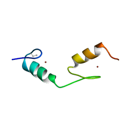
HIGH-RESOLUTION SOLUTION STRUCTURE OF THE DOUBLE CYS2*HIS2 ZINC FINGER FROM THE HUMAN ENHANCER BINDING PROTEIN MBP-1
Descriptor: HUMAN ENHANCER-BINDING PROTEIN MBP-1, ZINC ION
Authors:Clore, G.M, Omichinski, J.G, Gronenborn, A.M.
Deposit date:1992-05-01
Release date:1993-10-31
Last modified:2023-11-15
Method:SOLUTION NMR
Cite:High-resolution solution structure of the double Cys2His2 zinc finger from the human enhancer binding protein MBP-1.
Biochemistry, 31, 1992
1BBL
DownloadVisualize
BU of 1bbl by Molmil
THREE-DIMENSIONAL SOLUTION STRUCTURE OF THE E3-BINDING DOMAIN OF THE DIHYDROLIPOAMIDE SUCCINYLTRANSFERASE CORE FROM THE 2-OXOGLUTARATE DEHYDROGENASE MULTIENZYME COMPLEX OF ESCHERICHIA COLI
Descriptor: DIHYDROLIPOAMIDE SUCCINYLTRANSFERASE
Authors:Clore, G.M, Robien, M.A, Gronenborn, A.M.
Deposit date:1992-02-20
Release date:1994-01-31
Last modified:2022-02-16
Method:SOLUTION NMR
Cite:Three-dimensional solution structure of the E3-binding domain of the dihydrolipoamide succinyltransferase core from the 2-oxoglutarate dehydrogenase multienzyme complex of Escherichia coli.
Biochemistry, 31, 1992
1BAL
DownloadVisualize
BU of 1bal by Molmil
THREE-DIMENSIONAL SOLUTION STRUCTURE OF THE E3-BINDING DOMAIN OF THE DIHYDROLIPOAMIDE SUCCINYLTRANSFERASE CORE FROM THE 2-OXOGLUTARATE DEHYDROGENASE MULTIENZYME COMPLEX OF (ESCHERICHIA COLI)
Descriptor: DIHYDROLIPOAMIDE SUCCINYLTRANSFERASE
Authors:Clore, G.M, Robien, M.A, Gronenborn, A.M.
Deposit date:1992-02-20
Release date:1994-01-31
Last modified:2022-02-16
Method:SOLUTION NMR
Cite:Three-dimensional solution structure of the E3-binding domain of the dihydrolipoamide succinyltransferase core from the 2-oxoglutarate dehydrogenase multienzyme complex of Escherichia coli.
Biochemistry, 31, 1992
1CBH
DownloadVisualize
BU of 1cbh by Molmil
DETERMINATION OF THE THREE-DIMENSIONAL STRUCTURE OF THE C-TERMINAL DOMAIN OF CELLOBIOHYDROLASE I FROM TRICHODERMA REESEI. A STUDY USING NUCLEAR MAGNETIC RESONANCE AND HYBRID DISTANCE GEOMETRY-DYNAMICAL SIMULATED ANNEALING
Descriptor: C-TERMINAL DOMAIN OF CELLOBIOHYDROLASE I
Authors:Clore, G.M, Gronenborn, A.M.
Deposit date:1989-05-30
Release date:1990-01-15
Last modified:2017-11-29
Method:SOLUTION NMR
Cite:Determination of the three-dimensional solution structure of the C-terminal domain of cellobiohydrolase I from Trichoderma reesei. A study using nuclear magnetic resonance and hybrid distance geometry-dynamical simulated annealing.
Biochemistry, 28, 1989
3LHC
DownloadVisualize
BU of 3lhc by Molmil
Crystal structure of cyanovirin-n swapping domain b mutant
Descriptor: Cyanovirin-N, PHOSPHATE ION, SODIUM ION
Authors:Matei, E, Zheng, A, Furey, W, Rose, J, Aiken, C, Gronenborn, A.M.
Deposit date:2010-01-21
Release date:2010-02-09
Last modified:2023-09-06
Method:X-RAY DIFFRACTION (1.34 Å)
Cite:Anti-HIV activity of defective cyanovirin-N mutants is restored by dimerization.
J.Biol.Chem., 285, 2010
3OBL
DownloadVisualize
BU of 3obl by Molmil
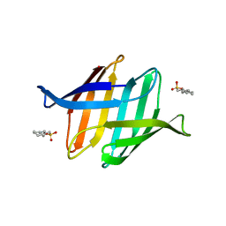
Crystal structure of the potent anti-HIV cyanobacterial lectin from Oscillatoria Agardhii
Descriptor: 3-CYCLOHEXYL-1-PROPYLSULFONIC ACID, Lectin
Authors:Koharudin, L.M.I, Furey, W, Gronenborn, A.M.
Deposit date:2010-08-06
Release date:2010-10-20
Last modified:2024-02-21
Method:X-RAY DIFFRACTION (1.2 Å)
Cite:Novel fold and carbohydrate specificity of the potent anti-HIV cyanobacterial lectin from Oscillatoria agardhii.
J.Biol.Chem., 286, 2011
4BZC
DownloadVisualize
BU of 4bzc by Molmil
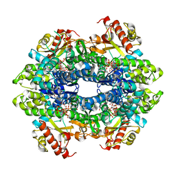
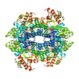
Crystal structure of the tetrameric dGTP-bound wild type SAMHD1 catalytic core
Descriptor: 2'-deoxyguanosine-5'-O-(1-thiotriphosphate), DEOXYNUCLEOSIDE TRIPHOSPHATE TRIPHOSPHOHYDROLASE SAMHD1, MAGNESIUM ION, ...
Authors:Ji, X, Yang, H, Wu, Y, Yan, J, Mehrens, J, DeLucia, M, Hao, C, Gronenborn, A.M, Skowronski, J, Ahn, J, Xiong, Y.
Deposit date:2013-07-25
Release date:2013-10-23
Last modified:2023-12-20
Method:X-RAY DIFFRACTION (2.88 Å)
Cite:Mechanism of Allosteric Activation of Samhd1 by Dgtp
Nat.Struct.Mol.Biol., 20, 2013
4BZB
DownloadVisualize
BU of 4bzb by Molmil
Crystal structure of the tetrameric dGTP-bound SAMHD1 mutant catalytic core
Descriptor: 2'-DEOXYGUANOSINE-5'-TRIPHOSPHATE, DEOXYNUCLEOSIDE TRIPHOSPHATE TRIPHOSPHOHYDROLASE SAMHD1, MAGNESIUM ION
Authors:Ji, X, Yang, H, Wu, Y, Yan, J, Mehrens, J, DeLucia, M, Hao, C, Gronenborn, A.M, Skowronski, J, Ahn, J, Xiong, Y.
Deposit date:2013-07-25
Release date:2013-10-23
Last modified:2023-12-20
Method:X-RAY DIFFRACTION (1.83 Å)
Cite:Mechanism of Allosteric Activation of Samhd1 by Dgtp
Nat.Struct.Mol.Biol., 20, 2013
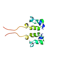
1WJB
DownloadVisualize
BU of 1wjb by Molmil
SOLUTION STRUCTURE OF THE N-TERMINAL ZN BINDING DOMAIN OF HIV-1 INTEGRASE (D FORM), NMR, 40 STRUCTURES
Descriptor: HIV-1 INTEGRASE, ZINC ION
Authors:Clore, G.M, Cai, M, Caffrey, M, Gronenborn, A.M.
Deposit date:1997-05-13
Release date:1998-05-13
Last modified:2022-03-02
Method:SOLUTION NMR
Cite:Solution structure of the N-terminal zinc binding domain of HIV-1 integrase.
Nat.Struct.Biol., 4, 1997
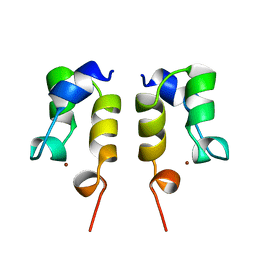
1WJA
DownloadVisualize
BU of 1wja by Molmil
SOLUTION STRUCTURE OF THE N-TERMINAL ZN BINDING DOMAIN OF HIV-1 INTEGRASE (D FORM), NMR, REGULARIZED MEAN STRUCTURE
Descriptor: HIV-1 INTEGRASE, ZINC ION
Authors:Clore, G.M, Cai, M, Caffrey, M, Gronenborn, A.M.
Deposit date:1997-05-13
Release date:1998-05-13
Last modified:2022-03-02
Method:SOLUTION NMR
Cite:Solution structure of the N-terminal zinc binding domain of HIV-1 integrase.
Nat.Struct.Biol., 4, 1997

219869

건을2024-05-15부터공개중

PDB statisticsPDBj update infoContact PDBjnumon