6DCX
 
 | |
5U6K
 | |
1T2U
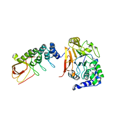
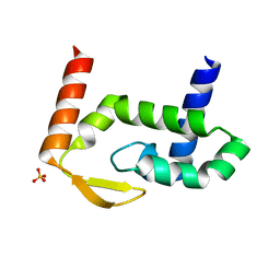
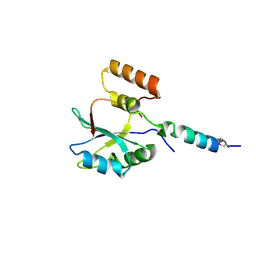
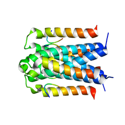
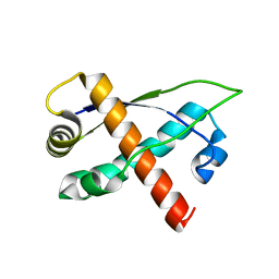
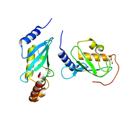
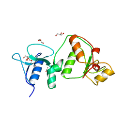
 | | Structural basis of phosphopeptide recognition by the BRCT domain of BRCA1: structure of BRCA1 missense variant V1809F | | 分子名称: | Breast cancer type 1 susceptibility protein, COBALT (II) ION, SULFATE ION | | 著者 | Williams, R.S, Lee, M.S, Duong, D.D, Glover, J.N.M. | | 登録日 | 2004-04-22 | | 公開日 | 2004-05-11 | | 最終更新日 | 2024-02-14 | | 実験手法 | X-RAY DIFFRACTION (2.8 Å) | | 主引用文献 | Structural Basis of Phosphopeptide recognition by the BRCT domain of BRCA1
Nat.Struct.Mol.Biol., 11, 2004
|
|
1T2V
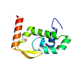
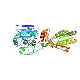
 | | Structural basis of phospho-peptide recognition by the BRCT domain of BRCA1, structure with phosphopeptide | | 分子名称: | BRCTide-7PS, Breast cancer type 1 susceptibility protein | | 著者 | Williams, R.S, Lee, M.S, Hau, D.D, Glover, J.N.M. | | 登録日 | 2004-04-22 | | 公開日 | 2004-05-11 | | 最終更新日 | 2011-07-13 | | 実験手法 | X-RAY DIFFRACTION (3.3 Å) | | 主引用文献 | Structural basis of phosphopeptide recognition by the BRCT domain of BRCA1
Nat.Struct.Mol.Biol., 11, 2004
|
|
7RGT
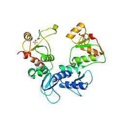
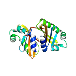
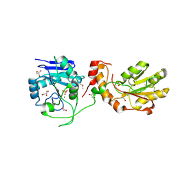
 | | The crystal structure of RocC, containing FinO domain, 1-126 | | 分子名称: | Repressor of competence, RNA Chaperone, SULFATE ION | | 著者 | Kim, H.J, Edwards, R.A, Glover, J.N.M. | | 登録日 | 2021-07-15 | | 公開日 | 2022-11-09 | | 最終更新日 | 2024-04-03 | | 実験手法 | X-RAY DIFFRACTION (2.02 Å) | | 主引用文献 | Structural basis for recognition of transcriptional terminator structures by ProQ/FinO domain RNA chaperones.
Nat Commun, 13, 2022
|
|
7RGS
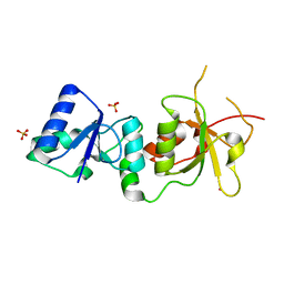
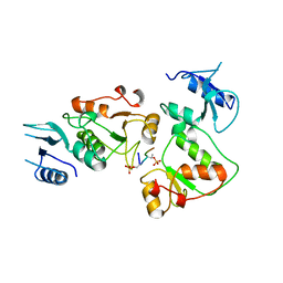
 | | The crystal structure of RocC, containing FinO domain, 24-126 | | 分子名称: | Repressor of competence, RNA Chaperone | | 著者 | Kim, H.J, Edwards, R.A, Glover, J.N.M. | | 登録日 | 2021-07-15 | | 公開日 | 2022-11-09 | | 最終更新日 | 2023-10-25 | | 実験手法 | X-RAY DIFFRACTION (2.1 Å) | | 主引用文献 | Structural basis for recognition of transcriptional terminator structures by ProQ/FinO domain RNA chaperones.
Nat Commun, 13, 2022
|
|
7RGU
 | |
4KQD
 | |
8D34
 | |
2F9D
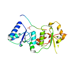
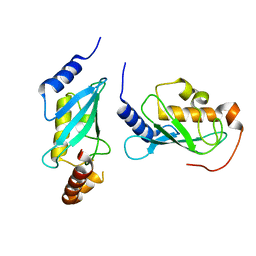
 | | 2.5 angstrom resolution structure of the spliceosomal protein p14 bound to region of SF3b155 | | 分子名称: | Pre-mRNA branch site protein p14, Splicing factor 3B subunit 1 | | 著者 | Schellenberg, M.J, Edwards, R.A, Ritchie, D.B, Glover, J.N.M, Macmillan, A.M. | | 登録日 | 2005-12-05 | | 公開日 | 2006-01-24 | | 最終更新日 | 2017-10-18 | | 実験手法 | X-RAY DIFFRACTION (2.5 Å) | | 主引用文献 | Crystal structure of a core spliceosomal protein interface
Proc.Natl.Acad.Sci.Usa, 103, 2006
|
|
1MN4
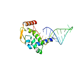
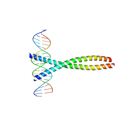
 | | Structure of Ndt80 (Residues 59-340) DNA-binding domain core | | 分子名称: | NDT80 PROTEIN | | 著者 | Lamoureux, J.S, Stuart, D, Tsang, R, Wu, C, Glover, J.N.M. | | 登録日 | 2002-09-04 | | 公開日 | 2002-11-06 | | 最終更新日 | 2024-02-14 | | 実験手法 | X-RAY DIFFRACTION (2.2 Å) | | 主引用文献 | Structure of the sporulation-specific transcription factor Ndt80 bound to DNA
Embo J., 21, 2002
|
|
1N5O
 | |
3D8A
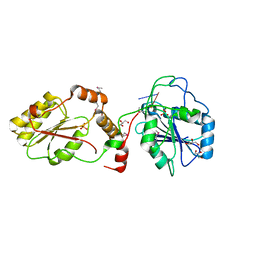
 | | Co-crystal structure of TraM-TraD complex. | | 分子名称: | Protein traD, Relaxosome protein TraM | | 著者 | Glover, J.N.M, Lu, J, Wong, J.J, Edwards, R.A. | | 登録日 | 2008-05-22 | | 公開日 | 2008-09-09 | | 最終更新日 | 2023-08-30 | | 実験手法 | X-RAY DIFFRACTION (2.55 Å) | | 主引用文献 | Structural basis of specific TraD-TraM recognition during F plasmid-mediated bacterial conjugation.
Mol.Microbiol., 70, 2008
|
|
1FOS
 | | TWO HUMAN C-FOS:C-JUN:DNA COMPLEXES | | 分子名称: | C-JUN PROTO-ONCOGENE PROTEIN, DNA (5'-D(*AP*AP*TP*GP*GP*AP*TP*GP*AP*GP*TP*CP*AP*TP*AP*GP*GP*AP*GP*A)-3'), DNA (5'-D(*TP*TP*CP*TP*CP*CP*TP*AP*TP*GP*AP*CP*TP*CP*AP*TP*CP*CP*AP*T)-3'), ... | | 著者 | Glover, J.N.M, Harrison, S.C. | | 登録日 | 1995-03-07 | | 公開日 | 1995-07-10 | | 最終更新日 | 2024-02-07 | | 実験手法 | X-RAY DIFFRACTION (3.05 Å) | | 主引用文献 | Crystal structure of the heterodimeric bZIP transcription factor c-Fos-c-Jun bound to DNA.
Nature, 373, 1995
|
|
8UK7
 | |
4WHV
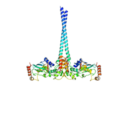 | | E3 ubiquitin-protein ligase RNF8 in complex with Ubiquitin-conjugating enzyme E2 N and Polyubiquitin-B | | 分子名称: | E3 ubiquitin-protein ligase RNF8, Polyubiquitin-B, Ubiquitin-conjugating enzyme E2 N, ... | | 著者 | Hodge, C.D, Edwards, R.A, Glover, J.N.M. | | 登録日 | 2014-09-23 | | 公開日 | 2015-09-30 | | 最終更新日 | 2023-09-27 | | 実験手法 | X-RAY DIFFRACTION (8.3 Å) | | 主引用文献 | RNF8 E3 Ubiquitin Ligase Stimulates Ubc13 E2 Conjugating Activity That Is Essential for DNA Double Strand Break Signaling and BRCA1 Tumor Suppressor Recruitment.
J.Biol.Chem., 291, 2016
|
|
2RH3
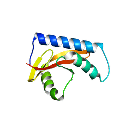
 | | Crystal structure of plasmid pTiC58 VirC2 | | 分子名称: | Protein virC2 | | 著者 | Lu, J, Glover, J.N.M. | | 登録日 | 2007-10-05 | | 公開日 | 2008-10-14 | | 最終更新日 | 2024-02-21 | | 実験手法 | X-RAY DIFFRACTION (1.7 Å) | | 主引用文献 | Agrobacterium tumefaciens VirC2 enhances T-DNA transfer and virulence through its C-terminal ribbon-helix-helix DNA-binding fold
Proc.Natl.Acad.Sci.USA, 106, 2009
|
|
4NRG
 | |
3U7G
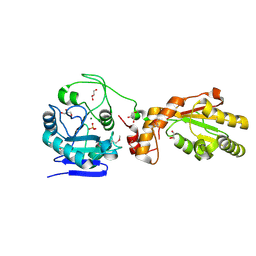
 | | Crystal structure of mPNKP catalytic fragment (D170A) bound to single-stranded DNA (TCCTAp) | | 分子名称: | Bifunctional polynucleotide phosphatase/kinase, DNA, GLYCEROL, ... | | 著者 | Coquelle, N, Havali, Z, Bernstein, N, Green, R, Glover, J.N.M. | | 登録日 | 2011-10-13 | | 公開日 | 2011-12-14 | | 最終更新日 | 2017-11-08 | | 実験手法 | X-RAY DIFFRACTION (2.1 Å) | | 主引用文献 | Structural basis for the phosphatase activity of polynucleotide kinase/phosphatase on single- and double-stranded DNA substrates.
Proc.Natl.Acad.Sci.USA, 108, 2011
|
|
3U7E
 | | Crystal structure of mPNKP catalytic fragment (D170A) | | 分子名称: | Bifunctional polynucleotide phosphatase/kinase, GLYCEROL, MAGNESIUM ION, ... | | 著者 | Coquelle, N, Havali, Z, Bernstein, N, Green, R, Glover, J.N.M. | | 登録日 | 2011-10-13 | | 公開日 | 2011-12-14 | | 最終更新日 | 2023-12-06 | | 実験手法 | X-RAY DIFFRACTION (1.7 Å) | | 主引用文献 | Structural basis for the phosphatase activity of polynucleotide kinase/phosphatase on single- and double-stranded DNA substrates.
Proc.Natl.Acad.Sci.USA, 108, 2011
|
|
4ONM
 | | Crystal structure of human Mms2/Ubc13 - NSC697923 | | 分子名称: | 2-[(4-methylphenyl)sulfonyl]-5-nitrofuran, GLYCEROL, Ubiquitin-conjugating enzyme E2 N, ... | | 著者 | Hodge, C.D, Edwards, R.A, Glover, J.N.M. | | 登録日 | 2014-01-28 | | 公開日 | 2015-05-06 | | 最終更新日 | 2017-11-22 | | 実験手法 | X-RAY DIFFRACTION (1.35 Å) | | 主引用文献 | Covalent Inhibition of Ubc13 Affects Ubiquitin Signaling and Reveals Active Site Elements Important for Targeting.
Acs Chem.Biol., 10, 2015
|
|
3U7F
 | | Crystal structure of mPNKP catalytic fragment (D170A) bound to single-stranded DNA (TCCTCp) | | 分子名称: | Bifunctional polynucleotide phosphatase/kinase, DNA, GLYCEROL, ... | | 著者 | Coquelle, N, Havali, Z, Bernstein, N, Green, R, Glover, J.N.M. | | 登録日 | 2011-10-13 | | 公開日 | 2011-12-14 | | 最終更新日 | 2017-11-08 | | 実験手法 | X-RAY DIFFRACTION (1.8 Å) | | 主引用文献 | Structural basis for the phosphatase activity of polynucleotide kinase/phosphatase on single- and double-stranded DNA substrates.
Proc.Natl.Acad.Sci.USA, 108, 2011
|
|
3U7H
 | | Crystal structure of mPNKP catalytic fragment (D170A) bound to single-stranded DNA (TCCTTp) | | 分子名称: | Bifunctional polynucleotide phosphatase/kinase, DNA, GLYCEROL, ... | | 著者 | Coquelle, N, Havali, Z, Bernstein, N, Green, R, Glover, J.N.M. | | 登録日 | 2011-10-13 | | 公開日 | 2011-12-14 | | 最終更新日 | 2017-11-08 | | 実験手法 | X-RAY DIFFRACTION (2 Å) | | 主引用文献 | Structural basis for the phosphatase activity of polynucleotide kinase/phosphatase on single- and double-stranded DNA substrates.
Proc.Natl.Acad.Sci.USA, 108, 2011
|
|
3UEO
 | |
3UEN
 | |


