4U0W
 
 | |
4U0Y
 | |
4UAI
 | |
7RU7
 | | Crystal structure of BtrK, a decarboxylase involved in butirosin biosynthesis | | 分子名称: | DI(HYDROXYETHYL)ETHER, L-glutamyl-[BtrI acyl-carrier protein] decarboxylase, PYRIDOXAL-5'-PHOSPHATE | | 著者 | Arenas, L.A.R, Paiva, F.C.R, Huang, F, Leadlay, P, Dias, M.V.B. | | 登録日 | 2021-08-16 | | 公開日 | 2022-08-17 | | 最終更新日 | 2023-10-18 | | 実験手法 | X-RAY DIFFRACTION (1.4 Å) | | 主引用文献 | Crystal structure of BtrK, a decarboxylase involved in the (S)-4-amino-2-hydroxybutyrate (AHBA) formation during butirosin biosynthesis
J.Mol.Struct., 1267, 2022
|
|
6D25
 | | Crystal structure of the GH51 arabinofuranosidase from Xanthomonas axonopodis pv. citri | | 分子名称: | Alpha-L-arabinosidase, GLYCEROL | | 著者 | Santos, C.R, Morais, M.A.B, Tonoli, C.C.C, Giuseppe, P.O, Murakami, M.T. | | 登録日 | 2018-04-13 | | 公開日 | 2019-02-20 | | 最終更新日 | 2023-10-04 | | 実験手法 | X-RAY DIFFRACTION (1.91 Å) | | 主引用文献 | The mechanism by which a distinguishing arabinofuranosidase can cope with internal di-substitutions in arabinoxylans.
Biotechnol Biofuels, 11, 2018
|
|
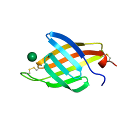
1A7S
 | | ATOMIC RESOLUTION STRUCTURE OF HBP | | 分子名称: | 2-acetamido-2-deoxy-beta-D-glucopyranose, 2-acetamido-2-deoxy-beta-D-glucopyranose-(1-4)-2-acetamido-2-deoxy-beta-D-glucopyranose, CHLORIDE ION, ... | | 著者 | Karlsen, S, Iversen, L.F, Larsen, I.K, Flodgaard, H.J, Kastrup, J.S. | | 登録日 | 1998-03-17 | | 公開日 | 1999-03-23 | | 最終更新日 | 2020-07-29 | | 実験手法 | X-RAY DIFFRACTION (1.12 Å) | | 主引用文献 | Atomic resolution structure of human HBP/CAP37/azurocidin.
Acta Crystallogr.,Sect.D, 54, 1998
|
|
7P4U
 | |
1AWC
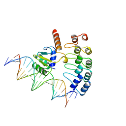 | | MOUSE GABP ALPHA/BETA DOMAIN BOUND TO DNA | | 分子名称: | DNA (5'-D(*AP*AP*(BRU)P*GP*AP*CP*CP*GP*GP*AP*AP*GP*TP*AP*(CBR)P*AP*CP*(CBR)P*GP*GP*A)-3'), DNA (5'-D(*TP*TP*CP*CP*GP*GP*(BRU)P*GP*(BRU)P*AP*CP*TP*TP*CP*CP*GP*GP*TP*CP*AP*T)-3'), PROTEIN (GA BINDING PROTEIN ALPHA), ... | | 著者 | Batchelor, A.H, Wolberger, C. | | 登録日 | 1997-10-01 | | 公開日 | 1998-03-18 | | 最終更新日 | 2024-02-07 | | 実験手法 | X-RAY DIFFRACTION (2.15 Å) | | 主引用文献 | The structure of GABPalpha/beta: an ETS domain- ankyrin repeat heterodimer bound to DNA.
Science, 279, 1998
|
|
7SKC
 | |
4XQM
 | |
7SOL
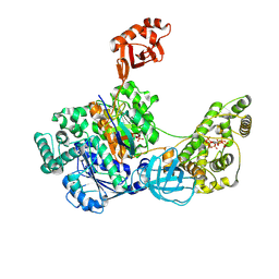 | | Crystal Structures of the bispecific ubiquitin/FAT10 activating enzyme, Uba6 | | 分子名称: | ADENOSINE MONOPHOSPHATE, INOSITOL HEXAKISPHOSPHATE, Ubiquitin, ... | | 著者 | Olsen, S.K, Gao, F, Lv, Z. | | 登録日 | 2021-10-31 | | 公開日 | 2022-11-02 | | 最終更新日 | 2024-04-17 | | 実験手法 | X-RAY DIFFRACTION (2.25000644 Å) | | 主引用文献 | Crystal structures reveal catalytic and regulatory mechanisms of the dual-specificity ubiquitin/FAT10 E1 enzyme Uba6.
Nat Commun, 13, 2022
|
|
7PON
 | |
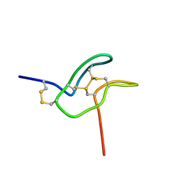
7PNO
 | | C terminal domain of Nipah Virus Phosphoprotein fused to the Ntail alpha more of the Nucleoprotein. | | 分子名称: | Phosphoprotein, alpha MoRE of Nipah virus Nucleoprotein tail | | 著者 | Bourhis, J.M, Yabukaski, F, Tarbouriech, N, Jamin, M. | | 登録日 | 2021-09-07 | | 公開日 | 2022-04-20 | | 最終更新日 | 2022-04-27 | | 実験手法 | X-RAY DIFFRACTION (2.79 Å) | | 主引用文献 | Structural Dynamics of the C-terminal X Domain of Nipah and Hendra Viruses Controls the Attachment to the C-terminal Tail of the Nucleocapsid Protein.
J.Mol.Biol., 434, 2022
|
|
4YNK
 | | Crystal structure of vitamin D receptor ligand binding domain complexed with a 19-norvitamin D compound | | 分子名称: | (1R,3R,7E,17beta)-17-{(5S)-5-hydroxy-5-[(3R,5R,7R)-tricyclo[3.3.1.1~3,7~]dec-1-yl]penta-1,3-diyn-1-yl}-2-methylidene-9,10-secoestra-5,7-diene-1,3-diol, Coactivator peptide drip from cDNA FLJ50196, highly similar to Peroxisome proliferator-activated receptor-binding protein, ... | | 著者 | Watarai, Y, Ikura, T, Ito, N. | | 登録日 | 2015-03-10 | | 公開日 | 2016-01-20 | | 最終更新日 | 2023-11-08 | | 実験手法 | X-RAY DIFFRACTION (2.3 Å) | | 主引用文献 | Synthesis, Biological Activities, and X-ray Crystal Structural Analysis of 25-Hydroxy-25(or 26)-adamantyl-17-[20(22),23-diynyl]-21-norvitamin D Compounds
J.Med.Chem., 58, 2015
|
|
5D9G
 | | Crystal structure of TIPRL, TOR signaling pathway regulator-like, in complex with peptide | | 分子名称: | CHLORIDE ION, NICKEL (II) ION, PHOSPHATE ION, ... | | 著者 | Scorsato, V, Sandy, J, Brandao-Neto, J, Pereira, H.M, Smetana, J.H.C, Aparicio, R. | | 登録日 | 2015-08-18 | | 公開日 | 2016-08-10 | | 最終更新日 | 2020-01-01 | | 実験手法 | X-RAY DIFFRACTION (2.15 Å) | | 主引用文献 | Crystal structure of the human Tip41 orthologue, TIPRL, reveals a novel fold and a binding site for the PP2Ac C-terminus.
Sci Rep, 6, 2016
|
|
3U79
 | | AL-103 Y32F Y96F | | 分子名称: | 2-(N-MORPHOLINO)-ETHANESULFONIC ACID, ACETATE ION, Amyloidogenic immunoglobulin light chain protein AL-103 Y32F Y96F, ... | | 著者 | DiCostanzo, A.C, Thompson, J.R, Ramirez-Alvarado, M. | | 登録日 | 2011-10-13 | | 公開日 | 2012-07-04 | | 実験手法 | X-RAY DIFFRACTION (1.62 Å) | | 主引用文献 | Tyrosine Residues mediate crucial interactions in amyloid formation for immunoglobulin light chains
To be Published
|
|
5L16
 | | Crystal Structure of N-terminus truncated selenophosphate synthetase from Leishmania major | | 分子名称: | Putative selenophosphate synthetase, SULFATE ION | | 著者 | Faim, L.M, Silva, I.R, Pereira, H.M, Dias, M.B, Silva, M.T.A, Brandao-Neto, J, Thiemann, O.H. | | 登録日 | 2016-07-28 | | 公開日 | 2017-08-09 | | 最終更新日 | 2023-10-04 | | 実験手法 | X-RAY DIFFRACTION (1.882 Å) | | 主引用文献 | Trypanosomatid selenophosphate synthetase structure, function and interaction with selenocysteine lyase.
Plos Negl Trop Dis, 14, 2020
|
|
5M6Q
 | | Crystal Structure of Kutzneria albida transglutaminase | | 分子名称: | 2-(N-MORPHOLINO)-ETHANESULFONIC ACID, CHLORIDE ION, TETRAETHYLENE GLYCOL, ... | | 著者 | Steffen, W, Benz, J, Rudolph, M.G. | | 登録日 | 2016-10-25 | | 公開日 | 2017-07-26 | | 最終更新日 | 2024-01-17 | | 実験手法 | X-RAY DIFFRACTION (1.98 Å) | | 主引用文献 | Discovery of a microbial transglutaminase enabling highly site-specific labeling of proteins.
J. Biol. Chem., 292, 2017
|
|
5MQQ
 | |
3YPI
 | |
6WUX
 | |
1FDM
 | |
1G7F
 | | HUMAN PTP1B CATALYTIC DOMAIN COMPLEXED WITH PNU177496 | | 分子名称: | 2-{4-[(2S)-2-[({[(1S)-1-CARBOXY-2-PHENYLETHYL]AMINO}CARBONYL)AMINO]-3-OXO-3-(PENTYLAMINO)PROPYL]PHENOXY}MALONIC ACID, PROTEIN-TYROSINE PHOSPHATASE, NON-RECEPTOR TYPE 1 | | 著者 | Bleasdale, J.E, Ogg, D, Larsen, S.D. | | 登録日 | 2000-11-10 | | 公開日 | 2001-06-06 | | 最終更新日 | 2023-08-09 | | 実験手法 | X-RAY DIFFRACTION (1.8 Å) | | 主引用文献 | Small molecule peptidomimetics containing a novel phosphotyrosine bioisostere inhibit protein tyrosine phosphatase 1B and augment insulin action.
Biochemistry, 40, 2001
|
|
6XN0
 | | Crystal structure of GH43_1 enzyme from Xanthomonas citri | | 分子名称: | CALCIUM ION, DI(HYDROXYETHYL)ETHER, GLYCEROL, ... | | 著者 | Morais, M.A.B, Tonoli, C.C.C, Santos, C.R, Murakami, M.T. | | 登録日 | 2020-07-02 | | 公開日 | 2020-12-02 | | 最終更新日 | 2023-10-18 | | 実験手法 | X-RAY DIFFRACTION (1.709 Å) | | 主引用文献 | Two distinct catalytic pathways for GH43 xylanolytic enzymes unveiled by X-ray and QM/MM simulations.
Nat Commun, 12, 2021
|
|
6XN2
 | | Crystal structure of the GH43_1 enzyme from Xanthomonas citri complexed with xylotriose | | 分子名称: | CALCIUM ION, DI(HYDROXYETHYL)ETHER, GLYCEROL, ... | | 著者 | Morais, M.A.B, Tonoli, C.C.C, Santos, C.R, Murakami, M.T. | | 登録日 | 2020-07-02 | | 公開日 | 2020-12-02 | | 最終更新日 | 2023-10-18 | | 実験手法 | X-RAY DIFFRACTION (1.652 Å) | | 主引用文献 | Two distinct catalytic pathways for GH43 xylanolytic enzymes unveiled by X-ray and QM/MM simulations.
Nat Commun, 12, 2021
|
|


