3BV0
 
 | |
5ZZM
 | |
1YGY
 | |
8ABY
 | |
8AD1
 | |
8ACP
 | |
8ABZ
 | |
8AC2
 | |
8AC1
 | |
3DC2
 | |
3DDN
 | |
3FGN
 | |
3FMI
 | |
3DRD
 | |
3FMF
 | |
3DU4
 | |
3FPA
 | |
3DOD
 | |
3LV2
 | |
4QHT
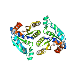
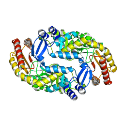
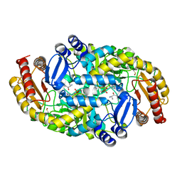
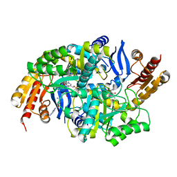
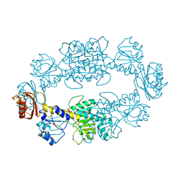
 | | Crystal structure of AAA+/ sigma 54 activator domain of the flagellar regulatory protein FlrC from Vibrio cholerae in ATP analog bound state | | 分子名称: | 1,2-ETHANEDIOL, Flagellar regulatory protein C, MAGNESIUM ION, ... | | 著者 | Dey, S, Biswas, M, Sen, U, Dasgupta, J. | | 登録日 | 2014-05-29 | | 公開日 | 2014-07-16 | | 最終更新日 | 2024-03-20 | | 実験手法 | X-RAY DIFFRACTION (2.559 Å) | | 主引用文献 | Unique ATPase site architecture triggers cis-mediated synchronized ATP binding in heptameric AAA+-ATPase domain of flagellar regulatory protein FlrC
J.Biol.Chem., 290, 2015
|
|
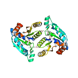
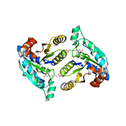
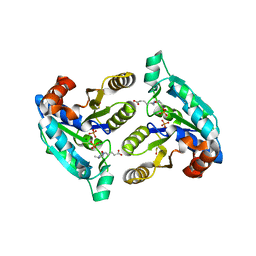
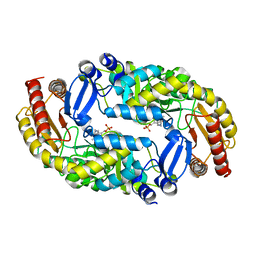
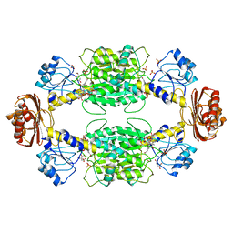
4QHS
 | | Crystal structure of AAA+sigma 54 activator domain of the flagellar regulatory protein FlrC of Vibrio cholerae in nucleotide free state | | 分子名称: | 1,2-ETHANEDIOL, Flagellar regulatory protein C | | 著者 | Dey, S, Biswas, M, Sen, U, Dasgupta, J. | | 登録日 | 2014-05-29 | | 公開日 | 2014-07-16 | | 最終更新日 | 2023-11-08 | | 実験手法 | X-RAY DIFFRACTION (2.3 Å) | | 主引用文献 | Unique ATPase site architecture triggers cis-mediated synchronized ATP binding in heptameric AAA+-ATPase domain of flagellar regulatory protein FlrC
J.Biol.Chem., 290, 2015
|
|
2P9C
 | |
2PA3
 | |
2P9G
 | |
2P9E
 | |


