Loading
PDBj
MenuPDBj@FacebookPDBj@TwitterPDBj@YouTubewwPDB FoundationwwPDB
RCSB PDBPDBeBMRBAdv. SearchSearch help
Search by PDB author
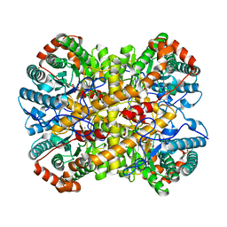
5NHM
DownloadVisualize
BU of 5nhm by Molmil
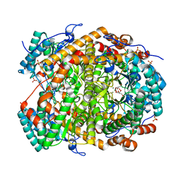
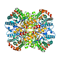
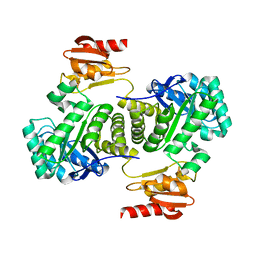
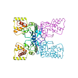
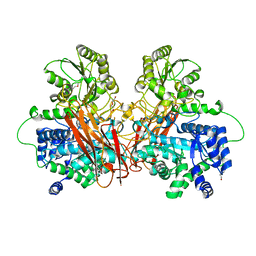
Crystal structure of apo xylose isomerase from Piromyces E2
Descriptor: ACETIC ACID, GLYCEROL, SULFATE ION, ...
Authors:Rozeboom, H.J, Janssen, D.B.
Deposit date:2017-03-21
Release date:2017-11-01
Last modified:2024-05-01
Method:X-RAY DIFFRACTION (1.67 Å)
Cite:Metal Dependence of the Xylose Isomerase from Piromyces sp. E2 Explored by Activity Profiling and Protein Crystallography.
Biochemistry, 56, 2017
5NH9
DownloadVisualize
BU of 5nh9 by Molmil
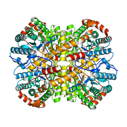
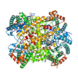
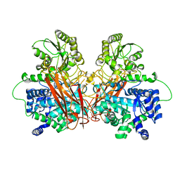
Crystal structure of xylose isomerase from Piromyces E2 in complex with two Mn2+ ions and xylose
Descriptor: D-xylose, MANGANESE (II) ION, SULFATE ION, ...
Authors:Rozeboom, H.J, Janssen, D.B.
Deposit date:2017-03-21
Release date:2017-11-01
Last modified:2024-05-01
Method:X-RAY DIFFRACTION (2.08 Å)
Cite:Metal Dependence of the Xylose Isomerase from Piromyces sp. E2 Explored by Activity Profiling and Protein Crystallography.
Biochemistry, 56, 2017
5NH7
DownloadVisualize
BU of 5nh7 by Molmil
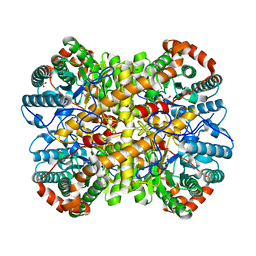
Crystal structure of xylose isomerase from Piromyces E2 in complex with two Mg2+ ions and xylose
Descriptor: D-xylose, MAGNESIUM ION, SULFATE ION, ...
Authors:Rozeboom, H.J, Janssen, D.B.
Deposit date:2017-03-21
Release date:2017-11-01
Last modified:2024-05-01
Method:X-RAY DIFFRACTION (1.9 Å)
Cite:Metal Dependence of the Xylose Isomerase from Piromyces sp. E2 Explored by Activity Profiling and Protein Crystallography.
Biochemistry, 56, 2017
5NHC
DownloadVisualize
BU of 5nhc by Molmil
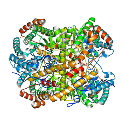
Crystal structure of xylose isomerase from Piromyces E2 in complex with two Co2+ ions and xylulose
Descriptor: 4-HYDROXYPROLINE, COBALT (II) ION, D-XYLULOSE, ...
Authors:Rozeboom, H.J, Janssen, D.B.
Deposit date:2017-03-21
Release date:2017-11-01
Last modified:2024-05-01
Method:X-RAY DIFFRACTION (1.93 Å)
Cite:Metal Dependence of the Xylose Isomerase from Piromyces sp. E2 Explored by Activity Profiling and Protein Crystallography.
Biochemistry, 56, 2017
5NH4
DownloadVisualize
BU of 5nh4 by Molmil
Crystal structure of xylose isomerase from Piromyces E2 in complex with one Mg2+ ions and glycerol
Descriptor: GLYCEROL, MAGNESIUM ION, SULFATE ION, ...
Authors:Rozeboom, H.J, Janssen, D.B.
Deposit date:2017-03-21
Release date:2017-11-01
Last modified:2024-01-17
Method:X-RAY DIFFRACTION (1.8 Å)
Cite:Metal Dependence of the Xylose Isomerase from Piromyces sp. E2 Explored by Activity Profiling and Protein Crystallography.
Biochemistry, 56, 2017
5NHD
DownloadVisualize
BU of 5nhd by Molmil
Crystal structure of xylose isomerase from Piromyces E2 in complex with 2 Ni2+ ions and xylose
Descriptor: D-xylose, NICKEL (II) ION, SULFATE ION, ...
Authors:Rozeboom, H.J, Janssen, D.B.
Deposit date:2017-03-21
Release date:2017-11-01
Last modified:2024-05-01
Method:X-RAY DIFFRACTION (1.8 Å)
Cite:Metal Dependence of the Xylose Isomerase from Piromyces sp. E2 Explored by Activity Profiling and Protein Crystallography.
Biochemistry, 56, 2017
5NHB
DownloadVisualize
BU of 5nhb by Molmil
Crystal structure of xylose isomerase from Piromyces E2 in complex with two Fe2+ ions
Descriptor: FE (II) ION, SULFATE ION, Xylose isomerase
Authors:Rozeboom, H.J, Janssen, D.B.
Deposit date:2017-03-21
Release date:2017-11-01
Last modified:2024-05-01
Method:X-RAY DIFFRACTION (2.4 Å)
Cite:Metal Dependence of the Xylose Isomerase from Piromyces sp. E2 Explored by Activity Profiling and Protein Crystallography.
Biochemistry, 56, 2017
5NHA
DownloadVisualize
BU of 5nha by Molmil
Crystal structure of xylose isomerase from Piromyces sp. E2 in complex with two Mn2+ ions and sorbitol
Descriptor: MANGANESE (II) ION, SULFATE ION, Xylose isomerase, ...
Authors:Rozeboom, H.J, Janssen, D.B.
Deposit date:2017-03-21
Release date:2017-11-01
Last modified:2024-05-01
Method:X-RAY DIFFRACTION (1.8 Å)
Cite:Metal Dependence of the Xylose Isomerase from Piromyces sp. E2 Explored by Activity Profiling and Protein Crystallography.
Biochemistry, 56, 2017
5NHE
DownloadVisualize
BU of 5nhe by Molmil
Crystal structure of xylose isomerase from Piromyces E2 in complex with two Cd2+ ions and xylose
Descriptor: CADMIUM ION, D-xylose, SULFATE ION, ...
Authors:Rozeboom, H.J, Janssen, D.B.
Deposit date:2017-03-21
Release date:2017-11-01
Last modified:2024-05-01
Method:X-RAY DIFFRACTION (1.86 Å)
Cite:Metal Dependence of the Xylose Isomerase from Piromyces sp. E2 Explored by Activity Profiling and Protein Crystallography.
Biochemistry, 56, 2017
5OHU
DownloadVisualize
BU of 5ohu by Molmil
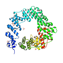
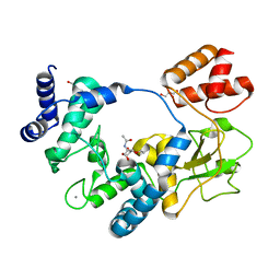
The X-ray Structure of Lytic Transglycosylase Slt from Pseudomonas aeruginosa
Descriptor: PHOSPHATE ION, Soluble lytic murein transglycosylase
Authors:Batuecas, M.T, Dominguez-Gil, T, Hermoso, J.A.
Deposit date:2017-07-18
Release date:2018-04-18
Last modified:2018-05-02
Method:X-RAY DIFFRACTION (2.2 Å)
Cite:Exolytic and endolytic turnover of peptidoglycan by lytic transglycosylase Slt ofPseudomonas aeruginosa.
Proc. Natl. Acad. Sci. U.S.A., 115, 2018
3NOY
DownloadVisualize
BU of 3noy by Molmil
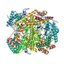
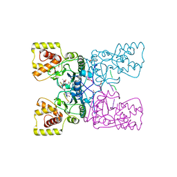
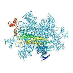
Crystal structure of IspG (gcpE)
Descriptor: 4-hydroxy-3-methylbut-2-en-1-yl diphosphate synthase, IRON/SULFUR CLUSTER
Authors:Groll, M, Graewert, T, Bacher, A.
Deposit date:2010-06-26
Release date:2010-11-17
Last modified:2023-12-27
Method:X-RAY DIFFRACTION (2.7 Å)
Cite:Biosynthesis of isoprenoids: crystal structure of the [4Fe-4S] cluster protein IspG.
J.Mol.Biol., 404, 2010
4BXE
DownloadVisualize
BU of 4bxe by Molmil
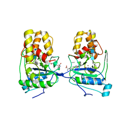
CRYSTAL STRUCTURE OF AMPDH3 FROM PSEUDOMONAS AERUGINOSA IN COMPLEX WITH ANHYDROMURAMIC PENTAPEPTIDE
Descriptor: ACETATE ION, AMPDH3, ANHYDROMURAMIC PEPTIDE, ...
Authors:Artola-Recolons, C, Hermoso, J.A.
Deposit date:2013-07-10
Release date:2013-10-09
Last modified:2024-05-01
Method:X-RAY DIFFRACTION (2.95 Å)
Cite:Cell-Wall Remodeling by the Zinc-Protease Ampdh3 from Pseudomonas Aeruginosa.
J.Am.Chem.Soc., 135, 2013
7LY9
DownloadVisualize
BU of 7ly9 by Molmil
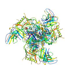
Cryo-EM structure of 2909 Fab in complex with 3BNC117 Fab and CAP256.wk34.c80 SOSIP.RnS2 N160K HIV-1 Env trimer
Descriptor: 2-acetamido-2-deoxy-beta-D-glucopyranose, 2-acetamido-2-deoxy-beta-D-glucopyranose-(1-4)-2-acetamido-2-deoxy-beta-D-glucopyranose, 2909 Heavy Chain, ...
Authors:Gorman, J, Kwong, P.D.
Deposit date:2021-03-06
Release date:2021-08-18
Last modified:2024-10-16
Method:ELECTRON MICROSCOPY (3.91 Å)
Cite:Extended antibody-framework-to-antigen distance observed exclusively with broad HIV-1-neutralizing antibodies recognizing glycan-dense surfaces.
Nat Commun, 12, 2021
4BXD
DownloadVisualize
BU of 4bxd by Molmil
CRYSTAL STRUCTURE OF AMPDH3 FROM PSEUDOMONAS AERUGINOSA IN COMPLEX WITH TETRASACCHARIDE PENTAPEPTIDE
Descriptor: 2-acetamido-2-deoxy-beta-D-glucopyranose-(1-4)-2-acetamido-2-deoxy-beta-D-glucopyranose-(1-4)-methyl 2-acetamido-2-deoxy-beta-D-glucopyranoside, AMPDH3, PEPTIDE, ...
Authors:Artola-Recolons, C, Hermoso, J.A.
Deposit date:2013-07-10
Release date:2013-10-09
Last modified:2024-10-16
Method:X-RAY DIFFRACTION (3.1 Å)
Cite:Cell-Wall Remodeling by the Zinc-Protease Ampdh3 from Pseudomonas Aeruginosa.
J.Am.Chem.Soc., 135, 2013
4BXJ
DownloadVisualize
BU of 4bxj by Molmil
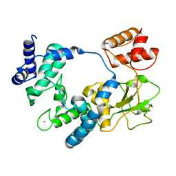
CRYSTAL STRUCTURE OF AMPDH3 FROM PSEUDOMONAS AERUGINOSA
Descriptor: AMPDH3, GLYCEROL
Authors:Carrasco-Lopez, C, Hermoso, J.A.
Deposit date:2013-07-12
Release date:2013-10-09
Last modified:2023-12-20
Method:X-RAY DIFFRACTION (2.35 Å)
Cite:Cell-Wall Remodeling by the Zinc-Protease Ampdh3 from Pseudomonas Aeruginosa.
J.Am.Chem.Soc., 135, 2013
5AO7
DownloadVisualize
BU of 5ao7 by Molmil
Crystal Structure of SltB3 from Pseudomonas aeruginosa in complex with NAG-anhNAM-pentapeptide
Descriptor: 2-(2-ACETYLAMINO-4-HYDROXY-6,8-DIOXA-BICYCLO[3.2.1]OCT-3-YLOXY)-PROPIONIC ACID, 2-AMINO-2-HYDROXYMETHYL-PROPANE-1,3-DIOL, 2-acetamido-2-deoxy-beta-D-glucopyranose, ...
Authors:Dominguez-Gil, T, Hermoso, J.A.
Deposit date:2015-09-09
Release date:2016-07-20
Last modified:2024-01-10
Method:X-RAY DIFFRACTION (2.09 Å)
Cite:Turnover of Bacterial Cell Wall by Sltb3, a Multidomain Lytic Transglycosylase of Pseudomonas Aeruginosa.
Acs Chem.Biol., 11, 2016
5AO8
DownloadVisualize
BU of 5ao8 by Molmil
Crystal Structure of SltB3 from Pseudomonas aeruginosa in complex with NAG-NAM-pentapeptide
Descriptor: 2-acetamido-2-deoxy-beta-D-glucopyranose, 2-acetamido-2-deoxy-beta-D-glucopyranose-(1-4)-methyl 2-acetamido-3-O-[(2R)-1-amino-1-oxopropan-2-yl]-2-deoxy-beta-D-glucopyranoside, CALCIUM ION, ...
Authors:Dominguez-Gil, T, Hermoso, J.A.
Deposit date:2015-09-09
Release date:2016-07-20
Last modified:2024-01-10
Method:X-RAY DIFFRACTION (2.23 Å)
Cite:Turnover of Bacterial Cell Wall by Sltb3, a Multidomain Lytic Transglycosylase of Pseudomonas Aeruginosa.
Acs Chem.Biol., 11, 2016
1YFM
DownloadVisualize
BU of 1yfm by Molmil
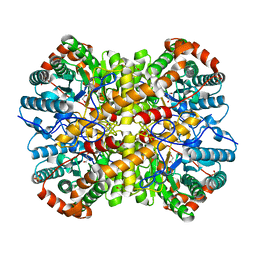
RECOMBINANT YEAST FUMARASE
Descriptor: FUMARASE
Authors:Weaver, T.M, Lees, M.R, Banaszak, L.J.
Deposit date:1998-01-07
Release date:1998-07-08
Last modified:2024-05-22
Method:X-RAY DIFFRACTION (2.6 Å)
Cite:Crystal structures of native and recombinant yeast fumarase.
J.Mol.Biol., 280, 1998
7O49
DownloadVisualize
BU of 7o49 by Molmil
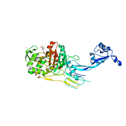
Crystal structure of Penicillin-Binding Protein 1 (PBP1) from Staphylococcus aureus
Descriptor: 4-(2-HYDROXYETHYL)-1-PIPERAZINE ETHANESULFONIC ACID, CADMIUM ION, CHLORIDE ION, ...
Authors:Martinez Caballero, S, Hermoso, J.A.
Deposit date:2021-04-05
Release date:2021-11-03
Last modified:2024-01-31
Method:X-RAY DIFFRACTION (3.03 Å)
Cite:Integrative structural biology of the penicillin-binding protein-1 from Staphylococcus aureus , an essential component of the divisome machinery.
Comput Struct Biotechnol J, 19, 2021
7O4A
DownloadVisualize
BU of 7o4a by Molmil
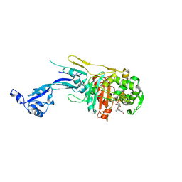
Crystal structure of Penicillin-Binding Protein 1 (PBP1) from Staphylococcus aureus in complex with piperacillin
Descriptor: Hydrolyzed piperacillin, Penicillin-binding protein 1
Authors:Martinez Caballero, S, Hermoso, J.A.
Deposit date:2021-04-05
Release date:2021-11-03
Last modified:2024-10-09
Method:X-RAY DIFFRACTION (3.028 Å)
Cite:Integrative structural biology of the penicillin-binding protein-1 from Staphylococcus aureus , an essential component of the divisome machinery.
Comput Struct Biotechnol J, 19, 2021
7O4C
DownloadVisualize
BU of 7o4c by Molmil
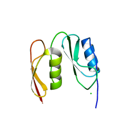
Crystal structure of PASTA domains of the Penicillin-Binding Protein 1 (PBP1) from Staphylococcus aureus
Descriptor: CHLORIDE ION, Penicillin-binding protein 1
Authors:Martinez Caballero, S, Hermoso, J.A.
Deposit date:2021-04-05
Release date:2021-11-03
Last modified:2024-01-31
Method:X-RAY DIFFRACTION (1.51 Å)
Cite:Integrative structural biology of the penicillin-binding protein-1 from Staphylococcus aureus , an essential component of the divisome machinery.
Comput Struct Biotechnol J, 19, 2021
4UW2
DownloadVisualize
BU of 4uw2 by Molmil
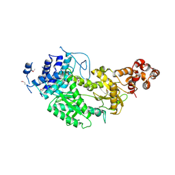
Crystal structure of Csm1 in T.onnurineus
Descriptor: CSM1
Authors:Jung, T.Y, An, Y, Park, K.H, Lee, M.H, Oh, B.H, Woo, E.J.
Deposit date:2014-08-08
Release date:2015-03-25
Last modified:2015-09-23
Method:X-RAY DIFFRACTION (2.632 Å)
Cite:Crystal Structure of the Csm1 Subunit of the Csm Complex and its Single-Stranded DNA-Specific Nuclease Activity.
Structure, 23, 2015
6R5V
DownloadVisualize
BU of 6r5v by Molmil
The crystal structure of Glycoside Hydrolase BglX inactive mutant D286N from P. aeruginosa in complex with xylotriose
Descriptor: DI(HYDROXYETHYL)ETHER, MAGNESIUM ION, Periplasmic beta-glucosidase, ...
Authors:Batuecas, M.T, Hermoso, J.A.
Deposit date:2019-03-25
Release date:2020-04-15
Last modified:2024-01-24
Method:X-RAY DIFFRACTION (1.8 Å)
Cite:Catalytic Cycle of Glycoside Hydrolase BglX fromPseudomonas aeruginosaand Its Implications for Biofilm Formation.
Acs Chem.Biol., 15, 2020
6R5P
DownloadVisualize
BU of 6r5p by Molmil
The crystal structure of Glycoside Hydrolase BglX inactive mutant D286N from P. aeruginosa in complex with glucose
Descriptor: MAGNESIUM ION, Periplasmic beta-glucosidase, beta-D-glucopyranose
Authors:Batuecas, M.T, Hermoso, J.A.
Deposit date:2019-03-25
Release date:2020-04-15
Last modified:2024-01-24
Method:X-RAY DIFFRACTION (2.85 Å)
Cite:Catalytic Cycle of Glycoside Hydrolase BglX fromPseudomonas aeruginosaand Its Implications for Biofilm Formation.
Acs Chem.Biol., 15, 2020
5C22
DownloadVisualize
BU of 5c22 by Molmil
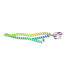
Crystal structure of Zn-bound HlyD from E. coli
Descriptor: Chromosomal hemolysin D, ZINC ION
Authors:Ha, N.C, Kim, J.S.
Deposit date:2015-06-15
Release date:2016-02-17
Last modified:2024-03-20
Method:X-RAY DIFFRACTION (2.302 Å)
Cite:Crystal Structure of a Soluble Fragment of the Membrane Fusion Protein HlyD in a Type I Secretion System of Gram-Negative Bacteria
Structure, 24, 2016

227111

PDB entries from 2024-11-06

PDB statisticsPDBj update infoContact PDBjnumon