Loading
PDBj
MenuPDBj@FacebookPDBj@TwitterPDBj@YouTubewwPDB FoundationwwPDB
RCSB PDBPDBeBMRBAdv. SearchSearch help
Search by PDB author
1ZKX
DownloadVisualize
BU of 1zkx by Molmil
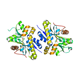
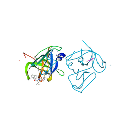
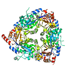
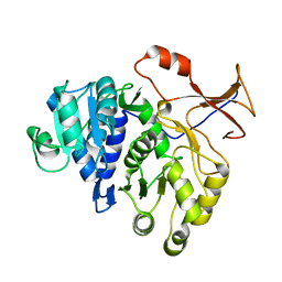
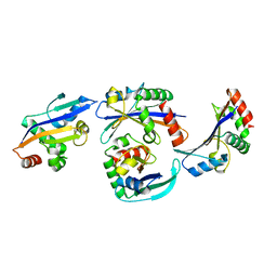
Crystal structure of Glu158Ala/Thr159Ala/Asn160Ala- a triple mutant of Clostridium botulinum neurotoxin E catalytic domain
Descriptor: CHLORIDE ION, ZINC ION, botulinum neurotoxin type E
Authors:Agarwal, R, Binz, T, Swaminathan, S.
Deposit date:2005-05-04
Release date:2005-07-05
Last modified:2023-08-23
Method:X-RAY DIFFRACTION (2.52 Å)
Cite:Analysis of Active Site Residues of Botulinum Neurotoxin E by Mutational, Functional, and Structural Studies: Glu335Gln Is an Apoenzyme.
Biochemistry, 44, 2005
2NXO
DownloadVisualize
BU of 2nxo by Molmil
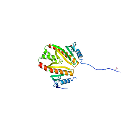
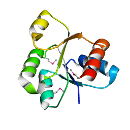
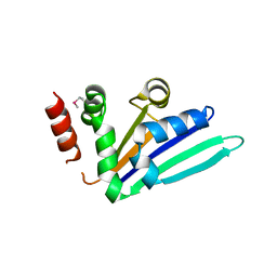
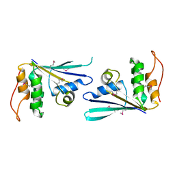
Crystal structure of protein SCO4506 from Streptomyces coelicolor, Pfam DUF178
Descriptor: Hypothetical protein SCO4506
Authors:Tyagi, R, Eswaramoorthy, S, Burley, S.K, Swaminathan, S, New York SGX Research Center for Structural Genomics (NYSGXRC)
Deposit date:2006-11-17
Release date:2006-12-19
Last modified:2023-12-27
Method:X-RAY DIFFRACTION (2.04 Å)
Cite:The crystal structure of a hypothetical protein SCO4506 (gene ID: Q9L0T8) from Streptomyces coelicolor to 2.04 Angstrom resolution
To be Published
1XCB
DownloadVisualize
BU of 1xcb by Molmil
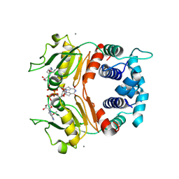
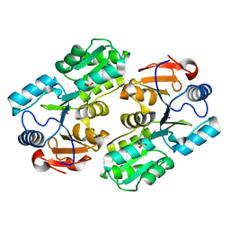
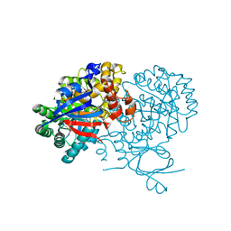
X-ray Structure of a Rex-Family Repressor/NADH Complex from Thermus Aquaticus
Descriptor: CALCIUM ION, NICOTINAMIDE-ADENINE-DINUCLEOTIDE, Redox-sensing transcriptional repressor rex
Authors:Sickmier, E.A, Brekasis, D, Paranawithana, S, Bonanno, J.B, Burley, S.K, Paget, M.S, Kielkopf, C.L, New York SGX Research Center for Structural Genomics (NYSGXRC)
Deposit date:2004-09-01
Release date:2004-09-28
Last modified:2021-02-03
Method:X-RAY DIFFRACTION (2.9 Å)
Cite:X-Ray Structure of a Rex-Family Repressor/NADH Complex: Insights into the Mechanism of Redox Sensing
Structure, 13, 2005
2PLG
DownloadVisualize
BU of 2plg by Molmil
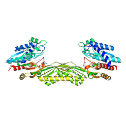
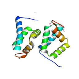
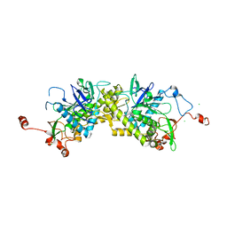
Crystal structure of T110839 protein from Synechococcus elongatus
Descriptor: Tll0839 protein
Authors:Madegowda, M, Eswaramoorthy, S, Burley, S.K, Swaminathan, S, New York SGX Research Center for Structural Genomics (NYSGXRC)
Deposit date:2007-04-19
Release date:2007-05-01
Last modified:2021-02-03
Method:X-RAY DIFFRACTION (2.6 Å)
Cite:Crystal structure of T110839 protein from Synechococcus elongatus.
To be Published
2POZ
DownloadVisualize
BU of 2poz by Molmil
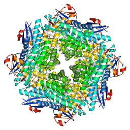
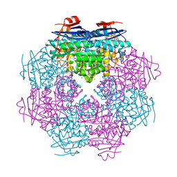
Crystal structure of a putative dehydratase from Mesorhizobium loti
Descriptor: Putative dehydratase
Authors:Sugadev, R, Eswaramoorthy, S, Burley, S.K, Swaminathan, S, New York SGX Research Center for Structural Genomics (NYSGXRC)
Deposit date:2007-04-27
Release date:2007-05-15
Last modified:2021-02-03
Method:X-RAY DIFFRACTION (2.04 Å)
Cite:Crystal structure of a putative dehydratase from Mesorhizobium loti.
To be Published
4IMZ
DownloadVisualize
BU of 4imz by Molmil
Structural Basis of Substrate Specificity and Protease Inhibition in Norwalk Virus
Descriptor: Genome polyprotein, SODIUM ION, THIOCYANATE ION, ...
Authors:Prasad, B.V.V, Muhaxhiri, Z, Deng, L, Shanker, S, Sankaran, B, Estes, M.K, Palzkill, T, Song, Y.
Deposit date:2013-01-03
Release date:2013-02-20
Last modified:2013-04-10
Method:X-RAY DIFFRACTION (1.7 Å)
Cite:Structural basis of substrate specificity and protease inhibition in norwalk virus.
J.Virol., 87, 2013
2QVG
DownloadVisualize
BU of 2qvg by Molmil
The crystal structure of a two-component response regulator from Legionella pneumophila
Descriptor: Two component response regulator
Authors:Tyagi, R, Eswaramoorthy, S, Burley, S.K, Swaminathan, S, New York SGX Research Center for Structural Genomics (NYSGXRC)
Deposit date:2007-08-08
Release date:2007-08-28
Last modified:2024-10-09
Method:X-RAY DIFFRACTION (1.5 Å)
Cite:The crystal structure of a two-component response regulator from Legionella pneumophila.
To be Published
1YV9
DownloadVisualize
BU of 1yv9 by Molmil
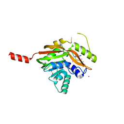
Crystal structure of a HAD-like phosphatase from Enterococcus faecalis V583
Descriptor: PHOSPHATE ION, hydrolase, haloacid dehalogenase family
Authors:Seetharaman, J, Swaminathan, S, Burley, S.K, New York SGX Research Center for Structural Genomics (NYSGXRC)
Deposit date:2005-02-15
Release date:2005-03-15
Last modified:2023-08-23
Method:X-RAY DIFFRACTION (2.8 Å)
Cite:Crystal Structure of a Hypothetical protein, hydrolase haloacid dehalogenase-like family
To be Published
1Z2L
DownloadVisualize
BU of 1z2l by Molmil
Crystal structure of Allantoate-amidohydrolase from E.coli K12 in complex with substrate Allantoate
Descriptor: ALLANTOATE ION, Allantoate amidohydrolase, SULFATE ION, ...
Authors:Agarwal, R, Swaminathan, S, Burley, S.K, New York SGX Research Center for Structural Genomics (NYSGXRC)
Deposit date:2005-03-08
Release date:2005-03-22
Last modified:2024-02-14
Method:X-RAY DIFFRACTION (2.25 Å)
Cite:Structural analysis of a ternary complex of allantoate amidohydrolase from Escherichia coli reveals its mechanics.
J.Mol.Biol., 368, 2007
1ZCC
DownloadVisualize
BU of 1zcc by Molmil
Crystal structure of glycerophosphodiester phosphodiesterase from Agrobacterium tumefaciens str.C58
Descriptor: ACETATE ION, SULFATE ION, glycerophosphodiester phosphodiesterase
Authors:Krishnamurthy, N.R, Kumaran, D, Swaminathan, S, Burley, S.K, New York SGX Research Center for Structural Genomics (NYSGXRC)
Deposit date:2005-04-11
Release date:2005-05-03
Last modified:2024-02-14
Method:X-RAY DIFFRACTION (2.5 Å)
Cite:Crystal structure of glycerophosphodiester phosphodiesterase from Agrobacterium tumefaciens by SAD with a large asymmetric unit.
Proteins, 65, 2006
1BNZ
DownloadVisualize
BU of 1bnz by Molmil
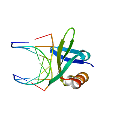
SSO7D HYPERTHERMOPHILE PROTEIN/DNA COMPLEX
Descriptor: 5'-D(*GP*TP*AP*AP*TP*TP*AP*C)-3', DNA-BINDING PROTEIN 7A
Authors:Gao, Y.-G, Su, S.-Y, Robinson, H, Padmanabhan, S, Lim, L, Mccrary, B.S, Edmondos, S.P, Shrive, J.W, Wang, A.H.-J.
Deposit date:1998-07-31
Release date:1998-11-11
Last modified:2024-02-07
Method:X-RAY DIFFRACTION (2 Å)
Cite:The crystal structure of the hyperthermophile chromosomal protein Sso7d bound to DNA.
Nat.Struct.Biol., 5, 1998
2A97
DownloadVisualize
BU of 2a97 by Molmil
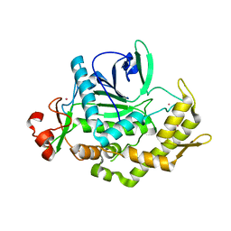
Crystal structure of catalytic domain of Clostridium botulinum neurotoxin serotype F
Descriptor: Botulinum neurotoxin type F, CADMIUM ION, ZINC ION
Authors:Agarwal, R, Binz, T, Swaminathan, S.
Deposit date:2005-07-11
Release date:2005-09-20
Last modified:2023-08-23
Method:X-RAY DIFFRACTION (1.8 Å)
Cite:Structural analysis of botulinum neurotoxin serotype f light chain: implications on substrate binding and inhibitor design
Biochemistry, 44, 2005
2PBE
DownloadVisualize
BU of 2pbe by Molmil
Crystal structure of an aminoglycoside 6-adenyltransferase from Bacillus subtilis
Descriptor: Aminoglycoside 6-adenylyltransferase
Authors:Tyagi, R, Eswaramoorthy, S, Burley, S.K, Swaminathan, S, New York SGX Research Center for Structural Genomics (NYSGXRC)
Deposit date:2007-03-28
Release date:2007-04-10
Last modified:2021-02-03
Method:X-RAY DIFFRACTION (2.65 Å)
Cite:The crystal structure of an aminoglycoside 6-adenyltransferase from Bacillus subtilis
To be Published
4M46
DownloadVisualize
BU of 4m46 by Molmil
Crystal structure of a green-emitter native of Lampyris turkestanicus luciferase
Descriptor: Luciferase
Authors:Sharafian, Z, Hosseinkhani, S, Naderi-manesh, H.
Deposit date:2013-08-06
Release date:2013-10-30
Last modified:2023-09-20
Method:X-RAY DIFFRACTION (2.7 Å)
Cite:Crystal structure of native and a mutant of Lampyris turkestanicus luciferase implicate in bioluminescence color shift.
Biochim.Biophys.Acta, 1834, 2013
3VYJ
DownloadVisualize
BU of 3vyj by Molmil
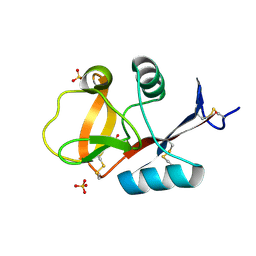
Crystal structure of C-type lectin domain of murine dendritic cell inhibitory receptor 2 (apo form)
Descriptor: C-type lectin domain family 4, member a4, SULFATE ION
Authors:Nagae, M, Yamanaka, K, Hanashima, S, Ikeda, A, Satoh, T, Matsumoto, N, Yamamoto, K, Yamaguchi, Y.
Deposit date:2012-09-26
Release date:2013-10-02
Last modified:2013-12-11
Method:X-RAY DIFFRACTION (2.15 Å)
Cite:Recognition of Bisecting N-Acetylglucosamine: STRUCTURAL BASIS FOR ASYMMETRIC INTERACTION WITH THE MOUSE LECTIN DENDRITIC CELL INHIBITORY RECEPTOR 2
J.Biol.Chem., 288, 2013
2N3O
DownloadVisualize
BU of 2n3o by Molmil
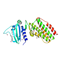
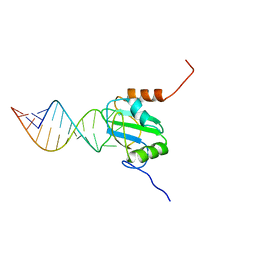
Structure of PTB RRM1(41-163) bound to an RNA stemloop containing a structured loop derived from viral internal ribosomal entry site RNA
Descriptor: Polypyrimidine tract-binding protein 1, RNA (5'-R(*GP*GP*GP*AP*CP*CP*UP*GP*GP*UP*CP*UP*UP*UP*CP*CP*AP*GP*GP*UP*CP*CP*C)-3')
Authors:Maris, C, Jayne, S.F, Damberger, F.F, Ravindranathan, S, Allain, F.H.-T.
Deposit date:2015-06-08
Release date:2016-08-10
Last modified:2024-05-01
Method:SOLUTION NMR
Cite:C-terminal helix folding upon pyrimidine-rich hairpin binding to PTB RRM1. Implications for PTB function in Encephalomyocarditis virus IRES activity.
To be Published
3VYK
DownloadVisualize
BU of 3vyk by Molmil
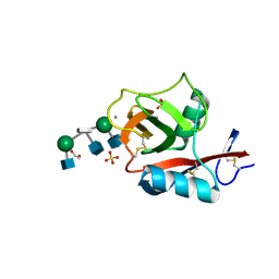
Crystal structure of C-type lectin domain of murine dendritic cell inhibitory receptor 2 in complex with N-glycan
Descriptor: 1,2-ETHANEDIOL, 2-acetamido-2-deoxy-beta-D-glucopyranose-(1-2)-alpha-D-mannopyranose-(1-3)-[2-acetamido-2-deoxy-beta-D-glucopyranose-(1-2)-alpha-D-mannopyranose-(1-6)][2-acetamido-2-deoxy-beta-D-glucopyranose-(1-4)]methyl alpha-D-mannopyranoside, C-type lectin domain family 4, ...
Authors:Nagae, M, Yamanaka, K, Hanashima, S, Ikeda, A, Satoh, T, Matsumoto, N, Yamamoto, K, Yamaguchi, Y.
Deposit date:2012-09-26
Release date:2013-10-02
Last modified:2020-07-29
Method:X-RAY DIFFRACTION (1.5 Å)
Cite:Recognition of Bisecting N-Acetylglucosamine: STRUCTURAL BASIS FOR ASYMMETRIC INTERACTION WITH THE MOUSE LECTIN DENDRITIC CELL INHIBITORY RECEPTOR 2
J.Biol.Chem., 288, 2013
2NRQ
DownloadVisualize
BU of 2nrq by Molmil
Crystal structure of protein SSO0741 from Sulfolobus solfataricus, Pfam DUF54
Descriptor: Hypothetical protein ORF-c20_032
Authors:Rao, K.N, Burley, S.K, Swaminathan, S, New York SGX Research Center for Structural Genomics (NYSGXRC)
Deposit date:2006-11-02
Release date:2006-11-28
Last modified:2023-12-27
Method:X-RAY DIFFRACTION (2.6 Å)
Cite:UPF201 archaeal specific family members reveal structural similarity to RNA-binding proteins but low likelihood for RNA-binding function.
Plos One, 3, 2008
2OY9
DownloadVisualize
BU of 2oy9 by Molmil
Crystal structure of an unknown conserved protein- Pfam: UPF0223
Descriptor: MAGNESIUM ION, UPF0223 protein BH2638
Authors:Sugadev, R, Burley, S.K, Swaminathan, S, New York SGX Research Center for Structural Genomics (NYSGXRC)
Deposit date:2007-02-21
Release date:2007-03-20
Last modified:2021-02-03
Method:X-RAY DIFFRACTION (1.6 Å)
Cite:Crystal structure of an unknown conserved protein - Pfam: UPF0223
To be Published
2P0L
DownloadVisualize
BU of 2p0l by Molmil
Crystal structure of a Lipoate-protein ligase A
Descriptor: Lipoate-protein ligase A
Authors:Sugadev, R, Burley, S.K, Swaminathan, S, New York SGX Research Center for Structural Genomics (NYSGXRC)
Deposit date:2007-02-28
Release date:2007-03-20
Last modified:2024-10-09
Method:X-RAY DIFFRACTION (2.04 Å)
Cite:Crystal structure of a Lipoate-protein ligase A
To be Published
4NEC
DownloadVisualize
BU of 4nec by Molmil
Conversion of a Disulfide Bond into a Thioacetal Group during Echinomycin Biosynthesis
Descriptor: 2-CARBOXYQUINOXALINE, ACETATE ION, Echinomycin, ...
Authors:Hotta, K, Keegan, R.M, Ranganathan, S, Fang, M, Bibby, J, Winn, M.D, Sato, M, Lian, M, Watanabe, K, Rigden, D.J, Kim, C.-Y.
Deposit date:2013-10-29
Release date:2014-01-15
Last modified:2024-04-03
Method:X-RAY DIFFRACTION (1.5 Å)
Cite:Conversion of a disulfide bond into a thioacetal group during echinomycin biosynthesis.
Angew.Chem.Int.Ed.Engl., 53, 2014
2OG9
DownloadVisualize
BU of 2og9 by Molmil
Crystal Structure of mandelate racemase/muconate lactonizing enzyme from Polaromonas sp. JS666
Descriptor: CALCIUM ION, Mandelate racemase/muconate lactonizing enzyme
Authors:Kumaran, D, Burley, S.K, Swaminathan, S, New York SGX Research Center for Structural Genomics (NYSGXRC)
Deposit date:2007-01-05
Release date:2007-02-27
Last modified:2023-12-27
Method:X-RAY DIFFRACTION (1.9 Å)
Cite:Crystal Structure of mandelate racemase/muconate lactonizing enzyme from Polaromonas sp. JS666
To be Published
2OGK
DownloadVisualize
BU of 2ogk by Molmil
Crystal structure of protein AF2318 from Archaeglobus fulgidus, Pfam DUF54
Descriptor: Hypothetical protein
Authors:Rao, K.N, Burley, S.K, Swaminathan, S, New York SGX Research Center for Structural Genomics (NYSGXRC)
Deposit date:2007-01-05
Release date:2007-01-30
Last modified:2023-12-27
Method:X-RAY DIFFRACTION (3 Å)
Cite:UPF201 archaeal specific family members reveal structural similarity to RNA-binding proteins but low likelihood for RNA-binding function.
Plos One, 3, 2008
2NWU
DownloadVisualize
BU of 2nwu by Molmil
Crystal structure of protein SSO1042 from Sulfolobus solfataricus, Pfam DUF54
Descriptor: UPF0201 protein SSO1042
Authors:Rao, K.N, Burley, S.K, Swaminathan, S, New York SGX Research Center for Structural Genomics (NYSGXRC)
Deposit date:2006-11-16
Release date:2006-12-12
Last modified:2024-10-16
Method:X-RAY DIFFRACTION (2.4 Å)
Cite:UPF201 archaeal specific family members reveal structural similarity to RNA-binding proteins but low likelihood for RNA-binding function.
Plos One, 3, 2008
2OOD
DownloadVisualize
BU of 2ood by Molmil
Crystal structure of guanine deaminase from Bradyrhizobium japonicum
Descriptor: Blr3880 protein, GUANINE, ZINC ION
Authors:Agarwal, R, Burley, S.K, Swaminathan, S, New York SGX Research Center for Structural Genomics (NYSGXRC)
Deposit date:2007-01-25
Release date:2007-02-20
Last modified:2024-10-09
Method:X-RAY DIFFRACTION (2.62 Å)
Cite:Crystal structure of guanine deaminase from Bradyrhizobium japonicum
To be Published

226262

PDB entries from 2024-10-16

PDB statisticsPDBj update infoContact PDBjnumon