3EDI
 
 | |
2HD5
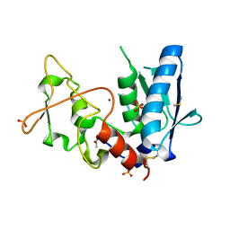
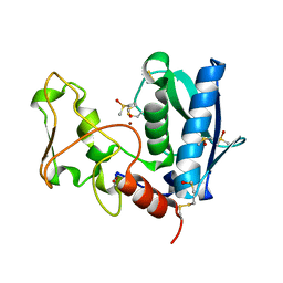
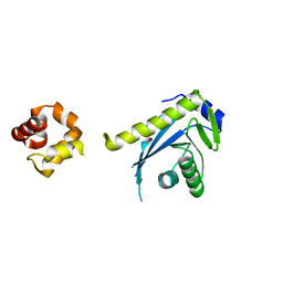
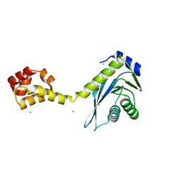
 | | USP2 in complex with ubiquitin | | Descriptor: | Polyubiquitin, Ubiquitin carboxyl-terminal hydrolase 2, ZINC ION | | Authors: | Renatus, M, Kroemer, M. | | Deposit date: | 2006-06-20 | | Release date: | 2006-08-15 | | Last modified: | 2023-08-30 | | Method: | X-RAY DIFFRACTION (1.85 Å) | | Cite: | Structural Basis of Ubiquitin Recognition by the Deubiquitinating Protease USP2.
Structure, 14, 2006
|
|
3EDH
 | |
2FDC
 | |
6MLB
 | |
6MR0
 | |
6MOX
 | |
6MKV
 | |
6MQX
 | |
6MQI
 | |
6MQZ
 | |
6MOR
 | |
6MQW
 | |
6ON5
 | |
6ON8
 | |
2NRW
 | |
2NRZ
 | |
2NRR
 | |
2NRX
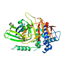
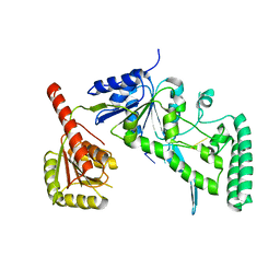
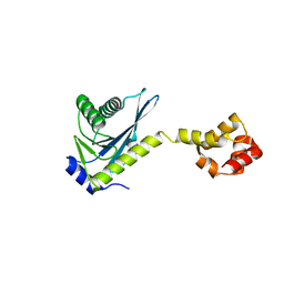
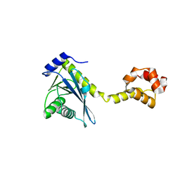
 | | Crystal structure of the C-terminal half of UvrC, in the presence of sulfate molecules | | Descriptor: | GLYCEROL, SULFATE ION, UvrABC system protein C | | Authors: | Karakas, E, Truglio, J.J, Kisker, C. | | Deposit date: | 2006-11-02 | | Release date: | 2007-02-06 | | Last modified: | 2023-08-30 | | Method: | X-RAY DIFFRACTION (1.9 Å) | | Cite: | Structure of the C-terminal half of UvrC reveals an RNase H endonuclease domain with an Argonaute-like catalytic triad.
Embo J., 26, 2007
|
|
2NRT
 | |
6ON7
 | |
2NRV
 | |
8VZX
 | |
8W00
 | |
4QZT
 | |


