Loading
PDBj
MenuPDBj@FacebookPDBj@TwitterPDBj@YouTubewwPDB FoundationwwPDB
RCSB PDBPDBeBMRBAdv. SearchSearch help
Search by PDB author
3E9M
DownloadVisualize
BU of 3e9m by Molmil
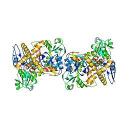
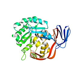
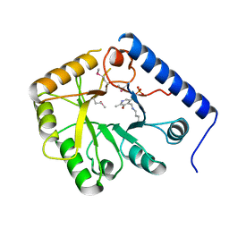
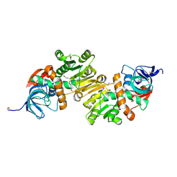
Crystal Structure of an oxidoreductase from Enterococcus faecalis
Descriptor: Oxidoreductase, Gfo/Idh/MocA family
Authors:Damodharan, L, Burley, S.K, Swaminathan, S, New York SGX Research Center for Structural Genomics (NYSGXRC)
Deposit date:2008-08-22
Release date:2008-10-14
Last modified:2021-02-10
Method:X-RAY DIFFRACTION (2.7 Å)
Cite:Crystal Structure of an oxidoreductase from Enterococcus faecalis
To be Published
3EUW
DownloadVisualize
BU of 3euw by Molmil
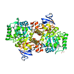
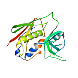
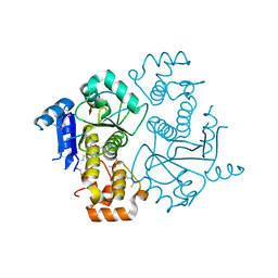
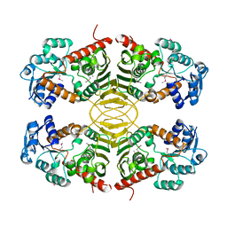
Crystal Structure of a Myo-inositol dehydrogenase from Corynebacterium glutamicum ATCC 13032
Descriptor: Myo-inositol dehydrogenase, SODIUM ION
Authors:Kumaran, D, Mahmood, A, Burley, S.K, Swaminathan, S, New York SGX Research Center for Structural Genomics (NYSGXRC)
Deposit date:2008-10-11
Release date:2008-10-21
Last modified:2023-12-27
Method:X-RAY DIFFRACTION (2.3 Å)
Cite:Crystal Structure of a Myo-inositol dehydrogenase from Corynebacterium glutamicum ATCC 13032
To be Published
2I6E
DownloadVisualize
BU of 2i6e by Molmil
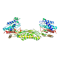
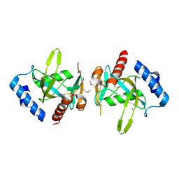
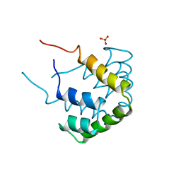
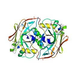
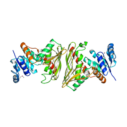
Crystal structure of protein DR0370 from Deinococcus radiodurans, Pfam DUF178
Descriptor: Hypothetical protein, SULFATE ION
Authors:Tyagi, R, Kumaran, D, Burley, S.K, Swaminathan, S, New York SGX Research Center for Structural Genomics (NYSGXRC)
Deposit date:2006-08-28
Release date:2006-09-05
Last modified:2021-02-03
Method:X-RAY DIFFRACTION (2.5 Å)
Cite:X-ray structures of two proteins belonging to Pfam DUF178 revealed unexpected structural similarity to the DUF191 Pfam family.
Bmc Struct.Biol., 7, 2007
2I9U
DownloadVisualize
BU of 2i9u by Molmil
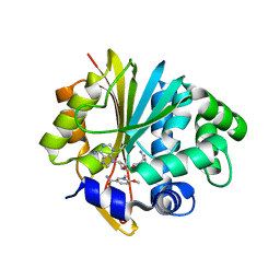
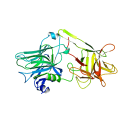
Crystal Structure of Guanine Deaminase from C. acetobutylicum with bound guanine in the active site
Descriptor: Cytosine/guanine deaminase related protein, FE (III) ION, GLYCEROL, ...
Authors:Kumaran, D, Burley, S.K, Swaminathan, S, New York SGX Research Center for Structural Genomics (NYSGXRC)
Deposit date:2006-09-06
Release date:2006-09-19
Last modified:2024-02-21
Method:X-RAY DIFFRACTION (2.05 Å)
Cite:Crystal Structure of Guanine Deaminase from C. acetobutylicum with bound guanine in the active site
To be Published
2IMO
DownloadVisualize
BU of 2imo by Molmil
Crystal structure of allantoate amidohydrolase from Escherichia coli at pH 4.6
Descriptor: Allantoate amidohydrolase
Authors:Agarwal, R, Burley, S.K, Swaminathan, S, New York SGX Research Center for Structural Genomics (NYSGXRC)
Deposit date:2006-10-04
Release date:2006-10-17
Last modified:2021-02-03
Method:X-RAY DIFFRACTION (2.8 Å)
Cite:Structural Analysis of a Ternary Complex of Allantoate Amidohydrolase from Escherichia coli Reveals its Mechanics.
J.Mol.Biol., 368, 2007
2IMG
DownloadVisualize
BU of 2img by Molmil
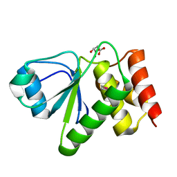
Crystal structure of dual specificity protein phosphatase 23 from Homo sapiens in complex with ligand malate ion
Descriptor: D-MALATE, Dual specificity protein phosphatase 23
Authors:Agarwal, R, Burley, S.K, Swaminathan, S, New York SGX Research Center for Structural Genomics (NYSGXRC)
Deposit date:2006-10-04
Release date:2006-10-17
Last modified:2021-02-03
Method:X-RAY DIFFRACTION (1.93 Å)
Cite:Structure of human dual specificity protein phosphatase 23, VHZ, enzyme-substrate/product complex.
J.Biol.Chem., 283, 2008
2ICS
DownloadVisualize
BU of 2ics by Molmil
Crystal structure of an adenine deaminase
Descriptor: ADENINE, Adenine Deaminase, ZINC ION
Authors:Sugadev, R, Kumaran, D, Burley, S.K, Swaminathan, S, New York SGX Research Center for Structural Genomics (NYSGXRC)
Deposit date:2006-09-13
Release date:2006-10-17
Last modified:2021-02-03
Method:X-RAY DIFFRACTION (2.3 Å)
Cite:Crystal structure of an adenine deaminase
TO BE PUBLISHED
1CQV
DownloadVisualize
BU of 1cqv by Molmil
CRYSTAL STRUCTURE OF STAPHYLOCOCCAL ENTEROTOXIN C2 AT 100K CRYSTALLIZED AT PH 5.0
Descriptor: PROTEIN (STAPHYLOCOCCAL ENTEROTOXIN C2), ZINC ION
Authors:Kumaran, D, Swaminathan, S.
Deposit date:1999-08-11
Release date:1999-08-22
Last modified:2023-08-09
Method:X-RAY DIFFRACTION (2.06 Å)
Cite:Structure of staphylococcal enterotoxin C2 at various pH levels.
Acta Crystallogr.,Sect.D, 57, 2001
3FCM
DownloadVisualize
BU of 3fcm by Molmil
Crystal structure of a NUDIX hydrolase from Clostridium perfringens
Descriptor: Hydrolase, NUDIX family, MANGANESE (II) ION
Authors:Palani, K, Burley, S.K, Swaninathan, S, New York SGX Research Center for Structural Genomics (NYSGXRC)
Deposit date:2008-11-21
Release date:2008-12-09
Last modified:2023-12-27
Method:X-RAY DIFFRACTION (2.2 Å)
Cite:Crystal structure of a NUDIX hydrolase from Clostridium perfringens
To be Published
5YJI
DownloadVisualize
BU of 5yji by Molmil
Co-crystal structure of Mouse Nicotinamide N-methyltransferase (NNMT) with small molecule analog of Nicotinamide
Descriptor: 6-methoxy-1-methyl-2H-pyridine-3-carboxamide, Nicotinamide N-methyltransferase, S-ADENOSYL-L-HOMOCYSTEINE
Authors:Birudukota, S, Swaminathan, S, Thakur, M.K, Parveen, R, Kandan, S, Hallur, M.S, Rajagopal, S, Ruf, S, Dhakshinamoorthy, S, Kannt, A, Gosu, R.
Deposit date:2017-10-10
Release date:2018-03-21
Last modified:2023-11-22
Method:X-RAY DIFFRACTION (1.99 Å)
Cite:A small molecule inhibitor of Nicotinamide N-methyltransferase for the treatment of metabolic disorders.
Sci Rep, 8, 2018
1I1E
DownloadVisualize
BU of 1i1e by Molmil
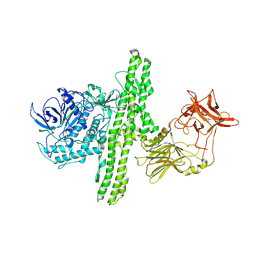
CRYSTAL STRUCTURE OF CLOSTRIDIUM BOTULINUM NEUROTOXIN B COMPLEXED WITH DOXORUBICIN
Descriptor: BOTULINUM NEUROTOXIN TYPE B, DOXORUBICIN, SULFATE ION, ...
Authors:Eswaramoorthy, S, Kumaran, D, Swaminathan, S.
Deposit date:2001-02-01
Release date:2001-11-21
Last modified:2023-08-09
Method:X-RAY DIFFRACTION (2.5 Å)
Cite:Crystallographic evidence for doxorubicin binding to the receptor-binding site in Clostridium botulinum neurotoxin B.
Acta Crystallogr.,Sect.D, 57, 2001
1G9C
DownloadVisualize
BU of 1g9c by Molmil
CRYSTAL STRUCTURE OF CLOSTRIDIUM BOTULINUM NEUROTOXIN B COMPLEXED WITH AN INHIBITOR (EXPERIMENT 4)
Descriptor: BIS(5-AMIDINO-BENZIMIDAZOLYL)METHANE, BOTULINUM NEUROTOXIN TYPE B, ZINC ION
Authors:Eswaramoorthy, S, Swaminathan, S.
Deposit date:2000-11-22
Release date:2002-11-13
Last modified:2023-08-09
Method:X-RAY DIFFRACTION (2.35 Å)
Cite:A Novel Mechanism for Clostridium botulinum Neurotoxin Inhibition
BIOCHEMISTRY, 41, 2002
1G9D
DownloadVisualize
BU of 1g9d by Molmil
CRYSTAL STRUCTURE OF CLOSTRIDIUM BOTULINUM NEUROTOXIN B COMPLEXED WITH AN INHIBITOR (EXPERIMENT 2)
Descriptor: BIS(5-AMIDINO-BENZIMIDAZOLYL)METHANE, BOTULINUM NEUROTOXIN TYPE B, ZINC ION
Authors:Eswaramoorthy, S, Swaminathan, S.
Deposit date:2000-11-22
Release date:2002-11-13
Last modified:2023-08-09
Method:X-RAY DIFFRACTION (2.2 Å)
Cite:A Novel Mechanism for Clostridium botulinum Neurotoxin Inhibition
BIOCHEMISTRY, 41, 2002
1G9B
DownloadVisualize
BU of 1g9b by Molmil
CRYSTAL STRUCTURE OF CLOSTRIDIUM BOTULINUM NEUROTOXIN B COMPLEXED WITH AN INHIBITOR (EXPERIMENT 1)
Descriptor: BIS(5-AMIDINO-BENZIMIDAZOLYL)METHANE, BOTULINUM NEUROTOXIN TYPE B, ZINC ION
Authors:Eswaramoorthy, S, Swaminathan, S.
Deposit date:2000-11-22
Release date:2002-11-13
Last modified:2023-08-09
Method:X-RAY DIFFRACTION (2 Å)
Cite:A Novel Mechanism for Clostridium botulinum Neurotoxin Inhibition
BIOCHEMISTRY, 41, 2002
1G9A
DownloadVisualize
BU of 1g9a by Molmil
CRYSTAL STRUCTURE OF CLOSTRIDIUM BOTULINUM NEUROTOXIN B COMPLEXED WITH AN INHIBITOR (EXPERIMENT 3)
Descriptor: BIS(5-AMIDINO-BENZIMIDAZOLYL)METHANE, BOTULINUM NEUROTOXIN TYPE B, ZINC ION
Authors:Eswaramoorthy, S, Swaminathan, S.
Deposit date:2000-11-22
Release date:2002-11-13
Last modified:2023-08-09
Method:X-RAY DIFFRACTION (2.1 Å)
Cite:A Novel Mechanism for Clostridium botulinum Neurotoxin Inhibition
BIOCHEMISTRY, 41, 2002
1CCD
DownloadVisualize
BU of 1ccd by Molmil
REFINED STRUCTURE OF RAT CLARA CELL 17 KDA PROTEIN AT 3.0 ANGSTROMS RESOLUTION
Descriptor: CLARA CELL 17 kD PROTEIN, SULFATE ION
Authors:Umland, T.C, Swaminathan, S, Furey, W, Singh, G, Pletcher, J, Sax, M.
Deposit date:1991-09-17
Release date:1994-01-31
Last modified:2017-11-29
Method:X-RAY DIFFRACTION (3 Å)
Cite:Refined structure of rat Clara cell 17 kDa protein at 3.0 A resolution.
J.Mol.Biol., 224, 1992
1I4P
DownloadVisualize
BU of 1i4p by Molmil
CRYSTAL STRUCTURE OF STAPHYLOCOCCAL ENTEROTOXIN C2 AT 100K CRYSTALLIZED AT PH 5.5
Descriptor: ENTEROTOXIN TYPE C-2, ZINC ION
Authors:Kumaran, D, Swaminathan, S.
Deposit date:2001-02-22
Release date:2001-03-14
Last modified:2023-08-09
Method:X-RAY DIFFRACTION (2 Å)
Cite:Structure of staphylococcal enterotoxin C2 at various pH levels.
Acta Crystallogr.,Sect.D, 57, 2001
1I4R
DownloadVisualize
BU of 1i4r by Molmil
CRYSTAL STRUCTURE OF STAPHYLOCOCCAL ENTEROTOXIN C2 AT 100K CRYSTALLIZED AT PH 6.5
Descriptor: ENTEROTOXIN TYPE C-2, ZINC ION
Authors:Kumaran, D, Swaminathan, S.
Deposit date:2001-02-22
Release date:2001-03-14
Last modified:2023-08-09
Method:X-RAY DIFFRACTION (2.1 Å)
Cite:Structure of staphylococcal enterotoxin C2 at various pH levels.
Acta Crystallogr.,Sect.D, 57, 2001
1I4Q
DownloadVisualize
BU of 1i4q by Molmil
CRYSTAL STRUCTURE OF STAPHYLOCOCCAL ENTEROTOXIN C2 AT 100K CRYSTALLIZED AT PH 6.0
Descriptor: ENTEROTOXIN TYPE C-2, ZINC ION
Authors:Kumaran, D, Swaminathan, S.
Deposit date:2001-02-22
Release date:2001-03-14
Last modified:2023-08-09
Method:X-RAY DIFFRACTION (2.2 Å)
Cite:Structure of staphylococcal enterotoxin C2 at various pH levels.
Acta Crystallogr.,Sect.D, 57, 2001
1CT5
DownloadVisualize
BU of 1ct5 by Molmil
CRYSTAL STRUCTURE OF YEAST HYPOTHETICAL PROTEIN YBL036C-SELENOMET CRYSTAL
Descriptor: PROTEIN (YEAST HYPOTHETICAL PROTEIN, SELENOMET), PYRIDOXAL-5'-PHOSPHATE
Authors:Eswaramoorthy, S, Swaminathan, S, Burley, S.K, New York SGX Research Center for Structural Genomics (NYSGXRC)
Deposit date:1999-08-18
Release date:1999-09-02
Last modified:2021-02-03
Method:X-RAY DIFFRACTION (2 Å)
Cite:Structure of a yeast hypothetical protein selected by a structural genomics approach.
Acta Crystallogr.,Sect.D, 59, 2003
1NJR
DownloadVisualize
BU of 1njr by Molmil
Crystal structure of yeast ymx7, an ADP-ribose-1''-monophosphatase
Descriptor: 32.1 kDa protein in ADH3-RCA1 intergenic region, Xylitol
Authors:Kumaran, D, Eswaramoorthy, S, Studier, F.W, Swaminathan, S, Burley, S.K, New York SGX Research Center for Structural Genomics (NYSGXRC)
Deposit date:2003-01-02
Release date:2004-08-17
Last modified:2021-02-03
Method:X-RAY DIFFRACTION (1.9 Å)
Cite:Structure and mechanism of ADP-ribose-1''-monophosphatase (Appr-1''-pase), a ubiquitous cellular processing enzyme
Protein Sci., 14, 2005
1NKQ
DownloadVisualize
BU of 1nkq by Molmil
Crystal structure of yeast ynq8, a fumarylacetoacetate hydrolase family protein
Descriptor: ACETIC ACID, CALCIUM ION, Hypothetical 28.8 kDa protein in PSD1-SKO1 intergenic region, ...
Authors:Eswaramoorthy, S, Kumaran, D, Daniels, B, Studier, F.W, Swaminathan, S, Burley, S.K, New York SGX Research Center for Structural Genomics (NYSGXRC)
Deposit date:2003-01-03
Release date:2004-06-15
Last modified:2021-02-03
Method:X-RAY DIFFRACTION (2.2 Å)
Cite:Crtystal Structure of Yeast Hypothetical Protein YNQ8_YEAST
To be Published
1AF9
DownloadVisualize
BU of 1af9 by Molmil
TETANUS NEUROTOXIN C FRAGMENT
Descriptor: TETANUS NEUROTOXIN
Authors:Umland, T.C, Wingert, L, Swaminathan, S, Furey, W.F, Schmidt, J.J, Sax, M.
Deposit date:1997-03-24
Release date:1998-04-29
Last modified:2024-02-07
Method:X-RAY DIFFRACTION (2.7 Å)
Cite:Structure of the receptor binding fragment HC of tetanus neurotoxin.
Nat.Struct.Biol., 4, 1997
4DUP
DownloadVisualize
BU of 4dup by Molmil
Crystal Structure of a quinone oxidoreductase from Rhizobium etli CFN 42
Descriptor: quinone oxidoreductase
Authors:Kumaran, D, Rice, S, Evans, B, Foti, R, Gizzi, A, Hillerich, B, Kar, A, Lafleur, J, Seidel, R, Villigas, G, Zencheck, W, Almo, S.C, Swaminathan, S, New York Structural Genomics Research Consortium (NYSGRC)
Deposit date:2012-02-22
Release date:2012-03-07
Method:X-RAY DIFFRACTION (2.45 Å)
Cite:Crystal Structure of a quinone oxidoreductase from Rhizobium etli CFN 42
To be Published
2MI1
DownloadVisualize
BU of 2mi1 by Molmil
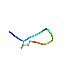
Somatostatin-14 solution structure in 5% D-mannitol
Descriptor: Somatostatin-14
Authors:Anoop, A, Ranganathan, S, Pratihar, S.
Deposit date:2013-12-08
Release date:2014-05-07
Last modified:2023-11-15
Method:SOLUTION NMR
Cite:Elucidating the role of disulfide bond on amyloid formation and fibril reversibility of somatostatin-14: relevance to its storage and secretion.
J.Biol.Chem., 289, 2014

222926

PDB entries from 2024-07-24

PDB statisticsPDBj update infoContact PDBjnumon