Loading
PDBj
MenuPDBj@FacebookPDBj@TwitterPDBj@YouTubewwPDB FoundationwwPDB
RCSB PDBPDBeBMRBAdv. SearchSearch help
Search by PDB author
1XGM
DownloadVisualize
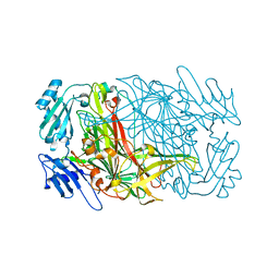
BU of 1xgm by Molmil
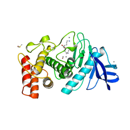
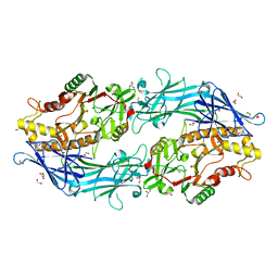
METHIONINE AMINOPEPTIDASE FROM HYPERTHERMOPHILE PYROCOCCUS FURIOSUS
Descriptor: COBALT (II) ION, METHIONINE AMINOPEPTIDASE
Authors:Tahirov, T.H, Tsukihara, T.
Deposit date:1997-11-17
Release date:1998-02-25
Last modified:2024-02-14
Method:X-RAY DIFFRACTION (2.8 Å)
Cite:Crystal structure of methionine aminopeptidase from hyperthermophile, Pyrococcus furiosus.
J.Mol.Biol., 284, 1998
5CTS
DownloadVisualize
BU of 5cts by Molmil
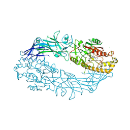
PROPOSED MECHANISM FOR THE CONDENSATION REACTION OF CITRATE SYNTHASE. 1.9-ANGSTROMS STRUCTURE OF THE TERNARY COMPLEX WITH OXALOACETATE AND CARBOXYMETHYL COENZYME A
Descriptor: CARBOXYMETHYL COENZYME *A, CITRATE SYNTHASE, OXALOACETATE ION
Authors:Karpusas, M, Branchaud, B, Remington, S.J.
Deposit date:1989-11-16
Release date:1990-10-15
Last modified:2024-03-06
Method:X-RAY DIFFRACTION (1.9 Å)
Cite:Proposed mechanism for the condensation reaction of citrate synthase: 1.9-A structure of the ternary complex with oxaloacetate and carboxymethyl coenzyme A.
Biochemistry, 29, 1990
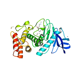
8TLN
DownloadVisualize
BU of 8tln by Molmil
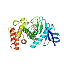
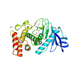
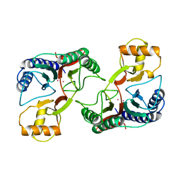
STRUCTURAL COMPARISON SUGGESTS THAT THERMOLYSIN AND RELATED NEUTRAL PROTEASES UNDERGO HINGE-BENDING MOTION DURING CATALYSIS
Descriptor: CALCIUM ION, DIMETHYL SULFOXIDE, LYSINE, ...
Authors:Tronrud, D, Matthews, B.W.
Deposit date:1993-09-01
Release date:1994-04-30
Last modified:2024-02-14
Method:X-RAY DIFFRACTION (1.6 Å)
Cite:Structural comparison suggests that thermolysin and related neutral proteases undergo hinge-bending motion during catalysis.
Biochemistry, 31, 1992
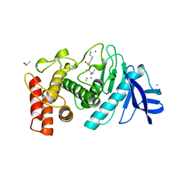
6TMN
DownloadVisualize
BU of 6tmn by Molmil
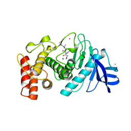
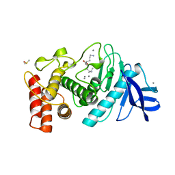
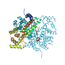
Structures of two thermolysin-inhibitor complexes that differ by a single hydrogen bond
Descriptor: CALCIUM ION, N-[(2R,4S)-4-hydroxy-2-(2-methylpropyl)-4-oxido-7-oxo-9-phenyl-3,8-dioxa-6-aza-4-phosphanonan-1-oyl]-L-leucine, THERMOLYSIN, ...
Authors:Tronrud, D.E, Holden, H.M, Matthews, B.W.
Deposit date:1987-06-29
Release date:1989-01-09
Last modified:2024-05-22
Method:X-RAY DIFFRACTION (1.6 Å)
Cite:Structures of two thermolysin-inhibitor complexes that differ by a single hydrogen bond.
Science, 235, 1987
8JT1
DownloadVisualize
BU of 8jt1 by Molmil
COLLAGENASE FROM GRIMONTIA (VIBRIO) HOLLISAE 1706B COMPLEXED WITH GLY-PRO-HYP-GLY-PRO-HYP
Descriptor: 1,2-ETHANEDIOL, 2-AMINO-2-HYDROXYMETHYL-PROPANE-1,3-DIOL, 6-mer peptide, ...
Authors:Ueshima, S, Yaskawa, K, Takita, T, Mikami, B.
Deposit date:2023-06-21
Release date:2024-02-07
Method:X-RAY DIFFRACTION (2 Å)
Cite:Insights into the catalytic mechanism of Grimontia hollisae collagenase through structural and mutational analyses.
Febs Lett., 597, 2023
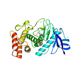
5TLN
DownloadVisualize
BU of 5tln by Molmil
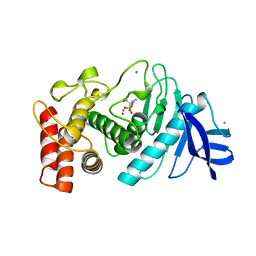
BINDING OF HYDROXAMIC ACID INHIBITORS TO CRYSTALLINE THERMOLYSIN SUGGESTS A PENTACOORDINATE ZINC INTERMEDIATE IN CATALYSIS
Descriptor: CALCIUM ION, HONH-BENZYLMALONYL-L-ALANYLGLYCINE-P-NITROANILIDE, THERMOLYSIN, ...
Authors:Matthews, B.W, Holmes, M.A.
Deposit date:1982-02-08
Release date:1982-05-26
Last modified:2024-05-22
Method:X-RAY DIFFRACTION (2.3 Å)
Cite:Binding of hydroxamic acid inhibitors to crystalline thermolysin suggests a pentacoordinate zinc intermediate in catalysis.
Biochemistry, 20, 1981
8J6G
DownloadVisualize
BU of 8j6g by Molmil
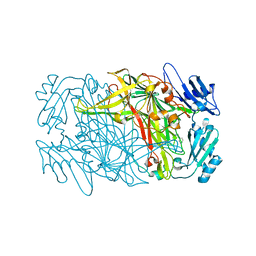
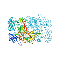
Neutron structure of copper amine oxidase from Arthrobacter globiformis anaerobically reduced by phenylethylamine at pD 9.0
Descriptor: 2-PHENYLETHYLAMINE, COPPER (II) ION, Phenylethylamine oxidase, ...
Authors:Murakawa, T, Okajima, T.
Deposit date:2023-04-25
Release date:2023-09-20
Method:NEUTRON DIFFRACTION (1.09 Å), X-RAY DIFFRACTION
Cite:Neutron Crystallography of a Semiquinone Radical Intermediate of Copper Amine Oxidase Reveals a Substrate-Assisted Conformational Change of the Peptidyl Quinone Cofactor
Acs Catalysis, 2023
1LMT
DownloadVisualize
BU of 1lmt by Molmil
STRUCTURE OF A CONFORMATIONALLY CONSTRAINED ARG-GLY-ASP SEQUENCE INSERTED INTO HUMAN LYSOZYME
Descriptor: 2-acetamido-2-deoxy-beta-D-glucopyranose-(1-4)-2-acetamido-2-deoxy-beta-D-glucopyranose-(1-4)-2-acetamido-2-deoxy-beta-D-glucopyranose, CHLORIDE ION, HUMAN LYSOZYME
Authors:Matsushima, M, Song, H.
Deposit date:1995-01-13
Release date:1995-03-31
Last modified:2020-07-29
Method:X-RAY DIFFRACTION (1.6 Å)
Cite:Structure of a conformationally constrained Arg-Gly-Asp sequence inserted into human lysozyme.
J.Biol.Chem., 270, 1995
2E89
DownloadVisualize
BU of 2e89 by Molmil
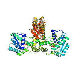
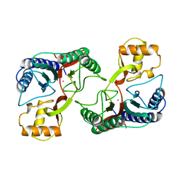
Crystal structure of Aquifex aeolicus TilS in a complex with ATP, Magnesium ion, and L-lysine
Descriptor: ADENOSINE-5'-TRIPHOSPHATE, LYSINE, MAGNESIUM ION, ...
Authors:Kuratani, M, Yoshikawa, Y, Takahashi, S, Yokoyama, S, RIKEN Structural Genomics/Proteomics Initiative (RSGI)
Deposit date:2007-01-19
Release date:2007-11-13
Last modified:2023-10-25
Method:X-RAY DIFFRACTION (2.5 Å)
Cite:Structural basis of the initial binding of tRNA(Ile) lysidine synthetase TilS with ATP and L-lysine
To be Published
7YNH
DownloadVisualize
BU of 7ynh by Molmil
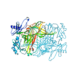
Catalytic intermediate of copper amine oxidase determined by serial femtosecond X-ray crystallography using a single-flow liquid jet system
Descriptor: COPPER (II) ION, PHENYLACETALDEHYDE, Phenylethylamine oxidase, ...
Authors:Murakawa, T, Okajima, T.
Deposit date:2022-07-31
Release date:2022-11-16
Last modified:2023-11-29
Method:X-RAY DIFFRACTION (1.94 Å)
Cite:Serial femtosecond X-ray crystallography of an anaerobically formed catalytic intermediate of copper amine oxidase.
Acta Crystallogr D Struct Biol, 78, 2022
2TMN
DownloadVisualize
BU of 2tmn by Molmil
CRYSTALLOGRAPHIC STRUCTURAL ANALYSIS OF PHOSPHORAMIDATES AS INHIBITORS AND TRANSITION-STATE ANALOGS OF THERMOLYSIN
Descriptor: CALCIUM ION, N~2~-phosphono-L-leucinamide, Thermolysin, ...
Authors:Tronrud, D.E, Monzingo, A.F, Matthews, B.W.
Deposit date:1987-06-29
Release date:1989-01-09
Last modified:2024-05-22
Method:X-RAY DIFFRACTION (1.6 Å)
Cite:Crystallographic structural analysis of phosphoramidates as inhibitors and transition-state analogs of thermolysin.
Eur.J.Biochem., 157, 1986
7WNP
DownloadVisualize
BU of 7wnp by Molmil
Crystallographic structure of copper amine oxidase from Arthrobacter glibiformis at pD 7.4 determined by both X-ray and neutron diffraction data at 1.72 angstrom resolution.
Descriptor: COPPER (II) ION, Phenylethylamine oxidase, SODIUM ION
Authors:Murakawa, T, Okajima, T.
Deposit date:2022-01-19
Release date:2022-04-20
Last modified:2022-05-25
Method:NEUTRON DIFFRACTION (1.72 Å), X-RAY DIFFRACTION
Cite:Re-evaluation of protein neutron crystallography with and without X-ray/neutron joint refinement.
Iucrj, 9, 2022
7WNO
DownloadVisualize
BU of 7wno by Molmil
Crystallographic structure of copper amine oxidase from Arthrobacter glibiformis at pD 7.4 determined by only neutron diffraction data.
Descriptor: COPPER (II) ION, Phenylethylamine oxidase
Authors:Murakawa, T, Okajima, T.
Deposit date:2022-01-19
Release date:2022-04-20
Last modified:2022-05-25
Method:NEUTRON DIFFRACTION (1.72 Å)
Cite:Re-evaluation of protein neutron crystallography with and without X-ray/neutron joint refinement.
Iucrj, 9, 2022
1XGS
DownloadVisualize
BU of 1xgs by Molmil
METHIONINE AMINOPEPTIDASE FROM HYPERTHERMOPHILE PYROCOCCUS FURIOSUS
Descriptor: COBALT (II) ION, METHIONINE AMINOPEPTIDASE
Authors:Tahirov, T.H, Tsukihara, T.
Deposit date:1997-04-25
Release date:1997-07-07
Last modified:2024-04-03
Method:X-RAY DIFFRACTION (1.75 Å)
Cite:Crystal structure of methionine aminopeptidase from hyperthermophile, Pyrococcus furiosus.
J.Mol.Biol., 284, 1998
2E21
DownloadVisualize
BU of 2e21 by Molmil
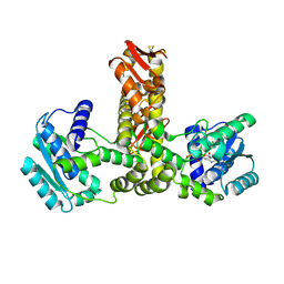
Crystal structure of TilS in a complex with AMPPNP from Aquifex aeolicus.
Descriptor: PHOSPHOAMINOPHOSPHONIC ACID-ADENYLATE ESTER, tRNA(Ile)-lysidine synthase
Authors:Kuratani, M, Yoshikawa, Y, Sekine, S, Ishii, T, Shibata, R, Bessho, Y, Yokoyama, S, RIKEN Structural Genomics/Proteomics Initiative (RSGI)
Deposit date:2006-11-06
Release date:2007-11-06
Last modified:2023-10-25
Method:X-RAY DIFFRACTION (2.7 Å)
Cite:Structural basis of the initial binding of tRNA(Ile) lysidine synthetase TilS with ATP and L-lysine
To be Published
1LNA
DownloadVisualize
BU of 1lna by Molmil
A STRUCTURAL ANALYSIS OF METAL SUBSTITUTIONS IN THERMOLYSIN
Descriptor: CALCIUM ION, COBALT (II) ION, DIMETHYL SULFOXIDE, ...
Authors:Holland, D.R, Hausrath, A.C, Juers, D, Matthews, B.W.
Deposit date:1994-05-13
Release date:1995-05-08
Last modified:2024-02-14
Method:X-RAY DIFFRACTION (1.9 Å)
Cite:Structural analysis of zinc substitutions in the active site of thermolysin.
Protein Sci., 4, 1995
1LNC
DownloadVisualize
BU of 1lnc by Molmil
A STRUCTURAL ANALYSIS OF METAL SUBSTITUTIONS IN THERMOLYSIN
Descriptor: CALCIUM ION, DIMETHYL SULFOXIDE, LYSINE, ...
Authors:Holland, D.R, Hausrath, A.C, Juers, D, Matthews, B.W.
Deposit date:1994-05-13
Release date:1995-05-08
Last modified:2024-02-14
Method:X-RAY DIFFRACTION (1.8 Å)
Cite:Structural analysis of zinc substitutions in the active site of thermolysin.
Protein Sci., 4, 1995
1LNE
DownloadVisualize
BU of 1lne by Molmil
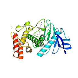
A STRUCTURAL ANALYSIS OF METAL SUBSTITUTIONS IN THERMOLYSIN
Descriptor: CADMIUM ION, CALCIUM ION, DIMETHYL SULFOXIDE, ...
Authors:Holland, D.R, Hausrath, A.C, Juers, D, Matthews, B.W.
Deposit date:1994-05-13
Release date:1995-05-08
Last modified:2024-02-14
Method:X-RAY DIFFRACTION (1.7 Å)
Cite:Structural analysis of zinc substitutions in the active site of thermolysin.
Protein Sci., 4, 1995
1LND
DownloadVisualize
BU of 1lnd by Molmil
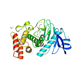
A STRUCTURAL ANALYSIS OF METAL SUBSTITUTIONS IN THERMOLYSIN
Descriptor: CALCIUM ION, DIMETHYL SULFOXIDE, LYSINE, ...
Authors:Holland, D.R, Hausrath, A.C, Juers, D, Matthews, B.W.
Deposit date:1994-05-13
Release date:1995-05-08
Last modified:2024-02-14
Method:X-RAY DIFFRACTION (1.7 Å)
Cite:Structural analysis of zinc substitutions in the active site of thermolysin.
Protein Sci., 4, 1995
1LNB
DownloadVisualize
BU of 1lnb by Molmil
A STRUCTURAL ANALYSIS OF METAL SUBSTITUTIONS IN THERMOLYSIN
Descriptor: CALCIUM ION, DIMETHYL SULFOXIDE, FE (III) ION, ...
Authors:Holland, D.R, Hausrath, A.C, Juers, D, Matthews, B.W.
Deposit date:1994-05-13
Release date:1995-05-08
Last modified:2024-02-14
Method:X-RAY DIFFRACTION (1.8 Å)
Cite:Structural analysis of zinc substitutions in the active site of thermolysin.
Protein Sci., 4, 1995
1LNF
DownloadVisualize
BU of 1lnf by Molmil
A structural analysis of metal substitutions in thermolysin
Descriptor: CALCIUM ION, DIMETHYL SULFOXIDE, LYSINE, ...
Authors:Holland, D.R, Matthews, B.W.
Deposit date:1994-05-13
Release date:1995-05-08
Last modified:2024-02-14
Method:X-RAY DIFFRACTION (1.7 Å)
Cite:Structural analysis of zinc substitutions in the active site of thermolysin.
Protein Sci., 4, 1995
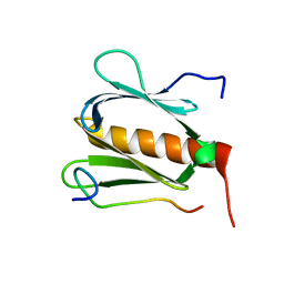
5GOW
DownloadVisualize
BU of 5gow by Molmil
Solution structure of the complex between DP1 acidic region and TFIIH p62 PH domain
Descriptor: DP1, General transcription factor IIH subunit 1
Authors:Okuda, M, Nishimura, Y.
Deposit date:2016-07-29
Release date:2016-12-07
Last modified:2024-05-01
Method:SOLUTION NMR
Cite:The Interaction Mode of the Acidic Region of the Cell Cycle Transcription Factor DP1 with TFIIH
J. Mol. Biol., 428, 2016
7TLN
DownloadVisualize
BU of 7tln by Molmil
STRUCTURAL ANALYSIS OF THE INHIBITION OF THERMOLYSIN BY AN ACTIVE-SITE-DIRECTED IRREVERSIBLE INHIBITOR
Descriptor: 2-(ACETYL-HYDROXY-AMINO)-4-METHYL-PENTANOIC ACID METHYL ESTER, CALCIUM ION, THERMOLYSIN, ...
Authors:Matthews, B.W, Holmes, M.A, Tronrud, D.E.
Deposit date:1983-01-27
Release date:1983-03-09
Last modified:2022-11-23
Method:X-RAY DIFFRACTION (2.3 Å)
Cite:Structural analysis of the inhibition of thermolysin by an active-site-directed irreversible inhibitor.
Biochemistry, 22, 1983
7D5V
DownloadVisualize
BU of 7d5v by Molmil
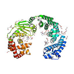
Structure of the C646A mutant of peptidylarginine deiminase type III (PAD3)
Descriptor: 1,2-ETHANEDIOL, GLYCEROL, Protein-arginine deiminase type-3
Authors:Akimoto, M, Mashimo, R, Unno, M.
Deposit date:2020-09-28
Release date:2021-06-02
Last modified:2023-11-29
Method:X-RAY DIFFRACTION (2.102 Å)
Cite:Structures of human peptidylarginine deiminase type III provide insights into substrate recognition and inhibitor design.
Arch.Biochem.Biophys., 708, 2021
7D56
DownloadVisualize
BU of 7d56 by Molmil
Structure of the peptidylarginine deiminase type III (PAD3) in complex with Cl-amidine
Descriptor: 1,2-ETHANEDIOL, CALCIUM ION, CHLORIDE ION, ...
Authors:Funabashi, K, Unno, M.
Deposit date:2020-09-25
Release date:2021-06-02
Last modified:2023-11-29
Method:X-RAY DIFFRACTION (3.175 Å)
Cite:Structures of human peptidylarginine deiminase type III provide insights into substrate recognition and inhibitor design.
Arch.Biochem.Biophys., 708, 2021

222926

PDB entries from 2024-07-24

PDB statisticsPDBj update infoContact PDBjnumon