Loading
PDBj
MenuPDBj@FacebookPDBj@TwitterPDBj@YouTubewwPDB FoundationwwPDB
RCSB PDBPDBeBMRBAdv. SearchSearch help
Search by PDB author
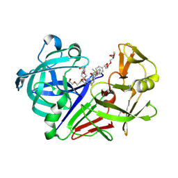
3PSY
DownloadVisualize
BU of 3psy by Molmil
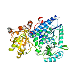
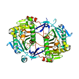
Endothiapepsin in complex with an inhibitor based on the Gewald reaction
Descriptor: DIMETHYL SULFOXIDE, Endothiapepsin, GLYCEROL, ...
Authors:Koester, H, Heine, A, Klebe, G.
Deposit date:2010-12-02
Release date:2011-12-07
Last modified:2023-11-01
Method:X-RAY DIFFRACTION (1.43 Å)
Cite:Tracing binding modes in hit-to-lead optimization: chameleon-like poses of aspartic protease inhibitors.
Angew.Chem.Int.Ed.Engl., 54, 2015
2GP4
DownloadVisualize
BU of 2gp4 by Molmil
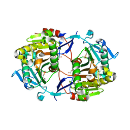
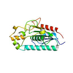
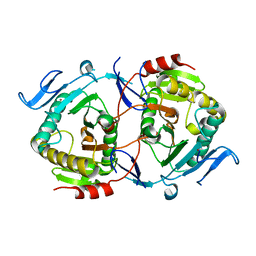
Structure of [FeS]cluster-free Apo Form of 6-Phosphogluconate Dehydratase from Shewanella oneidensis
Descriptor: 6-phosphogluconate dehydratase
Authors:Schormann, N, Symersky, J, Southeast Collaboratory for Structural Genomics (SECSG)
Deposit date:2006-04-16
Release date:2006-05-09
Last modified:2011-07-13
Method:X-RAY DIFFRACTION (2.49 Å)
Cite:Structure of [FeS]cluster-free Apo Form of 6-Phosphogluconate Dehydratase from Shewanella oneidensis
To be Published
1Z84
DownloadVisualize
BU of 1z84 by Molmil
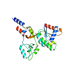
X-ray structure of galt-like protein from arabidopsis thaliana at5g18200
Descriptor: 1,2-ETHANEDIOL, ADENOSINE MONOPHOSPHATE, ZINC ION, ...
Authors:Mccoy, J.G, Bitto, E, Phillips Jr, G.N, Bingman, C.A, Center for Eukaryotic Structural Genomics (CESG)
Deposit date:2005-03-29
Release date:2005-04-19
Last modified:2023-08-23
Method:X-RAY DIFFRACTION (1.83 Å)
Cite:Structure and Mechanism of an ADP-Glucose Phosphorylase from Arabidopsis thaliana
Biochemistry, 45, 2006
8F6D
DownloadVisualize
BU of 8f6d by Molmil
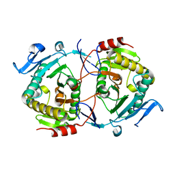
Crystal structure of the CNNM2 CBS-pair domain in complex with ARL15
Descriptor: ADP-ribosylation factor-like protein 15, Metal transporter CNNM2
Authors:Kozlov, G, Mahbub, L, Gehring, K.
Deposit date:2022-11-16
Release date:2023-07-12
Last modified:2024-01-24
Method:X-RAY DIFFRACTION (3.2 Å)
Cite:Structural insights into regulation of CNNM-TRPM7 divalent cation uptake by the small GTPase ARL15.
Elife, 12, 2023
1ZWJ
DownloadVisualize
BU of 1zwj by Molmil
X-ray structure of galt-like protein from arabidopsis thaliana AT5G18200
Descriptor: ZINC ION, putative galactose-1-phosphate uridyl transferase
Authors:Wesenberg, G.E, Smith, D.W, Phillips Jr, G.N, McCoy, J.G, Johnson, K.A, Bitto, E, Bingman, C.A, Allard, S.T.M, Center for Eukaryotic Structural Genomics (CESG)
Deposit date:2005-06-03
Release date:2005-06-14
Last modified:2023-08-23
Method:X-RAY DIFFRACTION (2.3 Å)
Cite:Structure and Mechanism of an ADP-Glucose Phosphorylase from Arabidopsis thaliana
Biochemistry, 45, 2006
2H39
DownloadVisualize
BU of 2h39 by Molmil
Crystal Structure of an ADP-Glucose Phosphorylase from Arabidopsis thaliana with bound ADP-Glucose
Descriptor: ADENOSINE-5'-DIPHOSPHATE-GLUCOSE, CHLORIDE ION, Probable galactose-1-phosphate uridyl transferase, ...
Authors:McCoy, J.G, Wesenberg, G.E, Phillips Jr, G.N, Bitto, E, Bingman, C.A, Center for Eukaryotic Structural Genomics (CESG)
Deposit date:2006-05-22
Release date:2006-06-13
Last modified:2023-08-30
Method:X-RAY DIFFRACTION (2.23 Å)
Cite:Crystal Structure of an ADP-Glucose Phosphorylase from Arabidopsis thaliana with bound ADP-Glucose
To be Published
3AJA
DownloadVisualize
BU of 3aja by Molmil
Crystal Structure of MSMEG_6394
Descriptor: Putative uncharacterized protein
Authors:Vivian, J.P, Scoble, J, Beddoe, T, Rossjohn, J.
Deposit date:2010-05-27
Release date:2010-08-11
Last modified:2011-07-13
Method:X-RAY DIFFRACTION (2.9 Å)
Cite:Functional characterisation of a lipase essential for mycobacterial viability
To be Published
5LGY
DownloadVisualize
BU of 5lgy by Molmil
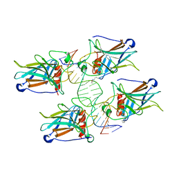
Lysine 120-acetylated P53 DNA binding domain in a complex with the BAX response element.
Descriptor: Cellular tumor antigen p53, DNA (5'-D(*AP*GP*GP*CP*TP*TP*GP*TP*CP*TP*CP*TP*AP*AP*CP*TP*TP*GP*TP*GP*A)-3'), DNA (5'-D(*TP*CP*AP*CP*AP*AP*GP*TP*TP*AP*GP*AP*GP*AP*CP*AP*AP*GP*CP*CP*T)-3'), ...
Authors:Arbely, E, Vainer, R.
Deposit date:2016-07-08
Release date:2016-07-27
Last modified:2024-01-10
Method:X-RAY DIFFRACTION (2.92 Å)
Cite:Structural Basis for p53 Lys120-Acetylation-Dependent DNA-Binding Mode.
J.Mol.Biol., 428, 2016
5BUA
DownloadVisualize
BU of 5bua by Molmil
Lysine 120-acetylated P53 DNA binding domain in a complex with DNA.
Descriptor: Cellular tumor antigen p53, DNA (5'-D(P*GP*GP*AP*CP*AP*TP*GP*TP*CP*C)-3'), ZINC ION
Authors:Arbely, E, Vainer, R.
Deposit date:2015-06-03
Release date:2016-07-06
Last modified:2024-01-10
Method:X-RAY DIFFRACTION (1.812 Å)
Cite:Structural Basis for p53 Lys120-Acetylation-Dependent DNA-Binding Mode.
J.Mol.Biol., 428, 2016
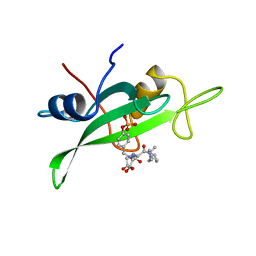
1SKJ
DownloadVisualize
BU of 1skj by Molmil
COCRYSTAL STRUCTURE OF UREA-SUBSTITUTED PHOSPHOPEPTIDE COMPLEX
Descriptor: 4-[3-CARBOXYMETHYL-3-(4-PHOSPHONOOXY-BENZYL)-UREIDO]-4-[(3-CYCLOHEXYL-PROPYL)-METHYL-CARBAMOYL]BUTYRIC ACID, PP60 V-SRC TYROSINE KINASE TRANSFORMING PROTEIN
Authors:Holland, D.R, Rubin, J.R.
Deposit date:1997-09-18
Release date:1998-02-25
Last modified:2024-05-22
Method:X-RAY DIFFRACTION (2 Å)
Cite:Design, synthesis, and cocrystal structure of a nonpeptide Src SH2 domain ligand.
J.Med.Chem., 40, 1997
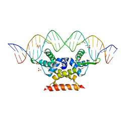
6QUA
DownloadVisualize
BU of 6qua by Molmil
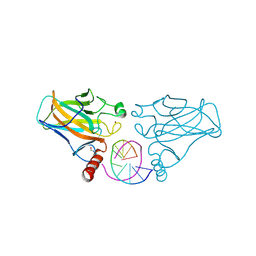
The complex structure of hsRosR-SG (vng0258/RosR-SG)
Descriptor: DNA (28-MER), MANGANESE (II) ION, SULFATE ION, ...
Authors:Shaanan, B, Kutnowski, N.
Deposit date:2019-02-27
Release date:2019-07-10
Last modified:2024-01-24
Method:X-RAY DIFFRACTION (2.681 Å)
Cite:Specificity of protein-DNA interactions in hypersaline environment: structural studies on complexes of Halobacterium salinarum oxidative stress-dependent protein hsRosR.
Nucleic Acids Res., 47, 2019
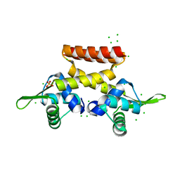
6F5C
DownloadVisualize
BU of 6f5c by Molmil
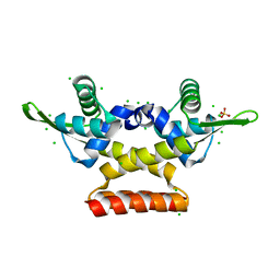
Structure of h. salinarum RosR (vng0258) grown from NaCl
Descriptor: CHLORIDE ION, HALOPHILIC WINGED-HELIX-TURN-HELIX DNA BINDING PROTEIN, SULFATE ION
Authors:Shaanan, B, Kutnowski, N, Shmuely, H.
Deposit date:2017-12-01
Release date:2018-08-22
Last modified:2024-05-01
Method:X-RAY DIFFRACTION (1.55 Å)
Cite:The 3-D structure of VNG0258H/RosR - A haloarchaeal DNA-binding protein in its ionic shell.
J. Struct. Biol., 204, 2018
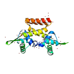
6EZ1
DownloadVisualize
BU of 6ez1 by Molmil
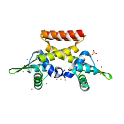
Structure of h. salinarum RosR (vng0258) grown from NaBr
Descriptor: BROMIDE ION, DNA binding protein, SULFATE ION
Authors:Shaanan, B, Kutnowski, N.
Deposit date:2017-11-13
Release date:2018-08-22
Last modified:2024-05-01
Method:X-RAY DIFFRACTION (1.75004315 Å)
Cite:The 3-D structure of VNG0258H/RosR - A haloarchaeal DNA-binding protein in its ionic shell.
J. Struct. Biol., 204, 2018
6FDH
DownloadVisualize
BU of 6fdh by Molmil
Structure of H. salinarum RosR (vng0258) grown from KCl
Descriptor: CHLORIDE ION, DNA binding protein, SULFATE ION
Authors:Shaanan, B, Kutnowski, N.
Deposit date:2017-12-23
Release date:2018-08-22
Last modified:2024-05-01
Method:X-RAY DIFFRACTION (1.85 Å)
Cite:The 3-D structure of VNG0258H/RosR - A haloarchaeal DNA-binding protein in its ionic shell.
J. Struct. Biol., 204, 2018
6FAQ
DownloadVisualize
BU of 6faq by Molmil
Structure of H. salinarum RosR (vng0258) grown from KBr
Descriptor: BROMIDE ION, DNA binding protein, SULFATE ION
Authors:Shaanan, B, Kutnowski, N.
Deposit date:2017-12-16
Release date:2018-08-22
Last modified:2024-05-01
Method:X-RAY DIFFRACTION (1.95 Å)
Cite:The 3-D structure of VNG0258H/RosR - A haloarchaeal DNA-binding protein in its ionic shell.
J. Struct. Biol., 204, 2018
6N3R
DownloadVisualize
BU of 6n3r by Molmil
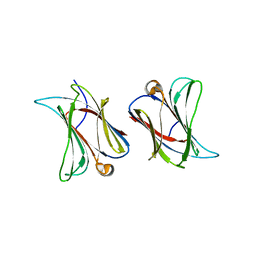
Sheep Galectin-11 (LGALS11) complex with galactose
Descriptor: Galectin, beta-D-galactopyranose
Authors:Beddoe, T.C, Sakthivel, D.
Deposit date:2018-11-16
Release date:2020-05-20
Last modified:2024-03-13
Method:X-RAY DIFFRACTION (2.397 Å)
Cite:Sheep Galectin-11 (LGALS11) complex with galactose
To be Published
2Q4L
DownloadVisualize
BU of 2q4l by Molmil
Ensemble refinement of the crystal structure of GALT-like protein from Arabidopsis thaliana At5g18200
Descriptor: Probable galactose-1-phosphate uridyl transferase, ZINC ION
Authors:Levin, E.J, Kondrashov, D.A, Wesenberg, G.E, Phillips Jr, G.N, Center for Eukaryotic Structural Genomics (CESG)
Deposit date:2007-05-31
Release date:2007-06-19
Last modified:2023-08-30
Method:X-RAY DIFFRACTION (2.3 Å)
Cite:Ensemble refinement of protein crystal structures: validation and application.
Structure, 15, 2007

222415

數據於2024-07-10公開中

PDB statisticsPDBj update infoContact PDBjnumon