Loading
PDBj
MenuPDBj@FacebookPDBj@TwitterPDBj@YouTubewwPDB FoundationwwPDB
RCSB PDBPDBeBMRBAdv. SearchSearch help
Search by PDB author
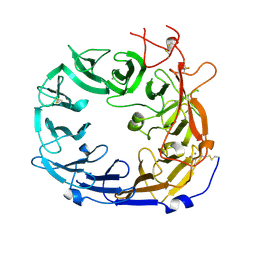
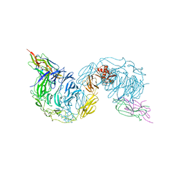
6FFY
DownloadVisualize
BU of 6ffy by Molmil
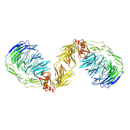
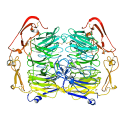
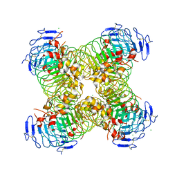
Structure of the mouse SorCS2-NGF complex
Descriptor: 2-acetamido-2-deoxy-beta-D-glucopyranose, Beta-nerve growth factor, VPS10 domain-containing receptor SorCS2, ...
Authors:Leloup, N.O.L, Janssen, B.J.C.
Deposit date:2018-01-09
Release date:2018-08-01
Last modified:2024-01-17
Method:X-RAY DIFFRACTION (3.9 Å)
Cite:Structural insights into SorCS2-Nerve Growth Factor complex formation.
Nat Commun, 9, 2018
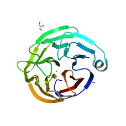
6FG9
DownloadVisualize
BU of 6fg9 by Molmil
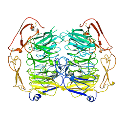
Mouse SORCS2 ectodomain (sortilin related VPS10 domain containing receptor 2)
Descriptor: 2-acetamido-2-deoxy-beta-D-glucopyranose, VPS10 domain-containing receptor SorCS2
Authors:Leloup, N.O.L, Janssen, B.J.C.
Deposit date:2018-01-10
Release date:2018-08-01
Last modified:2024-01-17
Method:X-RAY DIFFRACTION (4.2 Å)
Cite:Structural insights into SorCS2-Nerve Growth Factor complex formation.
Nat Commun, 9, 2018
6GRQ
DownloadVisualize
BU of 6grq by Molmil
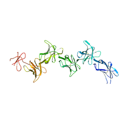
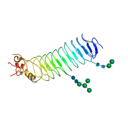
Paired immunoglobulin-like receptor B (PirB) or Leukocyte immunoglobulin-like receptor subfamily B member 3 (LILRB3) full extracellular domain
Descriptor: 2-acetamido-2-deoxy-beta-D-glucopyranose, 2-acetamido-2-deoxy-beta-D-glucopyranose-(1-4)-2-acetamido-2-deoxy-beta-D-glucopyranose, Paired immunoglobulin-like receptor B
Authors:Vlieg, H.C, Huizinga, E.G, Janssen, B.J.C.
Deposit date:2018-06-12
Release date:2019-02-06
Last modified:2024-01-17
Method:X-RAY DIFFRACTION (3.3 Å)
Cite:Structure and flexibility of the extracellular region of the PirB receptor.
J.Biol.Chem., 294, 2019
6GRT
DownloadVisualize
BU of 6grt by Molmil
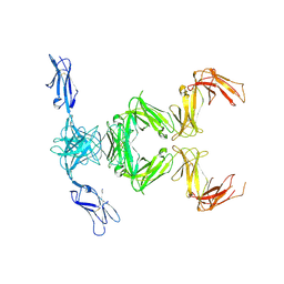
Paired immunoglobulin-like receptor B (PirB) or Leukocyte immunoglobulin-like receptor subfamily B member 3 (LILRB3) full extracellular domain
Descriptor: 2-acetamido-2-deoxy-beta-D-glucopyranose, Paired immunoglobulin-like receptor B, alpha-D-mannopyranose-(1-3)-[alpha-D-mannopyranose-(1-6)]beta-D-mannopyranose-(1-4)-2-acetamido-2-deoxy-beta-D-glucopyranose-(1-4)-2-acetamido-2-deoxy-beta-D-glucopyranose, ...
Authors:Vlieg, H.C, Huizinga, E.G, Janssen, B.J.C.
Deposit date:2018-06-12
Release date:2019-02-06
Last modified:2024-01-17
Method:X-RAY DIFFRACTION (4.504 Å)
Cite:Structure and flexibility of the extracellular region of the PirB receptor.
J.Biol.Chem., 294, 2019
6GRS
DownloadVisualize
BU of 6grs by Molmil
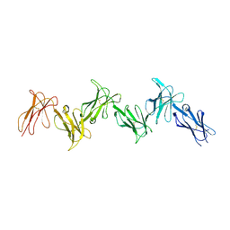
Paired immunoglobulin-like receptor B (PirB) or Leukocyte immunoglobulin-like receptor subfamily B member 3 (LILRB3) full extracellular domain
Descriptor: 2-acetamido-2-deoxy-beta-D-glucopyranose, 2-acetamido-2-deoxy-beta-D-glucopyranose-(1-4)-2-acetamido-2-deoxy-beta-D-glucopyranose, Paired immunoglobulin-like receptor B
Authors:Vlieg, H.C, Huizinga, E.G, Janssen, B.J.C.
Deposit date:2018-06-12
Release date:2019-02-06
Last modified:2024-01-17
Method:X-RAY DIFFRACTION (3.4 Å)
Cite:Structure and flexibility of the extracellular region of the PirB receptor.
J.Biol.Chem., 294, 2019
5NMT
DownloadVisualize
BU of 5nmt by Molmil
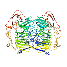
Dimer structure of Sortilin ectodomain crystal form 1, 2.3A
Descriptor: 2-acetamido-2-deoxy-beta-D-glucopyranose, 2-acetamido-2-deoxy-beta-D-glucopyranose-(1-4)-2-acetamido-2-deoxy-beta-D-glucopyranose, CHLORIDE ION, ...
Authors:Leloup, N.O.L, Janssen, B.J.C.
Deposit date:2017-04-07
Release date:2017-11-29
Last modified:2024-01-17
Method:X-RAY DIFFRACTION (2.3 Å)
Cite:Low pH-induced conformational change and dimerization of sortilin triggers endocytosed ligand release.
Nat Commun, 8, 2017
5O0K
DownloadVisualize
BU of 5o0k by Molmil
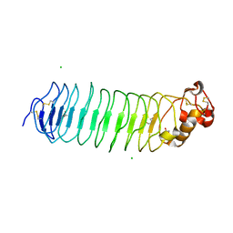
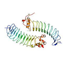
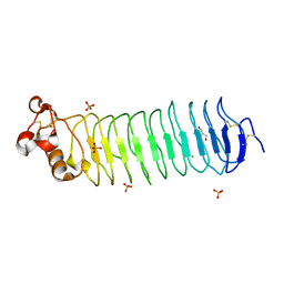
Deglycosylated Nogo Receptor with native disulfide structure
Descriptor: 2-acetamido-2-deoxy-beta-D-glucopyranose, CHLORIDE ION, GLYCEROL, ...
Authors:Pronker, M.F, Janssen, B.J.C.
Deposit date:2017-05-16
Release date:2017-10-18
Last modified:2024-01-17
Method:X-RAY DIFFRACTION (2.3 Å)
Cite:Nogo Receptor crystal structures with a native disulfide pattern suggest a novel mode of self-interaction.
Acta Crystallogr D Struct Biol, 73, 2017
5O0M
DownloadVisualize
BU of 5o0m by Molmil
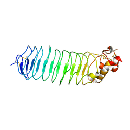
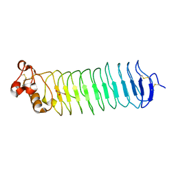
Deglycosylated Nogo Receptor with native disulfide structure 3
Descriptor: 2-acetamido-2-deoxy-beta-D-glucopyranose, Reticulon-4 receptor
Authors:Pronker, M.F, Janssen, B.J.C.
Deposit date:2017-05-16
Release date:2017-10-18
Last modified:2024-10-09
Method:X-RAY DIFFRACTION (1.9 Å)
Cite:Nogo Receptor crystal structures with a native disulfide pattern suggest a novel mode of self-interaction.
Acta Crystallogr D Struct Biol, 73, 2017
5O0R
DownloadVisualize
BU of 5o0r by Molmil
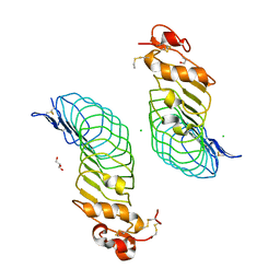
Deglycosylated Nogo Receptor with native disulfide structure
Descriptor: 2-acetamido-2-deoxy-beta-D-glucopyranose, CHLORIDE ION, GLYCEROL, ...
Authors:Pronker, M.F, Janssen, B.J.C.
Deposit date:2017-05-16
Release date:2017-11-08
Last modified:2024-10-23
Method:X-RAY DIFFRACTION (2.5 Å)
Cite:Nogo Receptor crystal structures with a native disulfide pattern suggest a novel mode of self-interaction.
Acta Crystallogr D Struct Biol, 73, 2017
5O0O
DownloadVisualize
BU of 5o0o by Molmil
Deglycosylated Nogo Receptor with native disulfide structure 5
Descriptor: 2-acetamido-2-deoxy-beta-D-glucopyranose, CHLORIDE ION, GLYCEROL, ...
Authors:Pronker, M.F, Janssen, B.J.C.
Deposit date:2017-05-16
Release date:2017-10-18
Last modified:2024-01-17
Method:X-RAY DIFFRACTION (2.2 Å)
Cite:Nogo Receptor crystal structures with a native disulfide pattern suggest a novel mode of self-interaction.
Acta Crystallogr D Struct Biol, 73, 2017
5NNI
DownloadVisualize
BU of 5nni by Molmil
Dimer structure of Sortilin ectodomain crystal form 2, 3.2 Angstrom
Descriptor: 2-acetamido-2-deoxy-beta-D-glucopyranose, 2-acetamido-2-deoxy-beta-D-glucopyranose-(1-4)-2-acetamido-2-deoxy-beta-D-glucopyranose, Sortilin, ...
Authors:Leloup, N.O.L, Janssen, B.J.C.
Deposit date:2017-04-09
Release date:2017-11-29
Last modified:2024-01-17
Method:X-RAY DIFFRACTION (3.21 Å)
Cite:Low pH-induced conformational change and dimerization of sortilin triggers endocytosed ligand release.
Nat Commun, 8, 2017
5O0L
DownloadVisualize
BU of 5o0l by Molmil
Deglycosylated Nogo Receptor with native disulfide structure 2
Descriptor: 2-acetamido-2-deoxy-beta-D-glucopyranose, CHLORIDE ION, Reticulon-4 receptor
Authors:Pronker, M.F, Janssen, B.J.C.
Deposit date:2017-05-16
Release date:2017-10-18
Last modified:2024-11-06
Method:X-RAY DIFFRACTION (2.511 Å)
Cite:Nogo Receptor crystal structures with a native disulfide pattern suggest a novel mode of self-interaction.
Acta Crystallogr D Struct Biol, 73, 2017
5O0P
DownloadVisualize
BU of 5o0p by Molmil
Deglycosylated Nogo Receptor with native disulfide structure 6
Descriptor: 2-acetamido-2-deoxy-beta-D-glucopyranose, GLYCEROL, Reticulon-4 receptor, ...
Authors:Pronker, M.F, Janssen, B.J.C.
Deposit date:2017-05-16
Release date:2017-10-18
Last modified:2024-10-09
Method:X-RAY DIFFRACTION (2 Å)
Cite:Nogo Receptor crystal structures with a native disulfide pattern suggest a novel mode of self-interaction.
Acta Crystallogr D Struct Biol, 73, 2017
5O0Z
DownloadVisualize
BU of 5o0z by Molmil
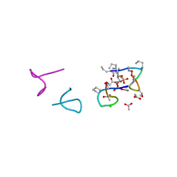
Structure of laspartomycin C in complex with geranyl-phosphate
Descriptor: ACETIC ACID, CALCIUM ION, CHLORIDE ION, ...
Authors:Vlieg, H.C, Kleijn, L.H.J, Martin, N.I, Janssen, B.J.C.
Deposit date:2017-05-17
Release date:2017-11-15
Last modified:2024-08-07
Method:X-RAY DIFFRACTION (1.28 Å)
Cite:A High-Resolution Crystal Structure that Reveals Molecular Details of Target Recognition by the Calcium-Dependent Lipopeptide Antibiotic Laspartomycin C.
Angew. Chem. Int. Ed. Engl., 56, 2017
5O0N
DownloadVisualize
BU of 5o0n by Molmil
Deglycosylated Nogo Receptor with native disulfide structure 4
Descriptor: 2-acetamido-2-deoxy-beta-D-glucopyranose-(1-4)-2-acetamido-2-deoxy-beta-D-glucopyranose, CHLORIDE ION, Reticulon-4 receptor, ...
Authors:Pronker, M.F, Tas, R.P, Vlieg, H.C, Janssen, B.J.C.
Deposit date:2017-05-16
Release date:2017-10-18
Last modified:2024-10-16
Method:X-RAY DIFFRACTION (2.5 Å)
Cite:Nogo Receptor crystal structures with a native disulfide pattern suggest a novel mode of self-interaction.
Acta Crystallogr D Struct Biol, 73, 2017
5O0Q
DownloadVisualize
BU of 5o0q by Molmil
Deglycosylated Nogo Receptor with native disulfide structure
Descriptor: 2-acetamido-2-deoxy-beta-D-glucopyranose, CHLORIDE ION, GLYCEROL, ...
Authors:Pronker, M.F, Janssen, B.J.C.
Deposit date:2017-05-16
Release date:2017-10-18
Last modified:2024-10-16
Method:X-RAY DIFFRACTION (2.5 Å)
Cite:Nogo Receptor crystal structures with a native disulfide pattern suggest a novel mode of self-interaction.
Acta Crystallogr D Struct Biol, 73, 2017
5NNJ
DownloadVisualize
BU of 5nnj by Molmil
Dimer structure of Sortilin ectodomain crystal form 3, 4.0 Angstrom
Descriptor: 2-acetamido-2-deoxy-beta-D-glucopyranose, Sortilin, beta-D-mannopyranose-(1-4)-2-acetamido-2-deoxy-beta-D-glucopyranose-(1-4)-2-acetamido-2-deoxy-beta-D-glucopyranose
Authors:Leloup, N.O.L, Janssen, B.J.C.
Deposit date:2017-04-09
Release date:2017-11-29
Last modified:2024-01-17
Method:X-RAY DIFFRACTION (4 Å)
Cite:Low pH-induced conformational change and dimerization of sortilin triggers endocytosed ligand release.
Nat Commun, 8, 2017
5NMR
DownloadVisualize
BU of 5nmr by Molmil
Monomeric mouse Sortilin extracellular domain
Descriptor: 2-acetamido-2-deoxy-beta-D-glucopyranose, 2-acetamido-2-deoxy-beta-D-glucopyranose-(1-4)-2-acetamido-2-deoxy-beta-D-glucopyranose, CALCIUM ION, ...
Authors:Leloup, N.O.L, Loessl, P, Meijer, D.H.M, Heck, A.J.R, Thies-Weesie, D.M.E, Janssen, B.J.C.
Deposit date:2017-04-07
Release date:2017-12-06
Last modified:2024-01-17
Method:X-RAY DIFFRACTION (2.1 Å)
Cite:Low pH-induced conformational change and dimerization of sortilin triggers endocytosed ligand release.
Nat Commun, 8, 2017
6QHJ
DownloadVisualize
BU of 6qhj by Molmil
High-resolution crystal structure of calcium- and sodium-bound mouse Olfactomedin-1 beta-propeller domain
Descriptor: 2-acetamido-2-deoxy-beta-D-glucopyranose, CALCIUM ION, GLYCEROL, ...
Authors:Pronker, M.F, van den Hoek, H.G, Janssen, B.J.C.
Deposit date:2019-01-16
Release date:2019-09-11
Last modified:2024-01-24
Method:X-RAY DIFFRACTION (1.25 Å)
Cite:Design and structural characterisation of olfactomedin-1 variants as tools for functional studies.
BMC Mol Cell Biol, 20, 2019
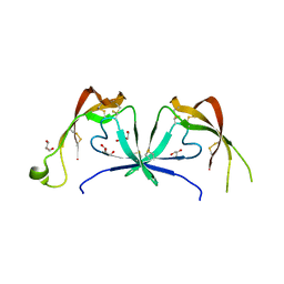
6STE
DownloadVisualize
BU of 6ste by Molmil
Crystal structure of the tick chemokine-binding protein Evasin-4 (SG 3)
Descriptor: ACETATE ION, DI(HYDROXYETHYL)ETHER, Evasin-4, ...
Authors:Ramirez-Escudero, M, Janssen, B.J.C.
Deposit date:2019-09-10
Release date:2020-09-02
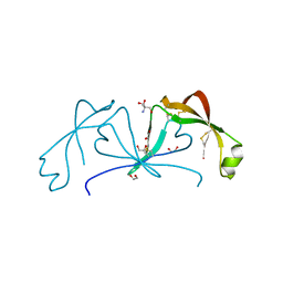
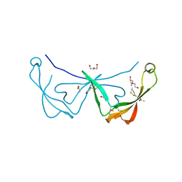
Last modified:2024-11-06
Method:X-RAY DIFFRACTION (1.79 Å)
Cite:Structural characterization of anti-CCL5 activity of the tick salivary protein evasin-4.
J.Biol.Chem., 295, 2020
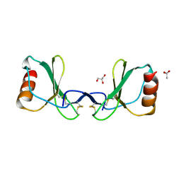
6STK
DownloadVisualize
BU of 6stk by Molmil
Crystal structure of the CC-chemokine 5 (CCL5) E66S mutation
Descriptor: ACETATE ION, C-C motif chemokine 5, GLYCEROL
Authors:Ramirez-Escudero, M, Janssen, B.J.C.
Deposit date:2019-09-10
Release date:2020-09-02
Last modified:2024-10-09
Method:X-RAY DIFFRACTION (1.52 Å)
Cite:Structural characterization of anti-CCL5 activity of the tick salivary protein evasin-4.
J.Biol.Chem., 295, 2020
6ST4
DownloadVisualize
BU of 6st4 by Molmil
Crystal structure of the tick chemokine-binding protein Evasin-4 (SG 1)
Descriptor: 1,2-ETHANEDIOL, 2-AMINO-2-HYDROXYMETHYL-PROPANE-1,3-DIOL, Evasin-4
Authors:Ramirez-Escudero, M, Janssen, B.J.C.
Deposit date:2019-09-10
Release date:2020-09-02
Last modified:2024-10-23
Method:X-RAY DIFFRACTION (1.29 Å)
Cite:Structural characterization of anti-CCL5 activity of the tick salivary protein evasin-4.
J.Biol.Chem., 295, 2020
6STC
DownloadVisualize
BU of 6stc by Molmil
Crystal structure of the tick chemokine-binding protein Evasin-4 (SG 2)
Descriptor: ACETATE ION, DI(HYDROXYETHYL)ETHER, Evasin-4
Authors:Ramirez-Escudero, M, Janssen, B.J.C.
Deposit date:2019-09-10
Release date:2020-09-02
Last modified:2024-01-24
Method:X-RAY DIFFRACTION (1.69 Å)
Cite:Structural characterization of anti-CCL5 activity of the tick salivary protein evasin-4.
J.Biol.Chem., 295, 2020
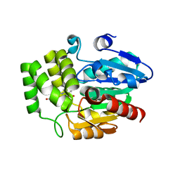
6AP7
DownloadVisualize
BU of 6ap7 by Molmil
Crystal Structure of DAD2 in complex with 2-(2-methyl-3-nitroanilino)benzoic acid
Descriptor: 2-[(2-methyl-3-nitrophenyl)amino]benzoic acid, GLYCEROL, Probable strigolactone esterase DAD2
Authors:Hamiaux, C.
Deposit date:2017-08-17
Release date:2018-03-21
Last modified:2023-10-04
Method:X-RAY DIFFRACTION (1.51 Å)
Cite:Inhibition of strigolactone receptors byN-phenylanthranilic acid derivatives: Structural and functional insights.
J. Biol. Chem., 293, 2018
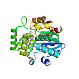
6AP6
DownloadVisualize
BU of 6ap6 by Molmil
Crystal Structure of DAD2 in complex with tolfenamic acid
Descriptor: 2-[(3-chloro-2-methylphenyl)amino]benzoic acid, Probable strigolactone esterase DAD2
Authors:Hamiaux, C.
Deposit date:2017-08-17
Release date:2018-03-21
Last modified:2023-10-04
Method:X-RAY DIFFRACTION (1.65 Å)
Cite:Inhibition of strigolactone receptors byN-phenylanthranilic acid derivatives: Structural and functional insights.
J. Biol. Chem., 293, 2018

227111

數據於2024-11-06公開中

PDB statisticsPDBj update infoContact PDBjnumon