6DVU
 
 | |
6NF3
 | |
1VZV
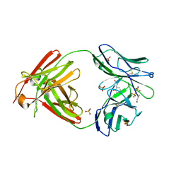
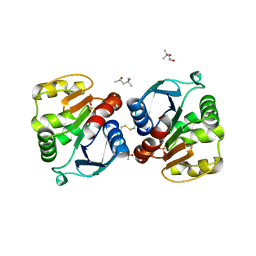
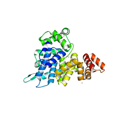
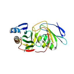
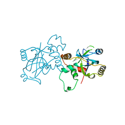 | | STRUCTURE OF VARICELLA-ZOSTER VIRUS PROTEASE | | Descriptor: | VARICELLA-ZOSTER VIRUS PROTEASE | | Authors: | Qiu, X, Jason, C.A, Culp, J.S, Richardson, S.B, Debouck, C, Smith, W.W, Abdel-Meguid, S.S. | | Deposit date: | 1997-02-10 | | Release date: | 1998-09-16 | | Last modified: | 2024-02-14 | | Method: | X-RAY DIFFRACTION (3 Å) | | Cite: | Crystal structure of varicella-zoster virus protease.
Proc.Natl.Acad.Sci.USA, 94, 1997
|
|
2OMB
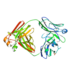
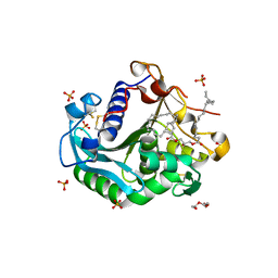
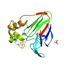
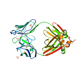 | | Bence Jones KWR Protein- Immunoglobulin Light Chain Dimer, P3(1)21 Crystal Form | | Descriptor: | Bence Jones KWR Protein - Immunoglobulin Light Chain, PHENOL, SULFATE ION | | Authors: | Makino, D.L, Larson, S.B, McPherson, A. | | Deposit date: | 2007-01-21 | | Release date: | 2007-07-17 | | Last modified: | 2023-08-30 | | Method: | X-RAY DIFFRACTION (2.9 Å) | | Cite: | Bence Jones KWR protein structures determined by X-ray crystallography.
Acta Crystallogr.,Sect.D, 63, 2007
|
|
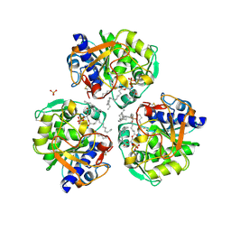
2OLD
 | | Bence Jones KWR Protein- Immunoglobulin Light Chain Dimer, P3(2)21 Crystal Form | | Descriptor: | Bence Jones KWR Protein - Immunoglobulin Light Chain, PHENOL, PHOSPHATE ION | | Authors: | Makino, D.L, Larson, S.B, McPherson, A. | | Deposit date: | 2007-01-19 | | Release date: | 2007-07-17 | | Last modified: | 2023-08-30 | | Method: | X-RAY DIFFRACTION (2.6 Å) | | Cite: | Bence Jones KWR protein structures determined by X-ray crystallography.
Acta Crystallogr.,Sect.D, 63, 2007
|
|
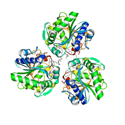
2OMN
 | | Bence Jones KWR Protein- Immunoglobulin Light Chain Dimer, P4(3)2(1)2 Crystal Form | | Descriptor: | Bence Jones KWR Protein - Immunoglobulin Light Chain, PHENOL, SULFATE ION | | Authors: | Makino, D.L, Larson, S.B, McPherson, A. | | Deposit date: | 2007-01-22 | | Release date: | 2007-07-17 | | Last modified: | 2023-08-30 | | Method: | X-RAY DIFFRACTION (2.2 Å) | | Cite: | Bence Jones KWR protein structures determined by X-ray crystallography.
Acta Crystallogr.,Sect.D, 63, 2007
|
|
1AUY
 | |
5CVZ
 | |
5CW0
 | |
1IGT
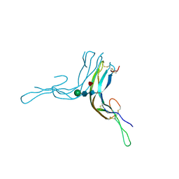
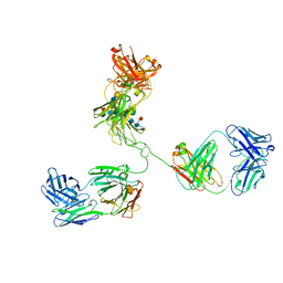 | | STRUCTURE OF IMMUNOGLOBULIN | | Descriptor: | IGG2A INTACT ANTIBODY - MAB231, beta-D-galactopyranose-(1-4)-2-acetamido-2-deoxy-beta-D-glucopyranose-(1-2)-alpha-D-mannopyranose-(1-6)-[2-acetamido-2-deoxy-beta-D-glucopyranose-(1-2)-alpha-D-mannopyranose-(1-3)]beta-D-mannopyranose-(1-4)-2-acetamido-2-deoxy-beta-D-glucopyranose-(1-4)-[alpha-L-fucopyranose-(1-6)]2-acetamido-2-deoxy-beta-D-glucopyranose, beta-D-galactopyranose-(1-4)-2-acetamido-2-deoxy-beta-D-glucopyranose-(1-2)-alpha-D-mannopyranose-(1-6)-[2-acetamido-2-deoxy-beta-D-glucopyranose-(1-2)-alpha-D-mannopyranose-(1-3)]beta-D-mannopyranose-(1-4)-2-acetamido-2-deoxy-beta-D-glucopyranose-(1-4)-[beta-L-fucopyranose-(1-6)]2-acetamido-2-deoxy-beta-D-glucopyranose | | Authors: | Harris, L.J, Larson, S.B, Hasel, K.W, McPherson, A. | | Deposit date: | 1996-10-25 | | Release date: | 1997-07-07 | | Last modified: | 2023-08-09 | | Method: | X-RAY DIFFRACTION (2.8 Å) | | Cite: | Refined structure of an intact IgG2a monoclonal antibody.
Biochemistry, 36, 1997
|
|
1JS9
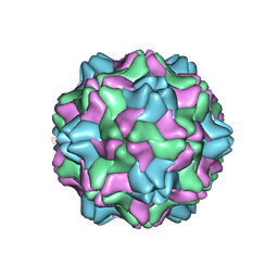
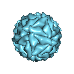
 | | Brome Mosaic Virus | | Descriptor: | Coat protein, HEXAETHYLENE GLYCOL, MAGNESIUM ION | | Authors: | Lucas, R.W, Larson, S.B, McPherson, A. | | Deposit date: | 2001-08-16 | | Release date: | 2002-04-03 | | Last modified: | 2023-08-16 | | Method: | X-RAY DIFFRACTION (3.4 Å) | | Cite: | The crystallographic structure of brome mosaic virus.
J.Mol.Biol., 317, 2002
|
|
1LAJ
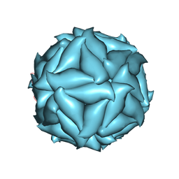
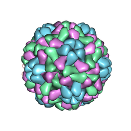
 | | The Structure of Tomato Aspermy Virus by X-Ray Crystallography | | Descriptor: | 5'-R(*AP*AP*A)-3', MAGNESIUM ION, PHOSPHATE ION, ... | | Authors: | Lucas, R.W, Larson, S.B, Canady, M.A, McPherson, A. | | Deposit date: | 2002-03-28 | | Release date: | 2002-11-27 | | Last modified: | 2023-08-16 | | Method: | X-RAY DIFFRACTION (3.4 Å) | | Cite: | The Structure of Tomato Aspermy Virus by X-Ray Crystallography
J.STRUCT.BIOL., 139, 2002
|
|
7R66
 | |
6P57
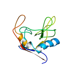
 | | Crystal Structure of the Beta Subunit of Luteinizing Hormone | | Descriptor: | 2-acetamido-2-deoxy-beta-D-glucopyranose-(1-2)-alpha-D-mannopyranose-(1-6)-beta-D-mannopyranose-(1-4)-2-acetamido-2-deoxy-beta-D-glucopyranose-(1-4)-[beta-L-fucopyranose-(1-6)]2-acetamido-2-deoxy-beta-D-glucopyranose, Lutropin subunit beta, alpha-D-mannopyranose, ... | | Authors: | McPherson, A. | | Deposit date: | 2019-05-29 | | Release date: | 2019-12-11 | | Last modified: | 2023-10-11 | | Method: | X-RAY DIFFRACTION (3.16 Å) | | Cite: | The Crystal Structure of the Beta Subunit of Luteinizing Hormone and a Model for the Intact Hormone
Curr Res Struct Biol, 2019
|
|
1CLH
 | |
6C6W
 | | Structure of a Complex Between Thaumatin and Thioflavin T | | Descriptor: | 2-[4-(dimethylamino)phenyl]-3,6-dimethyl-1,3-benzothiazol-3-ium, L(+)-TARTARIC ACID, Thaumatin I | | Authors: | McPherson, A. | | Deposit date: | 2018-01-19 | | Release date: | 2018-02-07 | | Last modified: | 2018-09-19 | | Method: | X-RAY DIFFRACTION (2.23 Å) | | Cite: | Investigation into the binding of dyes within protein crystals.
Acta Crystallogr F Struct Biol Commun, 74, 2018
|
|
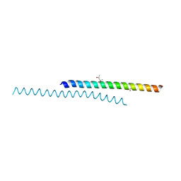
5UKH
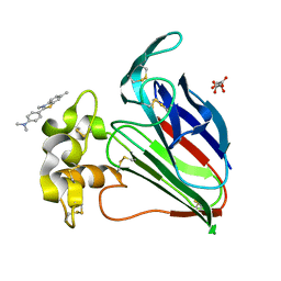
 | | Structure of TelC from Streptococcus intermedius B196 | | Descriptor: | CALCIUM ION, Uncharacterized protein | | Authors: | Whitney, J.C, Ching, M.Q, Bryant, D, Mougous, J.D. | | Deposit date: | 2017-01-22 | | Release date: | 2017-07-26 | | Last modified: | 2019-12-11 | | Method: | X-RAY DIFFRACTION (1.98 Å) | | Cite: | A broadly distributed toxin family mediates contact-dependent antagonism between gram-positive bacteria.
Elife, 6, 2017
|
|
6XOK
 | |
6XRV
 | |
6XS3
 | |
6U08
 | |
5LIP
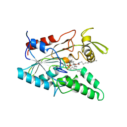 | | PSEUDOMONAS LIPASE COMPLEXED WITH RC-(RP, SP)-1,2-DIOCTYLCARBAMOYLGLYCERO-3-O-OCTYLPHOSPHONATE | | Descriptor: | CALCIUM ION, OCTYL-PHOSPHINIC ACID 1,2-BIS-OCTYLCARBAMOYLOXY-ETHYL ESTER, TRIACYL-GLYCEROL HYDROLASE | | Authors: | Lang, D.A, Dijkstra, B.W. | | Deposit date: | 1997-09-02 | | Release date: | 1998-08-19 | | Last modified: | 2023-08-09 | | Method: | X-RAY DIFFRACTION (2.9 Å) | | Cite: | Structural basis of the chiral selectivity of Pseudomonas cepacia lipase
Eur.J.Biochem., 254, 1998
|
|
6DXR
 | |
6DK5
 | |
6E0D
 | | X-ray structure of a complex of thaumatin with xylene cyanol | | Descriptor: | (1R,2R,3S,4R,6S)-4,6-diamino-2,3-dihydroxycyclohexyl 2,6-diamino-2,6-dideoxy-alpha-D-glucopyranoside, L(+)-TARTARIC ACID, Thaumatin I | | Authors: | McPherson, A. | | Deposit date: | 2018-07-06 | | Release date: | 2018-09-19 | | Last modified: | 2020-07-29 | | Method: | X-RAY DIFFRACTION (2.24 Å) | | Cite: | Investigation into the binding of dyes within protein crystals.
Acta Crystallogr F Struct Biol Commun, 74, 2018
|
|


