Loading
PDBj
MenuPDBj@FacebookPDBj@TwitterPDBj@YouTubewwPDB FoundationwwPDB
RCSB PDBPDBeBMRBAdv. SearchSearch help
Search by PDB author
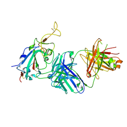
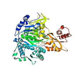
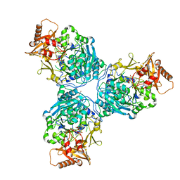
7S49
DownloadVisualize
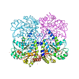
BU of 7s49 by Molmil
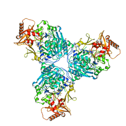
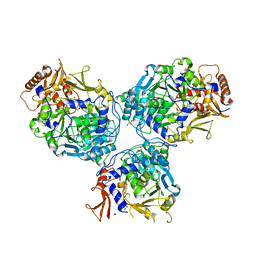
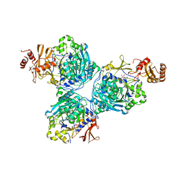
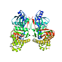
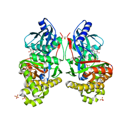
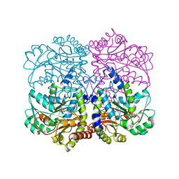
Crystal Structure of Inhibitor-bound Galactokinase
Descriptor: (4R)-2-[(1,3-benzoxazol-2-yl)amino]-4-(4-chloro-1H-pyrazol-5-yl)-4,6,7,8-tetrahydroquinazolin-5(1H)-one, Galactokinase, PHOSPHATE ION, ...
Authors:Whitby, F.G.
Deposit date:2021-09-08
Release date:2021-09-29
Last modified:2023-10-25
Method:X-RAY DIFFRACTION (2.2 Å)
Cite:Structure-Based Optimization of Small Molecule Human Galactokinase Inhibitors.
J.Med.Chem., 64, 2021
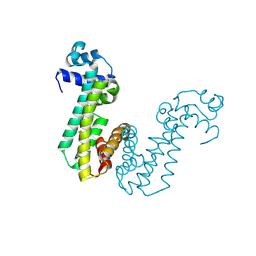
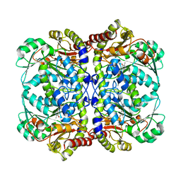
3DKE
DownloadVisualize
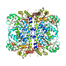
BU of 3dke by Molmil
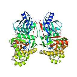
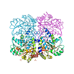
Polar and non-polar cavities in phage T4 lysozyme
Descriptor: 2-HYDROXYETHYL DISULFIDE, 4-(2-HYDROXYETHYL)-1-PIPERAZINE ETHANESULFONIC ACID, AZIDE ION, ...
Authors:Liu, L.J, Matthews, B.W.
Deposit date:2008-06-24
Release date:2008-11-25
Last modified:2021-10-20
Method:X-RAY DIFFRACTION (1.25 Å)
Cite:Use of experimental crystallographic phases to examine the hydration of polar and nonpolar cavities in T4 lysozyme
Proc.Natl.Acad.Sci.USA, 105, 2008
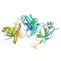
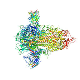
7TNI
DownloadVisualize
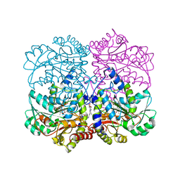
BU of 7tni by Molmil
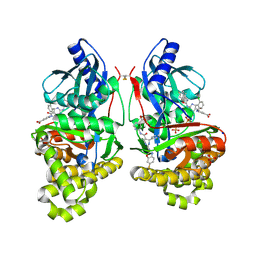
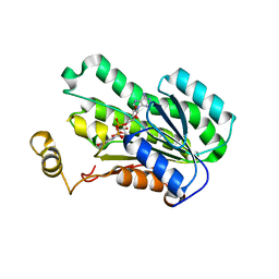
Structure of EC12 Y1392W variant of BT-R1 from Manduca sexta, a Cry1A toxin binding domain
Descriptor: Cadherin, MAGNESIUM ION
Authors:Fisher, A.J, Wilcox, X.E.
Deposit date:2022-01-21
Release date:2022-05-04
Last modified:2023-10-18
Method:X-RAY DIFFRACTION (1.9 Å)
Cite:Functional and Structural Analysis of the Toxin-Binding Site of the Cadherin G-Protein-Coupled Receptor, BT-R 1 , for Cry1A Toxins of Bacillus thuringiensis .
Biochemistry, 61, 2022
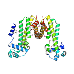
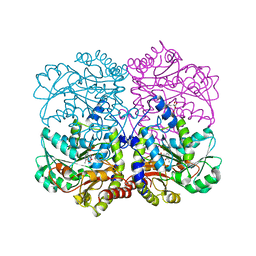
4MO7
DownloadVisualize
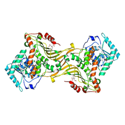
BU of 4mo7 by Molmil
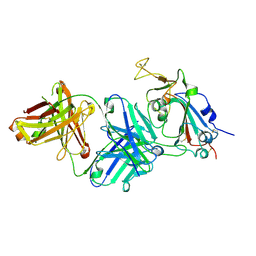
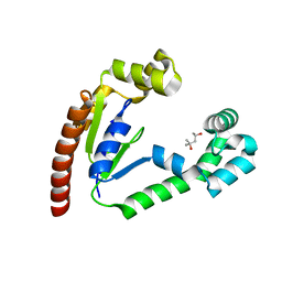
Crystal structure of superantigen PfiT
Descriptor: BETA-MERCAPTOETHANOL, MAGNESIUM ION, Transcriptional regulator I2
Authors:Liu, L.H, Chen, H, Li, H.M.
Deposit date:2013-09-11
Release date:2014-02-05
Method:X-RAY DIFFRACTION (1.701 Å)
Cite:Pfit is a structurally novel Crohn's disease-associated superantigen.
Plos Pathog., 9, 2013
8DQC
DownloadVisualize
BU of 8dqc by Molmil
Crystal structure of 3-dehydroquinate dehydratase I from Klebsiella oxytoca (I222 Form)
Descriptor: 3-dehydroquinate dehydratase I, ACETATE ION, ZINC ION
Authors:Seattle Structural Genomics Center for Infectious Disease, Seattle Structural Genomics Center for Infectious Disease (SSGCID)
Deposit date:2022-07-18
Release date:2022-07-27
Last modified:2023-11-22
Method:X-RAY DIFFRACTION (2.7 Å)
Cite:Crystal structure of 3-dehydroquinate dehydratase I from Klebsiella oxytoca (I222 Form)
To be published
8DQB
DownloadVisualize
BU of 8dqb by Molmil
Crystal structure of 3-dehydroquinate dehydratase I from Klebsiella oxytoca (I23 Form)
Descriptor: 3-dehydroquinate dehydratase I, ZINC ION
Authors:Seattle Structural Genomics Center for Infectious Disease, Seattle Structural Genomics Center for Infectious Disease (SSGCID)
Deposit date:2022-07-18
Release date:2022-07-27
Last modified:2023-11-22
Method:X-RAY DIFFRACTION (2.5 Å)
Cite:Crystal structure of 3-dehydroquinate dehydratase I from Klebsiella oxytoca (I23 Form)
To be published
8DOQ
DownloadVisualize
BU of 8doq by Molmil
Crystal structure of 2,3-diketo-5-methylthiopentyl-1-phosphate enolase-phosphatase from Klebsiella aerogenes (P21 Form)
Descriptor: (4S)-2-METHYL-2,4-PENTANEDIOL, 2,3-diketo-5-methylthiopentyl-1-phosphate enolase-phosphatase, BENZOIC ACID
Authors:Seattle Structural Genomics Center for Infectious Disease (SSGCID)
Deposit date:2022-07-14
Release date:2022-07-27
Last modified:2023-11-22
Method:X-RAY DIFFRACTION (1.65 Å)
Cite:Crystal structure of 2,3-diketo-5-methylthiopentyl-1-phosphate enolase-phosphatase from Klebsiella aerogenes (P1 Form)
To be published
8DQ9
DownloadVisualize
BU of 8dq9 by Molmil
Crystal structure of GDP bound 3-dehydroquinate dehydratase I from Klebsiella oxytoca
Descriptor: 3-dehydroquinate dehydratase I, CHLORIDE ION, GUANOSINE-5'-DIPHOSPHATE, ...
Authors:Seattle Structural Genomics Center for Infectious Disease, Seattle Structural Genomics Center for Infectious Disease (SSGCID)
Deposit date:2022-07-18
Release date:2022-07-27
Last modified:2023-11-22
Method:X-RAY DIFFRACTION (2.8 Å)
Cite:Crystal structure of GDP bound 3-dehydroquinate dehydratase I from Klebsiella oxytoca
To be published
8SA8
DownloadVisualize
BU of 8sa8 by Molmil
Crystal Structure of Cystathionine beta lyase from Klebsiella aerogenes, Covalently bound and free PLP (I2 form)
Descriptor: 1,2-ETHANEDIOL, CHLORIDE ION, Cystathionine beta-lyase, ...
Authors:Seattle Structural Genomics Center for Infectious Disease (SSGCID)
Deposit date:2023-03-31
Release date:2023-04-12
Method:X-RAY DIFFRACTION (1.3 Å)
Cite:Crystal Structure of Cystathionine beta lyase from Klebsiella aerogenes (I2 form)
To be published
8SA9
DownloadVisualize
BU of 8sa9 by Molmil
Crystal Structure of Cystathionine beta lyase from Klebsiella aerogenes, PLP-Oxamate Adduct (C2 form)
Descriptor: CITRATE ANION, Cystathionine beta-lyase, PYRIDOXAL-5'-PHOSPHATE, ...
Authors:Seattle Structural Genomics Center for Infectious Disease (SSGCID)
Deposit date:2023-03-31
Release date:2023-04-12
Method:X-RAY DIFFRACTION (1.1 Å)
Cite:Crystal Structure of Cystathionine beta lyase from Klebsiella aerogenes, PLP-Oxamate Adduct (C2 form)
To be published
8SBZ
DownloadVisualize
BU of 8sbz by Molmil
Crystal Structure of 2,3-dihydro-2,3-dihydroxybenzoate dehydrogenase from Klebsiella aerogenes (NAD bound, No sulfate hexagonal form)
Descriptor: 2,3-dihydroxybenzoate-2,3-dehydrogenase, NICOTINAMIDE-ADENINE-DINUCLEOTIDE
Authors:Seattle Structural Genomics Center for Infectious Disease (SSGCID)
Deposit date:2023-04-04
Release date:2023-04-12
Last modified:2024-05-22
Method:X-RAY DIFFRACTION (1.9 Å)
Cite:Crystal Structure of 2,3-dihydro-2,3-dihydroxybenzoate dehydrogenase from Klebsiella aerogenes (NAD bound, No sulfate hexagonal form)
To be published
8SAB
DownloadVisualize
BU of 8sab by Molmil
Crystal Structure of Cystathionine beta lyase from Klebsiella aerogenes, PLP adduct with Alanine (C2 form)
Descriptor: (4S)-2-METHYL-2,4-PENTANEDIOL, 2-AMINO-2-HYDROXYMETHYL-PROPANE-1,3-DIOL, ALANYL-PYRIDOXAL-5'-PHOSPHATE, ...
Authors:Seattle Structural Genomics Center for Infectious Disease (SSGCID)
Deposit date:2023-03-31
Release date:2023-04-12
Method:X-RAY DIFFRACTION (1.6 Å)
Cite:Crystal Structure of Cystathionine beta lyase from Klebsiella aerogenes, PLP adduct with Alanine (C2 form)
To be published
8SAD
DownloadVisualize
BU of 8sad by Molmil
Crystal Structure of Cystathionine beta lyase from Klebsiella aerogenes, PLP/Malonate complex (C2 form)
Descriptor: (4S)-2-METHYL-2,4-PENTANEDIOL, CHLORIDE ION, Cystathionine beta-lyase, ...
Authors:Seattle Structural Genomics Center for Infectious Disease (SSGCID)
Deposit date:2023-03-31
Release date:2023-04-12
Method:X-RAY DIFFRACTION (1.4 Å)
Cite:Crystal Structure of Cystathionine beta lyase from Klebsiella aerogenes, PLP/Malonate complex (C2 form)
To be published
8SA7
DownloadVisualize
BU of 8sa7 by Molmil
Crystal Structure of Cystathionine beta lyase from Klebsiella aerogenes (C2 form)
Descriptor: BROMIDE ION, CHLORIDE ION, Cystathionine beta-lyase, ...
Authors:Seattle Structural Genomics Center for Infectious Disease (SSGCID)
Deposit date:2023-03-31
Release date:2023-04-12
Method:X-RAY DIFFRACTION (1.4 Å)
Cite:Crystal Structure of Cystathionine beta lyase from Klebsiella aerogenes (C2 form)
To be published
8SAA
DownloadVisualize
BU of 8saa by Molmil
Crystal Structure of Cystathionine beta lyase from Klebsiella aerogenes, PLP and phosphate bound (C2 form)
Descriptor: Cystathionine beta-lyase, MAGNESIUM ION, PHOSPHATE ION, ...
Authors:Seattle Structural Genomics Center for Infectious Disease (SSGCID)
Deposit date:2023-03-31
Release date:2023-04-12
Method:X-RAY DIFFRACTION (1.45 Å)
Cite:Crystal Structure of Cystathionine beta lyase from Klebsiella aerogenes, PLP and phosphate bound (C2 form)
To be published
8SAE
DownloadVisualize
BU of 8sae by Molmil
Crystal Structure of Cystathionine beta lyase from Klebsiella aerogenes, PLP and Hepes bound (C2 form)
Descriptor: (4S)-2-METHYL-2,4-PENTANEDIOL, 4-(2-HYDROXYETHYL)-1-PIPERAZINE ETHANESULFONIC ACID, Cystathionine beta-lyase, ...
Authors:Seattle Structural Genomics Center for Infectious Disease (SSGCID)
Deposit date:2023-03-31
Release date:2023-04-12
Method:X-RAY DIFFRACTION (1.5 Å)
Cite:Crystal Structure of Cystathionine beta lyase from Klebsiella aerogenes, PLP and Hepes bound (C2 form)
To be published
8SAC
DownloadVisualize
BU of 8sac by Molmil
Crystal Structure of Cystathionine beta lyase from Klebsiella aerogenes (P21212 form)
Descriptor: Cystathionine beta-lyase, MAGNESIUM ION, PYRIDOXAL-5'-PHOSPHATE
Authors:Seattle Structural Genomics Center for Infectious Disease (SSGCID)
Deposit date:2023-03-31
Release date:2023-04-12
Method:X-RAY DIFFRACTION (1.8 Å)
Cite:Crystal Structure of Cystathionine beta lyase from Klebsiella aerogenes (P21212 form)
To be published
8SBO
DownloadVisualize
BU of 8sbo by Molmil
Crystal Structure of Dephospho-CoA kinase from Klebsiella aerogenes (P21 Form 2)
Descriptor: (4S)-2-METHYL-2,4-PENTANEDIOL, CHLORIDE ION, Dephospho-CoA kinase
Authors:Seattle Structural Genomics Center for Infectious Disease (SSGCID)
Deposit date:2023-04-03
Release date:2023-04-12
Last modified:2024-05-22
Method:X-RAY DIFFRACTION (1.4 Å)
Cite:Crystal Structure of Dephospho-CoA kinase from Klebsiella aerogenes (P21 Form 2)
To be published
8SC0
DownloadVisualize
BU of 8sc0 by Molmil
Crystal Structure of 2,3-dihydro-2,3-dihydroxybenzoate dehydrogenase from Klebsiella aerogenes (NAD bound, orthorhombic form)
Descriptor: 2,3-dihydroxybenzoate-2,3-dehydrogenase, GLYCEROL, NICOTINAMIDE-ADENINE-DINUCLEOTIDE
Authors:Seattle Structural Genomics Center for Infectious Disease (SSGCID)
Deposit date:2023-04-04
Release date:2023-04-12
Last modified:2024-05-22
Method:X-RAY DIFFRACTION (1.81 Å)
Cite:Crystal Structure of 2,3-dihydro-2,3-dihydroxybenzoate dehydrogenase from Klebsiella aerogenes (NAD bound, orthorhombic form)
To be published
8U2W
DownloadVisualize
BU of 8u2w by Molmil
Glucose-6-phosphate 1-dehydrogenase (G6PDH) from Crithidia fasciculata (NADP bound)
Descriptor: Acetyl-coenzyme A synthetase, NADP NICOTINAMIDE-ADENINE-DINUCLEOTIDE PHOSPHATE
Authors:Seattle Structural Genomics Center for Infectious Disease, Seattle Structural Genomics Center for Infectious Disease (SSGCID)
Deposit date:2023-09-06
Release date:2023-09-13
Method:X-RAY DIFFRACTION (2.35 Å)
Cite:Glucose-6-phosphate 1-dehydrogenase (G6PDH) from Crithidia fasciculata (NADP bound)
To be published
8U2S
DownloadVisualize
BU of 8u2s by Molmil
Crystal Structure of Acetyl-coenzyme A synthetase from Leishmania infantum (Ethyl AMP bound, P21 form)
Descriptor: 5'-O-[(S)-ethoxy(hydroxy)phosphoryl]adenosine, Acetyl-coenzyme A synthetase, CACODYLATE ION, ...
Authors:Seattle Structural Genomics Center for Infectious Disease, Seattle Structural Genomics Center for Infectious Disease (SSGCID)
Deposit date:2023-09-06
Release date:2023-09-13
Method:X-RAY DIFFRACTION (2.52 Å)
Cite:Crystal Structure of Acetyl-coenzyme A synthetase from Leishmania infantum (Ethyl AMP bound, P21 form)
To be published
8U2X
DownloadVisualize
BU of 8u2x by Molmil
Crystal Structure of NendoU (Uridylate-specific endoribonuclease, nsp15) from Betacoronavirus SARS-CoV-2 (H235A mutant)
Descriptor: 2-AMINO-2-HYDROXYMETHYL-PROPANE-1,3-DIOL, CHLORIDE ION, Uridylate-specific endoribonuclease nsp15
Authors:Seattle Structural Genomics Center for Infectious Disease, Seattle Structural Genomics Center for Infectious Disease (SSGCID)
Deposit date:2023-09-06
Release date:2023-09-13
Method:X-RAY DIFFRACTION (2.25 Å)
Cite:Crystal Structure of NendoU (Uridylate-specific endoribonuclease, nsp15) from Betacoronavirus SARS-CoV-2 (H235A mutant)
To be published
8U96
DownloadVisualize
BU of 8u96 by Molmil
Crystal Structure of Dephospho-CoA kinase from Klebsiella aerogenes (ATP bound)
Descriptor: (4S)-2-METHYL-2,4-PENTANEDIOL, ADENOSINE-5'-TRIPHOSPHATE, CHLORIDE ION, ...
Authors:Seattle Structural Genomics Center for Infectious Disease (SSGCID)
Deposit date:2023-09-18
Release date:2023-09-27
Method:X-RAY DIFFRACTION (1.5 Å)
Cite:Crystal Structure of Dephospho-CoA kinase from Klebsiella aerogenes (ATP bound)
To be published
8U98
DownloadVisualize
BU of 8u98 by Molmil
Crystal Structure of Cystathionine beta lyase from Klebsiella aerogenes (PLP-Glycine adduct)
Descriptor: 1,2-ETHANEDIOL, Cystathionine beta-lyase, GLYCINE, ...
Authors:Seattle Structural Genomics Center for Infectious Disease, Seattle Structural Genomics Center for Infectious Disease (SSGCID)
Deposit date:2023-09-18
Release date:2023-09-27
Method:X-RAY DIFFRACTION (1.4 Å)
Cite:Crystal Structure of Cystathionine beta lyase from Klebsiella aerogenes (PLP-Glycine adduct)
To be published
8U2U
DownloadVisualize
BU of 8u2u by Molmil
Crystal Structure of Acetyl-coenzyme A synthetase from Leishmania infantum (CoA, AMP and potassium bound)
Descriptor: ADENOSINE MONOPHOSPHATE, Acetyl-coenzyme A synthetase, CHLORIDE ION, ...
Authors:Seattle Structural Genomics Center for Infectious Disease, Seattle Structural Genomics Center for Infectious Disease (SSGCID)
Deposit date:2023-09-06
Release date:2023-09-13
Method:X-RAY DIFFRACTION (1.97 Å)
Cite:Crystal Structure of Acetyl-coenzyme A synthetase from Leishmania infantum (CoA, AMP and potassium bound)
To be published

221051

數據於2024-06-12公開中

PDB statisticsPDBj update infoContact PDBjnumon