Loading
PDBj
MenuPDBj@FacebookPDBj@TwitterPDBj@YouTubewwPDB FoundationwwPDB
RCSB PDBPDBeBMRBAdv. SearchSearch help
Search by PDB author
1ZL5
DownloadVisualize
BU of 1zl5 by Molmil
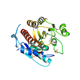
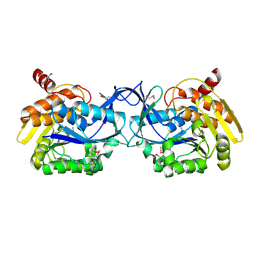
Crystal structure of Glu335Gln mutant of Clostridium botulinum neurotoxin E catalytic domain
Descriptor: CHLORIDE ION, botulinum neurotoxin type E
Authors:Agarwal, R, Binz, T, Swaminathan, S.
Deposit date:2005-05-05
Release date:2005-07-05
Last modified:2023-08-23
Method:X-RAY DIFFRACTION (2.6 Å)
Cite:Analysis of Active Site Residues of Botulinum Neurotoxin E by Mutational, Functional, and Structural Studies: Glu335Gln Is an Apoenzyme.
Biochemistry, 44, 2005
3KZH
DownloadVisualize
BU of 3kzh by Molmil
Crystal structure of a putative sugar kinase from Clostridium perfringens
Descriptor: Probable sugar kinase, beta-D-glucopyranose
Authors:Syed Ibrahim, B, Kumaran, D, Burley, S.K, Swaminathan, S, New York SGX Research Center for Structural Genomics (NYSGXRC)
Deposit date:2009-12-08
Release date:2009-12-22
Last modified:2021-02-10
Method:X-RAY DIFFRACTION (2.45 Å)
Cite:Crystal structure of a putative sugar kinase from Clostridium perfringens
To be Published
2A97
DownloadVisualize
BU of 2a97 by Molmil
Crystal structure of catalytic domain of Clostridium botulinum neurotoxin serotype F
Descriptor: Botulinum neurotoxin type F, CADMIUM ION, ZINC ION
Authors:Agarwal, R, Binz, T, Swaminathan, S.
Deposit date:2005-07-11
Release date:2005-09-20
Last modified:2023-08-23
Method:X-RAY DIFFRACTION (1.8 Å)
Cite:Structural analysis of botulinum neurotoxin serotype f light chain: implications on substrate binding and inhibitor design
Biochemistry, 44, 2005
4ZJX
DownloadVisualize
BU of 4zjx by Molmil
Crystal structure of the catalytic domain of botulinum neurotoxin serotype A with a Novel Cyclic Peptide Inhibitor
Descriptor: Botulinum neurotoxin type A, SULFATE ION, ZINC ION, ...
Authors:Kumaran, D, Swaminathan, S.
Deposit date:2015-04-29
Release date:2016-06-01
Last modified:2023-09-27
Method:X-RAY DIFFRACTION (1.94 Å)
Cite:Crystal structure of the catalytic domain of botulinum neurotoxin serotype A with a Novel Cyclic Peptide Inhibitor
To Be Published
3T69
DownloadVisualize
BU of 3t69 by Molmil
Crystal structure of a putative 2-dehydro-3-deoxygalactonokinase protein from Sinorhizobium meliloti
Descriptor: Putative 2-dehydro-3-deoxygalactonokinase, SULFATE ION
Authors:Agarwal, R, Chamala, S, Evans, B, Foti, R, Gizzi, A, Hillerich, B, Kar, A, LaFleur, J, Seidel, R, Villigas, G, Zencheck, W, Almo, S.C, Swaminathan, S, New York Structural Genomics Research Consortium (NYSGRC)
Deposit date:2011-07-28
Release date:2011-08-17
Last modified:2024-02-28
Method:X-RAY DIFFRACTION (2.55 Å)
Cite:Crystal structure of a putative 2-dehydro-3-deoxygalactonokinase protein from Sinorhizobium meliloti
To be Published
2AFA
DownloadVisualize
BU of 2afa by Molmil
Crystal Structure of putative NAG isomerase from Salmonella typhimurium
Descriptor: NAG isomerase
Authors:Kumaran, D, Swaminathan, S, Burley, S.K, New York SGX Research Center for Structural Genomics (NYSGXRC)
Deposit date:2005-07-25
Release date:2005-08-16
Last modified:2021-02-03
Method:X-RAY DIFFRACTION (2.15 Å)
Cite:Crystal Structure of putative NAG isomerase from Salmonella typhimurium
To be Published
1YIR
DownloadVisualize
BU of 1yir by Molmil
Crystal Structure of a Nicotinate Phosphoribosyltransferase
Descriptor: Nicotinate phosphoribosyltransferase 2, SULFATE ION
Authors:Seetharaman, J, Swaminathan, S, Burley, S.K, New York SGX Research Center for Structural Genomics (NYSGXRC)
Deposit date:2005-01-12
Release date:2005-02-01
Last modified:2024-02-14
Method:X-RAY DIFFRACTION (2.1 Å)
Cite:Crystal Structure of a Nicotinate Phosphoribosyltransferase
To be Published
3LJS
DownloadVisualize
BU of 3ljs by Molmil
Crystal structure of Fructokinase from Xylella fastidiosa
Descriptor: Fructokinase, PHOSPHATE ION
Authors:Satyanarayana, L, Burley, S.K, Swaminathan, S, New York SGX Research Center for Structural Genomics (NYSGXRC)
Deposit date:2010-01-26
Release date:2010-03-09
Last modified:2021-02-10
Method:X-RAY DIFFRACTION (1.97 Å)
Cite:Crystal structure of Fructokinase from Xylella fastidiosa
To be Published
3LM4
DownloadVisualize
BU of 3lm4 by Molmil
Crystal Structure of 2,3-Dihydroxy Biphenyl dioxygenase from Rhodococcus sp. (strain RHA1)
Descriptor: (2Z,4E)-2-HYDROXY-6-OXO-6-PHENYLHEXA-2,4-DIENOIC ACID, Catechol 2,3-dioxygenase, FE (III) ION, ...
Authors:Syed Ibrahim, B, Kumaran, D, Burley, S.K, Swaminathan, S, New York SGX Research Center for Structural Genomics (NYSGXRC)
Deposit date:2010-01-29
Release date:2010-02-23
Last modified:2021-02-10
Method:X-RAY DIFFRACTION (1.8 Å)
Cite:Crystal Structure of 2,3-Dihydroxy Biphenyl dioxygenase from Rhodococcus sp. (strain RHA1)
To be Published
3LDT
DownloadVisualize
BU of 3ldt by Molmil
Crystal structure of an Outer membrane protein(OmpA)from Legionella pneumophila
Descriptor: GLYCEROL, Outer membrane protein, OmpA family protein
Authors:Satyanarayana, L, Burley, S.K, Swaminathan, S, New York SGX Research Center for Structural Genomics (NYSGXRC)
Deposit date:2010-01-13
Release date:2010-02-02
Last modified:2021-02-10
Method:X-RAY DIFFRACTION (2.3 Å)
Cite:Crystal structure of an Outer membrane protein(OmpA)from Legionella pneumophila
To be Published
3T8Q
DownloadVisualize
BU of 3t8q by Molmil
Crystal structure of mandelate racemase/muconate lactonizing enzyme family protein from Hoeflea phototrophica
Descriptor: MAGNESIUM ION, MALONATE ION, Mandelate racemase/muconate lactonizing enzyme family protein
Authors:Agarwal, R, Chamala, S, Evans, B, Foti, R, Gizzi, A, Hillerich, B, Kar, A, LaFleur, J, Seidel, R, Villigas, G, Zencheck, W, Almo, S.C, Swaminathan, S, New York Structural Genomics Research Consortium (NYSGRC)
Deposit date:2011-08-01
Release date:2011-08-17
Last modified:2023-09-13
Method:X-RAY DIFFRACTION (2 Å)
Cite:Crystal structure of mandelate racemase/muconate lactonizing enzyme family protein from Hoeflea phototrophica
To be Published
3LHL
DownloadVisualize
BU of 3lhl by Molmil
Crystal structure of a putative agmatinase from Clostridium difficile
Descriptor: (4S)-2-METHYL-2,4-PENTANEDIOL, MANGANESE (II) ION, PHOSPHATE ION, ...
Authors:Palani, K, Burley, S.K, Swaminathan, S, New York SGX Research Center for Structural Genomics (NYSGXRC)
Deposit date:2010-01-22
Release date:2010-02-23
Last modified:2021-02-10
Method:X-RAY DIFFRACTION (2.3 Å)
Cite:Crystal structure of a putative agmatinase from Clostridium difficile
To be Published
3LKI
DownloadVisualize
BU of 3lki by Molmil
Crystal Structure of Fructokinase with bound ATP from Xylella fastidiosa
Descriptor: ADENOSINE-5'-TRIPHOSPHATE, Fructokinase, PHOSPHATE ION, ...
Authors:Satyanarayana, L, Burley, S.K, Swaminathan, S, New York SGX Research Center for Structural Genomics (NYSGXRC)
Deposit date:2010-01-27
Release date:2010-03-09
Last modified:2023-11-22
Method:X-RAY DIFFRACTION (2.04 Å)
Cite:Crystal Structure of Fructokinase with bound ATP from Xylella fastidiosa
To be Published
3LNV
DownloadVisualize
BU of 3lnv by Molmil
The crystal structure of fatty acyl-adenylate ligase from L. pneumophila in complex with acyl adenylate and pyrophosphate
Descriptor: 5'-O-[(S)-(dodecanoyloxy)(hydroxy)phosphoryl]adenosine, PYROPHOSPHATE 2-, Saframycin Mx1 synthetase B
Authors:Zhang, Z, Burley, S.K, Swaminathan, S, New York SGX Research Center for Structural Genomics (NYSGXRC)
Deposit date:2010-02-03
Release date:2010-04-07
Last modified:2023-11-22
Method:X-RAY DIFFRACTION (2 Å)
Cite:Structural and Functional Studies of Fatty Acyl Adenylate Ligases from E. coli and L. pneumophila.
J.Mol.Biol., 406, 2011
3LOP
DownloadVisualize
BU of 3lop by Molmil
Crystal structure of substrate-binding periplasmic protein (Pbp) from Ralstonia solanacearum
Descriptor: 1,2-ETHANEDIOL, LEUCINE, MAGNESIUM ION, ...
Authors:Palani, K, Burley, S.K, Swaminathan, S, New York SGX Research Center for Structural Genomics (NYSGXRC)
Deposit date:2010-02-04
Release date:2010-02-23
Last modified:2021-02-10
Method:X-RAY DIFFRACTION (1.55 Å)
Cite:Crystal structure of substrate-binding periplasmic protein (Pbp) from Ralstonia solanacearum
To be Published
3SSZ
DownloadVisualize
BU of 3ssz by Molmil
The crystal structure of Mandelate racemase/muconate lactonizing enzyme from Rhodobacteraceae bacterium
Descriptor: Mandelate racemase/muconate lactonizing enzyme, N-terminal domain protein, SULFATE ION
Authors:Zhang, Z, Chamala, S, Evans, B, Foti, R, Gizzi, A, Hillerich, B, Kar, A, LaFleur, J, Seidel, R, Villigas, G, Zencheck, W, Almo, S.C, Swaminathan, S, New York Structural Genomics Research Consortium (NYSGRC)
Deposit date:2011-07-08
Release date:2011-08-17
Last modified:2023-09-13
Method:X-RAY DIFFRACTION (2.392 Å)
Cite:The crystal structure of Mandelate racemase/muconate lactonizing enzyme from Rhodobacteraceae bacterium
To be Published
3T4W
DownloadVisualize
BU of 3t4w by Molmil
The crystal structure of mandelate racemase/muconate lactonizing enzyme from Sulfitobacter sp
Descriptor: Mandelate racemase/muconate lactonizing enzyme family protein
Authors:Zhang, Z, Chamala, S, Evans, B, Foti, R, Gizzi, A, Hillerich, B, Kar, A, LaFleur, J, Seidel, R, Villigas, G, Zencheck, W, Almo, S.C, Swaminathan, S, New York Structural Genomics Research Consortium (NYSGRC)
Deposit date:2011-07-26
Release date:2011-08-17
Last modified:2023-09-13
Method:X-RAY DIFFRACTION (2.522 Å)
Cite:The crystal structure of mandelate racemase/muconate lactonizing enzyme from Sulfitobacter sp
To be Published
3LTO
DownloadVisualize
BU of 3lto by Molmil
Crystal structure of a mevalonate diphosphate decarboxylase from Legionella pneumophila
Descriptor: Mevalonate diphosphate decarboxylase, SULFATE ION
Authors:Palani, K, Burley, S.K, Swaminathan, S, New York SGX Research Center for Structural Genomics (NYSGXRC)
Deposit date:2010-02-16
Release date:2010-02-23
Last modified:2021-02-10
Method:X-RAY DIFFRACTION (2.27 Å)
Cite:Crystal structure of a mevalonate diphosphate decarboxylase from Legionella pneumophila
To be Published
1YT8
DownloadVisualize
BU of 1yt8 by Molmil
Crystal Structure of Thiosulfate sulfurtransferase from Pseudomonas aeruginosa
Descriptor: GLYCEROL, SULFITE ION, thiosulfate sulfurtransferase
Authors:Kumaran, D, Swaminathan, S, Burley, S.K, New York SGX Research Center for Structural Genomics (NYSGXRC)
Deposit date:2005-02-10
Release date:2005-03-22
Last modified:2021-02-03
Method:X-RAY DIFFRACTION (1.9 Å)
Cite:Crystal Structure of Thiosulfate sulfurtransferase from Pseudomonas aeruginosa
To be Published
3LUA
DownloadVisualize
BU of 3lua by Molmil
Crystal structure of a Signal receiver domain of Two component Signal Transduction (Histidine Kinase) from Clostridium thermocellum
Descriptor: Response regulator receiver protein
Authors:Satyanarayana, L, Burley, S.K, Swaminathan, S, New York SGX Research Center for Structural Genomics (NYSGXRC)
Deposit date:2010-02-17
Release date:2010-03-23
Last modified:2021-02-10
Method:X-RAY DIFFRACTION (2.4 Å)
Cite:Crystal structure of a Signal receiver domain of Two component Signal Transduction (Histidine Kinase) from Clostridium thermocellum
To be Published
3LL3
DownloadVisualize
BU of 3ll3 by Molmil
The crystal structure of ligand bound xylulose kinase from Lactobacillus acidophilus
Descriptor: 1-DEOXY-D-XYLULOSE-5-PHOSPHATE, ADENOSINE-5'-DIPHOSPHATE, ADENOSINE-5'-TRIPHOSPHATE, ...
Authors:Zhang, Z, Burley, S.K, Swaminathan, S, New York SGX Research Center for Structural Genomics (NYSGXRC)
Deposit date:2010-01-28
Release date:2010-03-23
Last modified:2023-11-22
Method:X-RAY DIFFRACTION (2.002 Å)
Cite:The crystal structure of xylulose kinase from Lactobacillus acidophilus
To be Published
3LXT
DownloadVisualize
BU of 3lxt by Molmil
Crystal structure of Glutathione S Transferase from Pseudomonas fluorescens
Descriptor: CHLORIDE ION, GLYCEROL, Glutathione S Transferase
Authors:Agarwal, R, Burley, S.K, Swaminathan, S, New York SGX Research Center for Structural Genomics (NYSGXRC)
Deposit date:2010-02-25
Release date:2010-03-09
Last modified:2024-02-21
Method:X-RAY DIFFRACTION (1.76 Å)
Cite:Crystal structure of Glutathione S Transferase from Pseudomonas fluorescens
To be Published
3K85
DownloadVisualize
BU of 3k85 by Molmil
Crystal structure of a D-glycero-D-manno-heptose 1-phosphate kinase from Bacteriodes thetaiotaomicron
Descriptor: D-glycero-D-manno-heptose 1-phosphate kinase
Authors:Palani, K, Kumaran, D, Burley, S.K, Swaminathan, S, New York SGX Research Center for Structural Genomics (NYSGXRC)
Deposit date:2009-10-13
Release date:2009-12-08
Last modified:2021-02-10
Method:X-RAY DIFFRACTION (2.28 Å)
Cite:Crystal structure of a D-glycero-D-manno-heptose 1-phosphate kinase from Bacteriodes thetaiotaomicron
To be Published
3K9E
DownloadVisualize
BU of 3k9e by Molmil
Crystal structure of a putative Ribokinase II (Apo Form) from E.coli
Descriptor: PUTATIVE RIBOKINASE II
Authors:Satyanarayana, L, Burley, S.K, Swaminathan, S, New York SGX Research Center for Structural Genomics (NYSGXRC)
Deposit date:2009-10-15
Release date:2009-11-10
Last modified:2023-11-22
Method:X-RAY DIFFRACTION (2.05 Å)
Cite:Crystal structure of a putative Ribokinase II (Apo Form) from E.coli
To be Published
3JY6
DownloadVisualize
BU of 3jy6 by Molmil
Crystal structure of LacI Transcriptional regulator from Lactobacillus brevis
Descriptor: 1,2-ETHANEDIOL, CHLORIDE ION, Transcriptional regulator, ...
Authors:Syed Ibrahim, B, Kumaran, D, Burley, S.K, Swaminathan, S, New York SGX Research Center for Structural Genomics (NYSGXRC)
Deposit date:2009-09-21
Release date:2009-10-13
Last modified:2024-02-21
Method:X-RAY DIFFRACTION (1.97 Å)
Cite:Crystal structure of LacI Transcriptional regulator from Lactobacillus brevis
To be Published

222926

數據於2024-07-24公開中

PDB statisticsPDBj update infoContact PDBjnumon