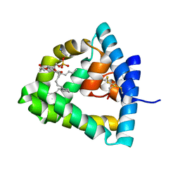
Loading
PDBj
MenuPDBj@FacebookPDBj@TwitterPDBj@YouTubewwPDB FoundationwwPDB
RCSB PDBPDBeBMRBAdv. SearchSearch help
Search by PDB author
1N69
DownloadVisualize
BU of 1n69 by Molmil
Crystal structure of human saposin B
Descriptor: 1,2-Distearoyl-sn-glycerophosphoethanolamine, SAPOSIN B
Authors:Ahn, V.E, Faull, K.F, Whitelegge, J.P, Fluharty, A.L, Prive, G.G.
Deposit date:2002-11-08
Release date:2003-01-07
Last modified:2021-06-30
Method:X-RAY DIFFRACTION (2.2 Å)
Cite:Crystal Structure of saposin B reveals a dimeric shell for lipid binding
Proc.Natl.Acad.Sci.USA, 100, 2003
3KPT
DownloadVisualize
BU of 3kpt by Molmil
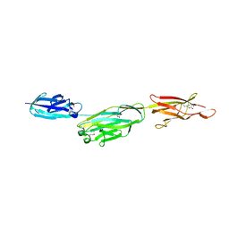
Crystal structure of BcpA, the major pilin subunit of Bacillus cereus
Descriptor: CALCIUM ION, Collagen adhesion protein
Authors:Poor, C.B, Budzik, J.M, Schneewind, O, He, C.
Deposit date:2009-11-16
Release date:2009-11-24
Last modified:2021-10-13
Method:X-RAY DIFFRACTION (2.102 Å)
Cite:Intramolecular amide bonds stabilize pili on the surface of bacilli.
Proc.Natl.Acad.Sci.USA, 106, 2009
4KCG
DownloadVisualize
BU of 4kcg by Molmil
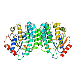
Human dCK C4S-S74E mutant in complex with UDP and the DI-39 inhibitor
Descriptor: Deoxycytidine kinase, N-{2-[5-(4-{[(4,6-diaminopyrimidin-2-yl)sulfanyl]methyl}-5-propyl-1,3-thiazol-2-yl)-2-methoxyphenoxy]ethyl}methanesulfonamide, URIDINE-5'-DIPHOSPHATE
Authors:Nomme, J, Lavie, A.
Deposit date:2013-04-24
Release date:2014-03-12
Last modified:2023-09-20
Method:X-RAY DIFFRACTION (2.09 Å)
Cite:Co-targeting of convergent nucleotide biosynthetic pathways for leukemia eradication.
J.Exp.Med., 211, 2014
3OVJ
DownloadVisualize
BU of 3ovj by Molmil
Structure of an amyloid forming peptide KLVFFA from amyloid beta in complex with orange G
Descriptor: 7-hydroxy-8-[(E)-phenyldiazenyl]naphthalene-1,3-disulfonic acid, KLVFFA hexapeptide segment from Amyloid beta
Authors:Landau, M, Eisenberg, D.
Deposit date:2010-09-16
Release date:2011-07-06
Last modified:2024-02-21
Method:X-RAY DIFFRACTION (1.8 Å)
Cite:Towards a pharmacophore for amyloid.
Plos Biol., 9, 2011
3OVL
DownloadVisualize
BU of 3ovl by Molmil
Structure of an amyloid forming peptide VQIVYK from the TAU protein in complex with orange G
Descriptor: 7-hydroxy-8-[(E)-phenyldiazenyl]naphthalene-1,3-disulfonic acid, ACETIC ACID, Microtubule-associated protein, ...
Authors:Landau, M, Eisenberg, D.
Deposit date:2010-09-16
Release date:2011-07-06
Last modified:2024-02-21
Method:X-RAY DIFFRACTION (1.81 Å)
Cite:Towards a pharmacophore for amyloid.
Plos Biol., 9, 2011
4RP7
DownloadVisualize
BU of 4rp7 by Molmil
Structure of the amyloid-forming segment TIITLE from p53 (residues 253-258)
Descriptor: TIITLE hexapeptide segment from p53, ZINC ION
Authors:Soriaga, A.B, Soragni, A, Eisenberg, D.
Deposit date:2014-10-29
Release date:2016-01-13
Last modified:2024-02-28
Method:X-RAY DIFFRACTION (1.576 Å)
Cite:A Designed Inhibitor of p53 Aggregation Rescues p53 Tumor Suppression in Ovarian Carcinomas.
Cancer Cell, 29, 2016
4RP6
DownloadVisualize
BU of 4rp6 by Molmil
Structure of the amyloid-forming segment LTIITLE from p53 (residues 252-258)
Descriptor: LTIITLE heptapeptide segment from p53
Authors:Soriaga, A.B, Soragni, A, Eisenberg, D.
Deposit date:2014-10-29
Release date:2016-01-13
Last modified:2024-02-28
Method:X-RAY DIFFRACTION (1.703 Å)
Cite:A Designed Inhibitor of p53 Aggregation Rescues p53 Tumor Suppression in Ovarian Carcinomas.
Cancer Cell, 29, 2016

222415

数据于2024-07-10公开中

PDB statisticsPDBj update infoContact PDBjnumon