Loading
PDBj
MenuPDBj@FacebookPDBj@TwitterPDBj@YouTubewwPDB FoundationwwPDB
RCSB PDBPDBeBMRBAdv. SearchSearch help
Search by PDB author
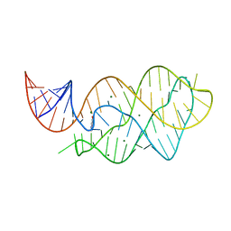
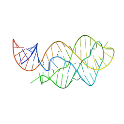
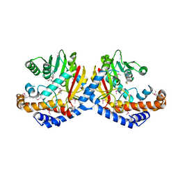
1Y39
DownloadVisualize
BU of 1y39 by Molmil
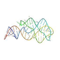
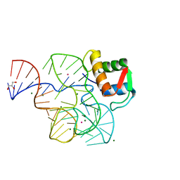
Co-evolution of protein and RNA structures within a highly conserved ribosomal domain
Descriptor: 50S ribosomal protein L11, 58 Nucleotide Ribosomal 23S RNA Domain, COBALT (III) ION, ...
Authors:Dunstan, M.S, GuhaThakurta, D, Draper, D.E, Conn, G.L.
Deposit date:2004-11-24
Release date:2005-03-22
Last modified:2023-10-25
Method:X-RAY DIFFRACTION (2.8 Å)
Cite:Coevolution of Protein and RNA Structures within a Highly Conserved Ribosomal Domain
Chem.Biol., 12, 2005
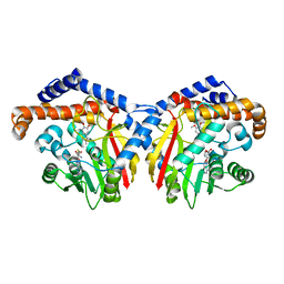
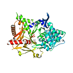
3GYQ
DownloadVisualize
BU of 3gyq by Molmil
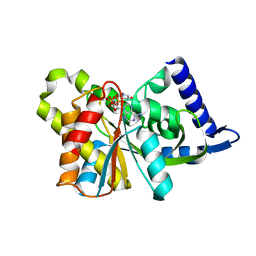
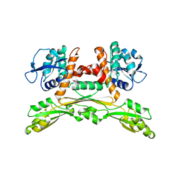
Structure of the Thiostrepton-Resistance Methyltransferase S-adenosyl-L-methionine Complex
Descriptor: S-ADENOSYLMETHIONINE, rRNA (adenosine-2'-O-)-methyltransferase
Authors:Dunstan, M.S, Conn, G.L.
Deposit date:2009-04-04
Release date:2009-04-14
Last modified:2023-11-01
Method:X-RAY DIFFRACTION (2.45 Å)
Cite:Structure of the thiostrepton-resistance methyltransferase-S-adenosyl-L-methionine complex and its interaction with ribosomal RNA
J.Biol.Chem., 284, 2009
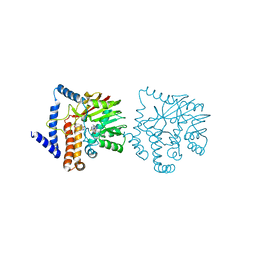
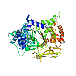
4EPP
DownloadVisualize
BU of 4epp by Molmil
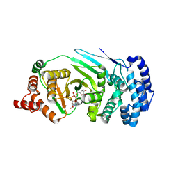
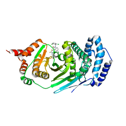
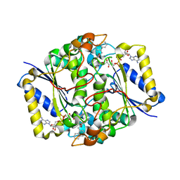
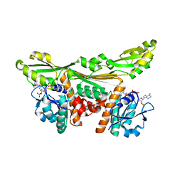
Canonical poly(ADP-ribose) glycohydrolase from Tetrahymena thermophila.
Descriptor: ADENOSINE-5-DIPHOSPHORIBOSE, Poly(ADP-ribose) glycohydrolase
Authors:Dunstan, M.S, Leys, D.
Deposit date:2012-04-17
Release date:2012-06-20
Last modified:2024-02-28
Method:X-RAY DIFFRACTION (1.95 Å)
Cite:Structure and mechanism of a canonical poly(ADP-ribose) glycohydrolase.
Nat Commun, 3, 2012
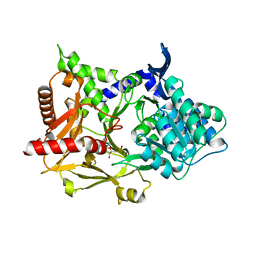
3TEM
DownloadVisualize
BU of 3tem by Molmil
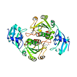
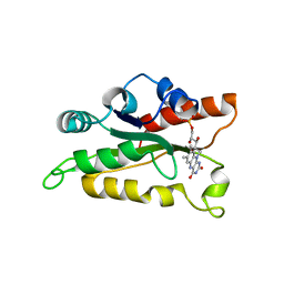
Quinone Oxidoreductase (NQ02) bound to the imidazoacridin-6-one 6a1
Descriptor: FLAVIN-ADENINE DINUCLEOTIDE, IMIDAZOLE, Ribosyldihydronicotinamide dehydrogenase [quinone], ...
Authors:Dunstan, M.S, Leys, D.
Deposit date:2011-08-15
Release date:2011-09-21
Last modified:2024-02-28
Method:X-RAY DIFFRACTION (1.45 Å)
Cite:Novel Inhibitors of NRH:Quinone Oxidoreductase 2 (NQO2): Crystal Structures, Biochemical Activity, and Intracellular Effects of Imidazoacridin-6-ones.
J.Med.Chem., 54, 2011
3TE7
DownloadVisualize
BU of 3te7 by Molmil
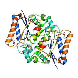
Quinone Oxidoreductase (NQ02) bound to the imidazoacridin-6-one 5a1
Descriptor: 5-{[2-(dimethylamino)ethyl]amino}-8-methoxy-6H-imidazo[4,5,1-de]acridin-6-one, FLAVIN-ADENINE DINUCLEOTIDE, IMIDAZOLE, ...
Authors:Dunstan, M.S.
Deposit date:2011-08-12
Release date:2011-09-21
Last modified:2023-09-13
Method:X-RAY DIFFRACTION (1.7 Å)
Cite:Novel Inhibitors of NRH:Quinone Oxidoreductase 2 (NQO2): Crystal Structures, Biochemical Activity, and Intracellular Effects of Imidazoacridin-6-ones.
J.Med.Chem., 54, 2011
4EPQ
DownloadVisualize
BU of 4epq by Molmil
canonical poly(ADP-ribose) glycohydrolase RBPI inhibitor complex from Tetrahymena thermophila
Descriptor: 3-{(5Z)-5-[5-chloro-1-(2,6-dichlorobenzyl)-2-oxo-1,2-dihydro-3H-indol-3-ylidene]-4-oxo-2-thioxo-1,3-thiazolidin-3-yl}propanoic acid, Poly(ADP-ribose) glycohydrolase
Authors:Dunstan, M.S, Leys, D.
Deposit date:2012-04-17
Release date:2012-06-13
Last modified:2023-09-13
Method:X-RAY DIFFRACTION (2.399 Å)
Cite:Structure and mechanism of a canonical poly(ADP-ribose) glycohydrolase.
Nat Commun, 3, 2012
3LA5
DownloadVisualize
BU of 3la5 by Molmil
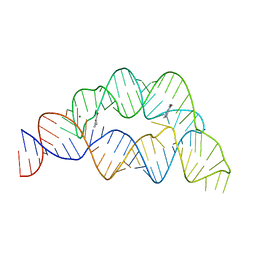
X-ray crystal structure of mc6 RNA Riboswitch bound to azacytosine
Descriptor: 6-amino-1,3,5-triazin-2(1H)-one, Adenosine RIboswitch, MAGNESIUM ION
Authors:Dunstan, M.S, Leys, D.
Deposit date:2010-01-06
Release date:2010-01-26
Last modified:2023-11-01
Method:X-RAY DIFFRACTION (1.7 Å)
Cite:Reengineering orthogonally selective riboswitches
Proc.Natl.Acad.Sci.USA, 107, 2010
6Q57
DownloadVisualize
BU of 6q57 by Molmil
X-ray crystal structure of the tetrahydrofolate riboswitch aptamer bound to 5-deazatetrahydropterin
Descriptor: 5-deazatetrahydropterin, MAGNESIUM ION, tetrahydrofolate riboswitch aptamer
Authors:Dunstan, M.S.
Deposit date:2018-12-07
Release date:2019-12-18
Last modified:2024-05-15
Method:X-RAY DIFFRACTION (2.72 Å)
Cite:Tetrahydrofolate Riboswitches Provide Distinct Genetic Outputs to Synthetic and Natural Signals.
To Be Published
5LV9
DownloadVisualize
BU of 5lv9 by Molmil
Crystal structure of thermophilic tryptophan halogenase (Th-Hal) enzyme from Streptomycin violaceusniger.
Descriptor: thermophilic tryptophan halogenase
Authors:Dunstan, M.S, Menon, B.
Deposit date:2016-09-13
Release date:2016-10-19
Last modified:2024-05-08
Method:X-RAY DIFFRACTION (2.33 Å)
Cite:Structure and biocatalytic scope of thermophilic flavin-dependent halogenase and flavin reductase enzymes.
Org.Biomol.Chem., 14, 2016
3JSX
DownloadVisualize
BU of 3jsx by Molmil
X-ray Crystal structure of NAD(P)H: Quinone Oxidoreductase-1 (NQO1) bound to the coumarin-based inhibitor AS1
Descriptor: 4-hydroxy-6,7-dimethyl-3-(naphthalen-1-ylmethyl)-2H-chromen-2-one, FLAVIN-ADENINE DINUCLEOTIDE, NAD(P)H dehydrogenase [quinone] 1
Authors:Dunstan, M.S, Levy, C, Leys, D.
Deposit date:2009-09-11
Release date:2010-01-12
Last modified:2023-11-01
Method:X-RAY DIFFRACTION (2.45 Å)
Cite:Synthesis and biological evaluation of coumarin-based inhibitors of NAD(P)H: quinone oxidoreductase-1 (NQO1).
J.Med.Chem., 52, 2009
3SII
DownloadVisualize
BU of 3sii by Molmil
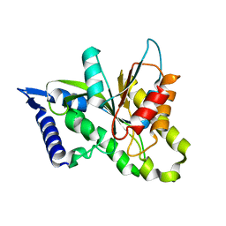
The X-ray crystal structure of poly(ADP-ribose) glycohydrolase bound to the inhibitor ADP-HPD from Thermomonospora curvata
Descriptor: 5'-O-[(S)-{[(S)-{[(2R,3R,4S)-3,4-DIHYDROXYPYRROLIDIN-2-YL]METHOXY}(HYDROXY)PHOSPHORYL]OXY}(HYDROXY)PHOSPHORYL]ADENOSINE, poly(ADP-ribose) glycohydrolase
Authors:Dunstan, M.S, Leys, D.
Deposit date:2011-06-18
Release date:2011-08-24
Last modified:2023-09-13
Method:X-RAY DIFFRACTION (1.48 Å)
Cite:The structure and catalytic mechanism of a poly(ADP-ribose) glycohydrolase.
Nature, 477, 2011
3SIH
DownloadVisualize
BU of 3sih by Molmil
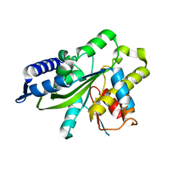
The X-ray crystal structure of poly(ADP-ribose) glycohydrolase (PARG) from Thermomonospora curvata
Descriptor: poly(ADP-ribose) glycohydrolase
Authors:Dunstan, M.S, Leys, D.
Deposit date:2011-06-18
Release date:2011-08-24
Last modified:2024-02-28
Method:X-RAY DIFFRACTION (1.5 Å)
Cite:The structure and catalytic mechanism of a poly(ADP-ribose) glycohydrolase.
Nature, 477, 2011
3SIJ
DownloadVisualize
BU of 3sij by Molmil
The X-ray crystal structure of poly(ADP-ribose) glycohydrolase E115A mutant from Thermomonospora curvata
Descriptor: poly(ADP-ribose) glycohydrolase
Authors:Dunstan, M.S, Leys, D.
Deposit date:2011-06-18
Release date:2011-08-24
Last modified:2023-09-13
Method:X-RAY DIFFRACTION (1.9 Å)
Cite:The structure and catalytic mechanism of a poly(ADP-ribose) glycohydrolase.
Nature, 477, 2011
3TZB
DownloadVisualize
BU of 3tzb by Molmil
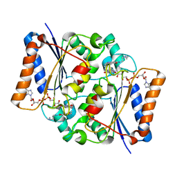
Quinone Oxidoreductase (NQ02) bound to NSC13000
Descriptor: 9-AMINOACRIDINE, FLAVIN-ADENINE DINUCLEOTIDE, Ribosyldihydronicotinamide dehydrogenase [quinone], ...
Authors:Dunstan, M.S, Leys, D.
Deposit date:2011-09-27
Release date:2011-11-02
Last modified:2024-02-28
Method:X-RAY DIFFRACTION (2.1901 Å)
Cite:In silico screening reveals structurally diverse, nanomolar inhibitors of NQO2 that are functionally active in cells and can modulate NF-kappa B signaling.
Mol.Cancer Ther., 11, 2012
4LX6
DownloadVisualize
BU of 4lx6 by Molmil
X-ray crystal structure of the M6C" riboswitch aptamer bound to 2-aminopyrimido[4,5-d]pyrimidin-4(3H)-one (PPAO)
Descriptor: 2-aminopyrimido[4,5-d]pyrimidin-4(3H)-one, MAGNESIUM ION, Mutated adenine riboswitch aptamer
Authors:Dunstan, M.S, Leys, D.
Deposit date:2013-07-29
Release date:2014-07-16
Last modified:2023-09-20
Method:X-RAY DIFFRACTION (2.15 Å)
Cite:Modular riboswitch toolsets for synthetic genetic control in diverse bacterial species.
J.Am.Chem.Soc., 136, 2014
4LX5
DownloadVisualize
BU of 4lx5 by Molmil
X-ray crystal structure of the M6" riboswitch aptamer bound to pyrimido[4,5-d]pyrimidine-2,4-diamine (PPDA)
Descriptor: MAGNESIUM ION, Mutated adenine riboswitch aptamer, pyrimido[4,5-d]pyrimidine-2,4-diamine
Authors:Dunstan, M.S, Leys, D.
Deposit date:2013-07-29
Release date:2014-07-16
Last modified:2023-09-20
Method:X-RAY DIFFRACTION (2.13 Å)
Cite:Modular riboswitch toolsets for synthetic genetic control in diverse bacterial species.
J.Am.Chem.Soc., 136, 2014
6GKV
DownloadVisualize
BU of 6gkv by Molmil
Crystal structure of Coclaurine N-Methyltransferase (CNMT) bound to N-methylheliamine and SAH
Descriptor: 6,7-dimethoxy-2-methyl-1,2,3,4-tetrahydroisoquinolin-2-ium, Coclaurine N-methyltransferase, S-ADENOSYL-L-HOMOCYSTEINE
Authors:Dunstan, M.S, Levy, C.W.
Deposit date:2018-05-22
Release date:2018-06-06
Last modified:2024-05-15
Method:X-RAY DIFFRACTION (2.35 Å)
Cite:Structure and Biocatalytic Scope of Coclaurine N-Methyltransferase.
Angew. Chem. Int. Ed. Engl., 57, 2018
6GKY
DownloadVisualize
BU of 6gky by Molmil
Crystal structure of Coclaurine N-Methyltransferase (CNMT) bound to N-methylheliamine and SAH
Descriptor: 6,7-dimethoxy-2,4-dihydro-1~{H}-isoquinolin-3-one, Coclaurine N-methyltransferase, S-ADENOSYL-L-HOMOCYSTEINE
Authors:Dunstan, M.S, Levy, C.W.
Deposit date:2018-05-22
Release date:2018-06-06
Last modified:2024-05-15
Method:X-RAY DIFFRACTION (2.847 Å)
Cite:Structure and Biocatalytic Scope of Coclaurine N-Methyltransferase.
Angew. Chem. Int. Ed. Engl., 57, 2018
6GKZ
DownloadVisualize
BU of 6gkz by Molmil
Crystal structure of Coclaurine N-Methyltransferase (CNMT) bound to N-methylheliamine and SAH
Descriptor: Coclaurine N-methyltransferase, S-ADENOSYL-L-HOMOCYSTEINE
Authors:Dunstan, M.S, Levy, C.W.
Deposit date:2018-05-22
Release date:2019-01-16
Last modified:2024-10-23
Method:X-RAY DIFFRACTION (2.43 Å)
Cite:Structure and Biocatalytic Scope of Coclaurine N-Methyltransferase.
Angew. Chem. Int. Ed. Engl., 57, 2018
5MSD
DownloadVisualize
BU of 5msd by Molmil
Structure of the A domain of carboxylic acid reductase (CAR) from Nocardia iowensis in complex with AMP and benzoic acid
Descriptor: ADENOSINE MONOPHOSPHATE, BENZOIC ACID, Carboxylic acid reductase
Authors:Dunstan, M.S, Leys, D.
Deposit date:2017-01-04
Release date:2017-07-05
Last modified:2024-01-17
Method:X-RAY DIFFRACTION (1.71 Å)
Cite:Structures of carboxylic acid reductase reveal domain dynamics underlying catalysis.
Nat. Chem. Biol., 13, 2017
5MSQ
DownloadVisualize
BU of 5msq by Molmil
Structure of the A domain of carboxylic acid reductase (CAR) from Nocardia iowensis in complex with AMP and iodide
Descriptor: ADENOSINE MONOPHOSPHATE, Carboxylic acid reductase, IODIDE ION
Authors:Dunstan, M.S, Leys, D.
Deposit date:2017-01-05
Release date:2018-01-17
Last modified:2024-05-08
Method:X-RAY DIFFRACTION (1.74 Å)
Cite:Structure and mechanism of carboxylic acid reductase
To be published
5MSC
DownloadVisualize
BU of 5msc by Molmil
Structure of the A domain of carboxylic acid reductase (CAR) from Nocardia iowensis in complex with AMP
Descriptor: ADENOSINE MONOPHOSPHATE, Carboxylic acid reductase
Authors:Dunstan, M.S, Leys, D.
Deposit date:2017-01-04
Release date:2017-07-05
Last modified:2024-05-08
Method:X-RAY DIFFRACTION (1.85 Å)
Cite:Structures of carboxylic acid reductase reveal domain dynamics underlying catalysis.
Nat. Chem. Biol., 13, 2017
5LVA
DownloadVisualize
BU of 5lva by Molmil
Crystal structure of thermophilic tryptophan halogenase (Th-Hal) enzyme from Streptomycin violaceusniger.
Descriptor: FLAVIN MONONUCLEOTIDE, NAD(P)H-FMN oxidoreductase
Authors:Dunstan, M.S, Menon, B.
Deposit date:2016-09-13
Release date:2016-10-19
Last modified:2024-05-08
Method:X-RAY DIFFRACTION (2.53 Å)
Cite:Structure and biocatalytic scope of thermophilic flavin-dependent halogenase and flavin reductase enzymes.
Org.Biomol.Chem., 14, 2016
5LOC
DownloadVisualize
BU of 5loc by Molmil
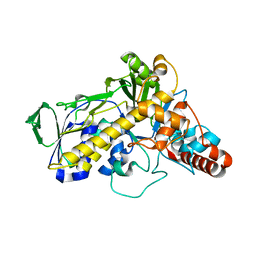
Crystal structure of the engineered D-Amino Acid Dehydrogenase (DAADH)
Descriptor: Meso-diaminopimelate D-dehydrogenase
Authors:Dunstan, M.S, Gahloth, D.
Deposit date:2016-08-09
Release date:2017-08-16
Last modified:2024-05-08
Method:X-RAY DIFFRACTION (2.04 Å)
Cite:Single-biocatalyst synthesis of enantiopure D-arylalanines exploiting an engineered D-amino acid dehydrogenase
To Be Published
5LOA
DownloadVisualize
BU of 5loa by Molmil
Crystal structure of the engineered D-Amino Acid Dehydrogenase (DAADH) bound to NADP+
Descriptor: Meso-diaminopimelate D-dehydrogenase, NADP NICOTINAMIDE-ADENINE-DINUCLEOTIDE PHOSPHATE
Authors:Dunstan, M.S, Gahloth, D.
Deposit date:2016-08-09
Release date:2017-08-16
Last modified:2024-05-08
Method:X-RAY DIFFRACTION (1.84 Å)
Cite:Single-biocatalyst synthesis of enantiopure D-arylalanines exploiting an engineered D-amino acid dehydrogenase
To Be Published


123>

226707

数据于2024-10-30公开中

PDB statisticsPDBj update infoContact PDBjnumon