Loading
PDBj
MenuPDBj@FacebookPDBj@TwitterPDBj@YouTubewwPDB FoundationwwPDB
RCSB PDBPDBeBMRBAdv. SearchSearch help
Search by PDB author
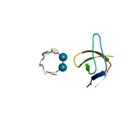
6XX2
DownloadVisualize
BU of 6xx2 by Molmil
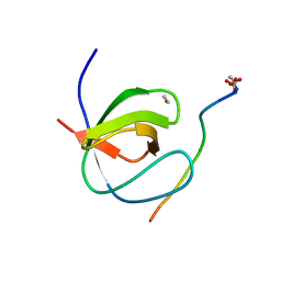
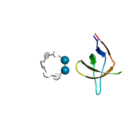
Crystal structure of the c-Src SH3 domain H122R-Q128K mutant in complex with Cu(II) at pH 7.5 co-crystallized with methyl beta-cyclodextrin
Descriptor: COPPER (II) ION, Cyclic 2,3-di-O-methyl-alpha-D-glucopyranose-(1-4)-2-O-methyl-alpha-D-glucopyranose-(1-4)-2,6-di-O-methyl-alpha-D-glucopyranose-(1-4)-2-O-methyl-alpha-D-glucopyranose-(1-4)-alpha-D-glucopyranose-(1-4)-alpha-D-glucopyranose-(1-4)-3-O-methyl-alpha-D-glucopyranose, Proto-oncogene tyrosine-protein kinase Src
Authors:Camara-Artigas, A.
Deposit date:2020-01-26
Release date:2020-04-22
Last modified:2024-01-24
Method:X-RAY DIFFRACTION (1.25 Å)
Cite:The effect of an engineered ATCUN motif on the structure and biophysical properties of the SH3 domain of c-Src tyrosine kinase.
J.Biol.Inorg.Chem., 25, 2020
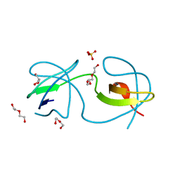
6XVO
DownloadVisualize
BU of 6xvo by Molmil
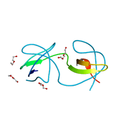
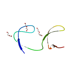
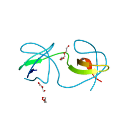
Crystal structure of the intertwined dimer of the c-Src SH3 domain without ATCUN motif
Descriptor: DI(HYDROXYETHYL)ETHER, Proto-oncogene tyrosine-protein kinase Src, TRIETHYLENE GLYCOL
Authors:Camara-Artigas, A, Plaza-Garrido, M.
Deposit date:2020-01-22
Release date:2020-04-22
Last modified:2024-01-24
Method:X-RAY DIFFRACTION (1.7 Å)
Cite:The effect of an engineered ATCUN motif on the structure and biophysical properties of the SH3 domain of c-Src tyrosine kinase.
J.Biol.Inorg.Chem., 25, 2020
6XX3
DownloadVisualize
BU of 6xx3 by Molmil
Crystal structure of the c-Src SH3 domain H122R-Q128E mutant in complex with Cu(II) at pH 6.5 co-crystallized with methyl beta-cyclodextrin
Descriptor: COPPER (II) ION, Cyclic 2,3-di-O-methyl-alpha-D-glucopyranose-(1-4)-2-O-methyl-alpha-D-glucopyranose-(1-4)-2,6-di-O-methyl-alpha-D-glucopyranose-(1-4)-2-O-methyl-alpha-D-glucopyranose-(1-4)-alpha-D-glucopyranose-(1-4)-alpha-D-glucopyranose-(1-4)-3-O-methyl-alpha-D-glucopyranose, Proto-oncogene tyrosine-protein kinase Src
Authors:Camara-Artigas, A.
Deposit date:2020-01-26
Release date:2020-04-22
Last modified:2024-01-24
Method:X-RAY DIFFRACTION (1.36 Å)
Cite:The effect of an engineered ATCUN motif on the structure and biophysical properties of the SH3 domain of c-Src tyrosine kinase.
J.Biol.Inorg.Chem., 25, 2020
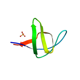
3M0R
DownloadVisualize
BU of 3m0r by Molmil
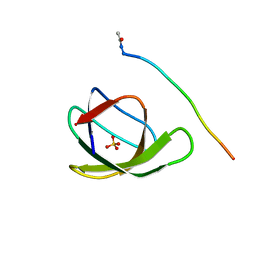
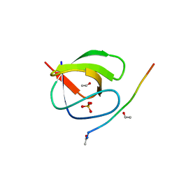
Crystal Structure of the R21D mutant of alpha-spectrin SH3 domain. Crystal obtained in ammonium sulphate at pH 6.
Descriptor: SULFATE ION, Spectrin alpha chain, brain
Authors:Gavira, J.A, Camara-Artigas, A.
Deposit date:2010-03-03
Release date:2011-01-19
Last modified:2023-09-06
Method:X-RAY DIFFRACTION (1.1 Å)
Cite:Understanding the polymorphic behaviour of a mutant of the alpha-spectrin SH3 domain by means of two 1.1 A structures
Acta Crystallogr.,Sect.D, 2011
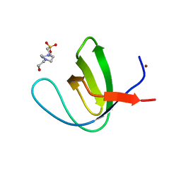
4OMO
DownloadVisualize
BU of 4omo by Molmil
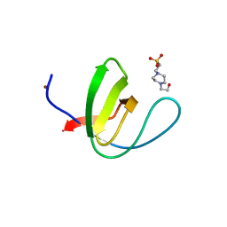
Crystal structure of the c-Src tyrosine kinase SH3 domain mutant Q128E
Descriptor: 4-(2-HYDROXYETHYL)-1-PIPERAZINE ETHANESULFONIC ACID, NICKEL (II) ION, Proto-oncogene tyrosine-protein kinase Src
Authors:Camara-Artigas, A, Bacarizo, J.
Deposit date:2014-01-27
Release date:2014-12-10
Last modified:2023-09-20
Method:X-RAY DIFFRACTION (1.04 Å)
Cite:Electrostatic Effects in the Folding of the SH3 Domain of the c-Src Tyrosine Kinase: pH-Dependence in 3D-Domain Swapping and Amyloid Formation.
Plos One, 9, 2014
4OMN
DownloadVisualize
BU of 4omn by Molmil
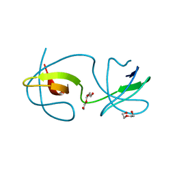
Crystal structure of the intertwined dimer of the c-Src tyrosine kinase SH3 domain mutant Q128E
Descriptor: DI(HYDROXYETHYL)ETHER, GLYCEROL, Proto-oncogene tyrosine-protein kinase Src, ...
Authors:Camara-Artigas, A, Bacarizo, J.
Deposit date:2014-01-27
Release date:2014-12-10
Last modified:2023-09-20
Method:X-RAY DIFFRACTION (1.5 Å)
Cite:Electrostatic Effects in the Folding of the SH3 Domain of the c-Src Tyrosine Kinase: pH-Dependence in 3D-Domain Swapping and Amyloid Formation.
Plos One, 9, 2014
4OMP
DownloadVisualize
BU of 4omp by Molmil
Crystal structure of the intertwined dimer of the c-Src tyrosine kinase SH3 domain mutant Q128K
Descriptor: DI(HYDROXYETHYL)ETHER, Proto-oncogene tyrosine-protein kinase Src, TRIETHYLENE GLYCOL
Authors:Camara-Artigas, A, Bacarizo, J.
Deposit date:2014-01-27
Release date:2014-12-10
Last modified:2023-09-20
Method:X-RAY DIFFRACTION (2 Å)
Cite:Electrostatic Effects in the Folding of the SH3 Domain of the c-Src Tyrosine Kinase: pH-Dependence in 3D-Domain Swapping and Amyloid Formation.
Plos One, 9, 2014
4OML
DownloadVisualize
BU of 4oml by Molmil
Crystal structure of the intertwined dimer of the c-Src tyrosine kinase SH3 domain mutant Q128R
Descriptor: DI(HYDROXYETHYL)ETHER, Proto-oncogene tyrosine-protein kinase Src, TRIETHYLENE GLYCOL
Authors:Camara-Artigas, A, Bacarizo, J.
Deposit date:2014-01-27
Release date:2014-12-10
Last modified:2023-09-20
Method:X-RAY DIFFRACTION (1.6 Å)
Cite:Electrostatic Effects in the Folding of the SH3 Domain of the c-Src Tyrosine Kinase: pH-Dependence in 3D-Domain Swapping and Amyloid Formation.
Plos One, 9, 2014
4QT7
DownloadVisualize
BU of 4qt7 by Molmil
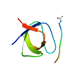
Crystal structure of the c-Src SH3 domain in complex with a peptide from the Hepatitis C virus NS5A-protein
Descriptor: NS5A, Proto-oncogene tyrosine-protein kinase Src
Authors:Camara-Artigas, A, Bacarizo, J.
Deposit date:2014-07-07
Release date:2014-12-24
Last modified:2023-11-08
Method:X-RAY DIFFRACTION (1.55 Å)
Cite:Structure of the c-Src-SH3 domain in complex with a proline-rich motif of NS5A protein from the hepatitis C virus.
J.Struct.Biol., 189, 2015
4HVV
DownloadVisualize
BU of 4hvv by Molmil
Crystal structure of the T98E c-Src-SH3 domain mutant in complex with the high affinity peptide APP12
Descriptor: Proto-oncogene tyrosine-protein kinase Src, SULFATE ION, SYNTHETIC PEPTIDE Acetyl-APPLPPRNRP
Authors:Camara-Artigas, A.
Deposit date:2012-11-07
Release date:2013-05-01
Last modified:2023-09-20
Method:X-RAY DIFFRACTION (1.1 Å)
Cite:Atomic resolution structures of the c-Src SH3 domain in complex with two high-affinity peptides from classes I and II.
Acta Crystallogr.,Sect.D, 69, 2013
4HVW
DownloadVisualize
BU of 4hvw by Molmil
Crystal structure of the T98E c-Src-SH3 domain mutant in complex with the high affinity peptide VSL12
Descriptor: ACETYL GROUP, Proto-oncogene tyrosine-protein kinase Src, SULFATE ION, ...
Authors:Camara-Artigas, A.
Deposit date:2012-11-07
Release date:2013-05-01
Last modified:2023-09-20
Method:X-RAY DIFFRACTION (0.98 Å)
Cite:Atomic resolution structures of the c-Src SH3 domain in complex with two high-affinity peptides from classes I and II.
Acta Crystallogr.,Sect.D, 69, 2013
4HVU
DownloadVisualize
BU of 4hvu by Molmil
Crystal structure of the T98D c-Src-SH3 domain mutant in complex with the high affinity peptide APP12
Descriptor: ACETYL GROUP, Proto-oncogene tyrosine-protein kinase Src, SULFATE ION, ...
Authors:Camara-Artigas, A.
Deposit date:2012-11-07
Release date:2013-05-01
Last modified:2023-09-20
Method:X-RAY DIFFRACTION (0.98 Å)
Cite:Atomic resolution structures of the c-Src SH3 domain in complex with two high-affinity peptides from classes I and II.
Acta Crystallogr.,Sect.D, 69, 2013
4JZ3
DownloadVisualize
BU of 4jz3 by Molmil
Crystal structure of the chicken c-Src-SH3 domain intertwined dimer
Descriptor: DI(HYDROXYETHYL)ETHER, Proto-oncogene tyrosine-protein kinase Src, TRIETHYLENE GLYCOL
Authors:Camara-Artigas, A.
Deposit date:2013-04-02
Release date:2014-04-23
Last modified:2023-09-20
Method:X-RAY DIFFRACTION (1.852 Å)
Cite:Electrostatic Effects in the Folding of the SH3 Domain of the c-Src Tyrosine Kinase: pH-Dependence in 3D-Domain Swapping and Amyloid Formation.
Plos One, 9, 2014
4JZ4
DownloadVisualize
BU of 4jz4 by Molmil
Crystal structure of chicken c-Src-SH3 domain: monomeric form
Descriptor: 4-(2-HYDROXYETHYL)-1-PIPERAZINE ETHANESULFONIC ACID, NICKEL (II) ION, Proto-oncogene tyrosine-protein kinase Src
Authors:Camara-Artigas, A.
Deposit date:2013-04-02
Release date:2014-04-23
Last modified:2023-09-20
Method:X-RAY DIFFRACTION (1.56 Å)
Cite:Electrostatic Effects in the Folding of the SH3 Domain of the c-Src Tyrosine Kinase: pH-Dependence in 3D-Domain Swapping and Amyloid Formation.
Plos One, 9, 2014
6F9Y
DownloadVisualize
BU of 6f9y by Molmil
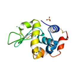
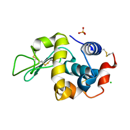
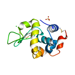
Lysozyme crystallized in presence of 10 mM lithium sulphate at pH 4.5
Descriptor: CHLORIDE ION, Lysozyme C, SULFATE ION
Authors:Camara-Artigas, A.
Deposit date:2017-12-15
Release date:2018-05-09
Last modified:2024-10-09
Method:X-RAY DIFFRACTION (1.2 Å)
Cite:Orthorhombic lysozyme crystallization at acidic pH values driven by phosphate binding.
Acta Crystallogr D Struct Biol, 74, 2018
6F1L
DownloadVisualize
BU of 6f1l by Molmil
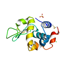
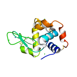
Lysozyme crystallized in presence of 100 mM sodium phosphate at pH 4.5
Descriptor: CHLORIDE ION, Lysozyme C, PHOSPHATE ION
Authors:Camara-Artigas, A.
Deposit date:2017-11-22
Release date:2018-05-09
Last modified:2024-01-17
Method:X-RAY DIFFRACTION (1.3 Å)
Cite:Orthorhombic lysozyme crystallization at acidic pH values driven by phosphate binding.
Acta Crystallogr D Struct Biol, 74, 2018
6F1P
DownloadVisualize
BU of 6f1p by Molmil
Tetragonal Lysozyme crystallized at 298 K and pH 4.5 with phosphate bound
Descriptor: CHLORIDE ION, Lysozyme C, PHOSPHATE ION, ...
Authors:Camara-Artigas, A.
Deposit date:2017-11-22
Release date:2018-05-09
Last modified:2024-01-17
Method:X-RAY DIFFRACTION (1.12 Å)
Cite:Orthorhombic lysozyme crystallization at acidic pH values driven by phosphate binding.
Acta Crystallogr D Struct Biol, 74, 2018
6F9Z
DownloadVisualize
BU of 6f9z by Molmil
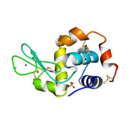
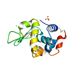
Lysozyme crystallized in presence of 5 mM ammonium sulphate at pH 4.5
Descriptor: CHLORIDE ION, Lysozyme C, SULFATE ION
Authors:Camara-Artigas, A.
Deposit date:2017-12-15
Release date:2018-05-09
Last modified:2024-10-16
Method:X-RAY DIFFRACTION (1.2 Å)
Cite:Orthorhombic lysozyme crystallization at acidic pH values driven by phosphate binding.
Acta Crystallogr D Struct Biol, 74, 2018
6F1O
DownloadVisualize
BU of 6f1o by Molmil
Orthorhombic Lysozyme crystallized at 298 K and pH 4.5
Descriptor: CHLORIDE ION, Lysozyme C, PHOSPHATE ION
Authors:Camara-Artigas, A.
Deposit date:2017-11-22
Release date:2018-05-09
Last modified:2024-01-17
Method:X-RAY DIFFRACTION (0.96 Å)
Cite:Orthorhombic lysozyme crystallization at acidic pH values driven by phosphate binding.
Acta Crystallogr D Struct Biol, 74, 2018
6F1R
DownloadVisualize
BU of 6f1r by Molmil
Tetragonal Lysozyme crystallized at 298 K and pH 4.5 with phosphate bound: control experiment
Descriptor: CHLORIDE ION, Lysozyme C, SODIUM ION
Authors:Camara-Artigas, A.
Deposit date:2017-11-22
Release date:2018-05-09
Last modified:2024-10-23
Method:X-RAY DIFFRACTION (1.1 Å)
Cite:Orthorhombic lysozyme crystallization at acidic pH values driven by phosphate binding.
Acta Crystallogr D Struct Biol, 74, 2018
6F1M
DownloadVisualize
BU of 6f1m by Molmil
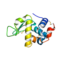
Lysozyme crystallized in presence of 100 mM sodium phosphate at pH 4.5: low-humidity form
Descriptor: Lysozyme C
Authors:Camara-Artigas, A.
Deposit date:2017-11-22
Release date:2018-05-09
Last modified:2024-10-23
Method:X-RAY DIFFRACTION (1.48 Å)
Cite:Orthorhombic lysozyme crystallization at acidic pH values driven by phosphate binding.
Acta Crystallogr D Struct Biol, 74, 2018
6FA0
DownloadVisualize
BU of 6fa0 by Molmil
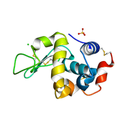
Lysozyme crystallized in presence of 100 mM ammonium sulphate at pH 4.5
Descriptor: CHLORIDE ION, Lysozyme C, SULFATE ION
Authors:Camara-Artigas, A.
Deposit date:2017-12-15
Release date:2018-05-09
Last modified:2024-01-17
Method:X-RAY DIFFRACTION (1.3 Å)
Cite:Orthorhombic lysozyme crystallization at acidic pH values driven by phosphate binding.
Acta Crystallogr D Struct Biol, 74, 2018
6F9X
DownloadVisualize
BU of 6f9x by Molmil
Lysozyme crystallized in presence of 100 mM lithium sulphate at pH 4.5
Descriptor: CHLORIDE ION, Lysozyme C, SULFATE ION
Authors:Camara-Artigas, A.
Deposit date:2017-12-15
Release date:2018-05-09
Last modified:2024-01-17
Method:X-RAY DIFFRACTION (1.25 Å)
Cite:Orthorhombic lysozyme crystallization at acidic pH values driven by phosphate binding.
Acta Crystallogr D Struct Biol, 74, 2018
2OLP
DownloadVisualize
BU of 2olp by Molmil
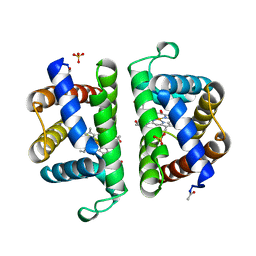
Structure and ligand selection of hemoglobin II from Lucina pectinata
Descriptor: Hemoglobin II, OXYGEN MOLECULE, PROTOPORPHYRIN IX CONTAINING FE, ...
Authors:Gavira, J.A, Camara-Artigas, A, de Jesus, W, Lopez-Garriga, J, Garcia-Ruiz, J.M.
Deposit date:2007-01-19
Release date:2007-12-18
Last modified:2023-08-30
Method:X-RAY DIFFRACTION (1.932 Å)
Cite:Structure and Ligand Selection of Hemoglobin II from Lucina pectinata
J.Biol.Chem., 283, 2008
2CDT
DownloadVisualize
BU of 2cdt by Molmil
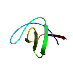
alpha-SPECTRIN SH3 DOMAIN A56S MUTANT
Descriptor: SPECTRIN ALPHA CHAIN
Authors:Casares, S, Camara-Artigas, A, Vega, M.C, Lopez-Mayorga, O, Conejero-Lara, F.
Deposit date:2006-01-27
Release date:2007-02-20
Last modified:2023-12-13
Method:X-RAY DIFFRACTION (2.54 Å)
Cite:Cooperative Propagation of Local Stability Changes from Low-Stability and High-Stability Regions in a SH3 Domain.
Proteins: Struct., Funct., Bioinf., 67, 2007

226707

数据于2024-10-30公开中

PDB statisticsPDBj update infoContact PDBjnumon