Loading
PDBj
MenuPDBj@FacebookPDBj@TwitterPDBj@YouTubewwPDB FoundationwwPDB
RCSB PDBPDBeBMRBAdv. SearchSearch help
Search by PDB author
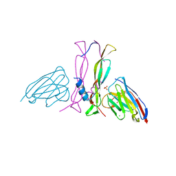
8B7W
DownloadVisualize
BU of 8b7w by Molmil
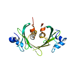
Complex IL-17A/anti-IL-17A-76
Descriptor: CHLORIDE ION, GLYCEROL, Interleukin-17A, ...
Authors:Kostareva, O.S, Svoeglazova, A, Kolyadenko, I.A, Dzhus, U.F, Tishchenko, S.V, Gabdulkhakov, A.G.
Deposit date:2022-10-03
Release date:2022-12-28
Last modified:2024-01-31
Method:X-RAY DIFFRACTION (2.85 Å)
Cite:Two Epitope Regions Revealed in the Complex of IL-17A and Anti-IL-17A V H H Domain.
Int J Mol Sci, 23, 2022
1AN7
DownloadVisualize
BU of 1an7 by Molmil
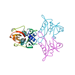
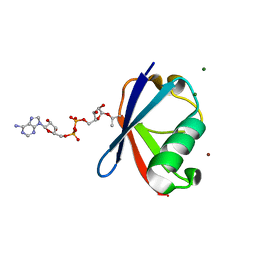
RIBOSOMAL PROTEIN S8 FROM THERMUS THERMOPHILUS
Descriptor: RIBOSOMAL PROTEIN S8
Authors:Nevskaya, N, Nikonov, S, Al-Karadaghi, S.
Deposit date:1997-06-27
Release date:1998-07-01
Last modified:2024-02-07
Method:X-RAY DIFFRACTION (2.9 Å)
Cite:Crystal structure of ribosomal protein S8 from Thermus thermophilus reveals a high degree of structural conservation of a specific RNA binding site.
J.Mol.Biol., 279, 1998
5LHL
DownloadVisualize
BU of 5lhl by Molmil
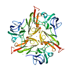
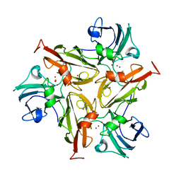
Crystal Structure of Two-Domain Laccase from Streptomyces griseoflavus
Descriptor: CHLORIDE ION, COPPER (II) ION, OXYGEN MOLECULE, ...
Authors:Gabdulkhakov, A.G.
Deposit date:2016-07-12
Release date:2017-08-16
Last modified:2024-01-10
Method:X-RAY DIFFRACTION (2 Å)
Cite:Crystal Structure of Two-Domain Laccase from Streptomyces griseoflavus
To Be Published
6YC5
DownloadVisualize
BU of 6yc5 by Molmil
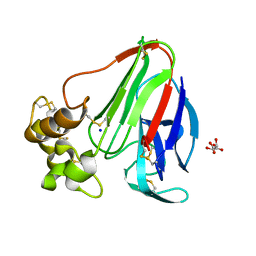
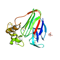
RT structure of Thaumatin obtained at 1.35 A resolution from crystal grown in a Kapton microchip.
Descriptor: L(+)-TARTARIC ACID, SODIUM ION, Thaumatin-1
Authors:Gavira, J.A, Martinez-Rodriguez, S.
Deposit date:2020-03-18
Release date:2020-08-12
Last modified:2024-01-24
Method:X-RAY DIFFRACTION (1.35 Å)
Cite:Attaining atomic resolution from in situ data collection at room temperature using counter-diffusion-based low-cost microchips.
Acta Crystallogr D Struct Biol, 76, 2020
6YBR
DownloadVisualize
BU of 6ybr by Molmil
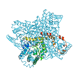
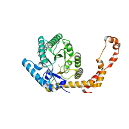
RT structure of Glucose Isomerase obtained at 1.20 A resolution from crystal grown in a Mylar microchip.
Descriptor: MAGNESIUM ION, SODIUM ION, Xylose isomerase
Authors:Gavira, J.A, Martinez-Rodriguez, S.
Deposit date:2020-03-17
Release date:2020-08-12
Last modified:2024-01-24
Method:X-RAY DIFFRACTION (1.2 Å)
Cite:Attaining atomic resolution from in situ data collection at room temperature using counter-diffusion-based low-cost microchips.
Acta Crystallogr D Struct Biol, 76, 2020
6YBF
DownloadVisualize
BU of 6ybf by Molmil
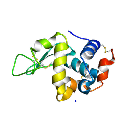
RT structure of HEW Lysozyme obtained at 1.13 A resolution from crystal grown in a Kapton microchip.
Descriptor: CHLORIDE ION, Lysozyme, SODIUM ION
Authors:Gavira, J, Martinez-Rodriguez, S.
Deposit date:2020-03-17
Release date:2020-08-12
Last modified:2024-01-24
Method:X-RAY DIFFRACTION (1.13 Å)
Cite:Attaining atomic resolution from in situ data collection at room temperature using counter-diffusion-based low-cost microchips.
Acta Crystallogr D Struct Biol, 76, 2020
6YBI
DownloadVisualize
BU of 6ybi by Molmil
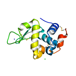
RT structure of HEW Lysozyme obtained at 1.12 A resolution from crystal grown in a Mylar microchip.
Descriptor: CHLORIDE ION, Lysozyme, SODIUM ION
Authors:Gavira, J.A, Martinez-Rodriguez, S.
Deposit date:2020-03-17
Release date:2020-08-12
Last modified:2024-01-24
Method:X-RAY DIFFRACTION (1.12 Å)
Cite:Attaining atomic resolution from in situ data collection at room temperature using counter-diffusion-based low-cost microchips.
Acta Crystallogr D Struct Biol, 76, 2020
6YBO
DownloadVisualize
BU of 6ybo by Molmil
RT structure of Glucose Isomerase obtained at 1.06 A resolution from crystal grown in a Kapton microchip.
Descriptor: MAGNESIUM ION, SODIUM ION, Xylose isomerase
Authors:Gavira, J.A, Martinez-Rodriguez, S.
Deposit date:2020-03-17
Release date:2020-08-12
Last modified:2024-01-24
Method:X-RAY DIFFRACTION (1.06 Å)
Cite:Attaining atomic resolution from in situ data collection at room temperature using counter-diffusion-based low-cost microchips.
Acta Crystallogr D Struct Biol, 76, 2020
6YBX
DownloadVisualize
BU of 6ybx by Molmil
RT structure of Thaumatin obtained at 1.14 A resolution from crystal grown in a Mylar microchip.
Descriptor: L(+)-TARTARIC ACID, SODIUM ION, Thaumatin-1
Authors:Gavira, J.A, Martinez-Rodriguez, S.
Deposit date:2020-03-18
Release date:2020-08-12
Last modified:2024-01-24
Method:X-RAY DIFFRACTION (1.14 Å)
Cite:Attaining atomic resolution from in situ data collection at room temperature using counter-diffusion-based low-cost microchips.
Acta Crystallogr D Struct Biol, 76, 2020
1XS0
DownloadVisualize
BU of 1xs0 by Molmil
Structure of the E. coli Ivy protein
Descriptor: Inhibitor of vertebrate lysozyme
Authors:Abergel, C, Monchois, V, Byrn, D, Lazzaroni, J.C, Claverie, J.M.
Deposit date:2004-10-18
Release date:2004-11-02
Last modified:2011-07-13
Method:X-RAY DIFFRACTION (1.58 Å)
Cite:Structure and evolution of the Ivy protein family, unexpected lysozyme inhibitors in Gram-negative bacteria.
Proc.Natl.Acad.Sci.USA, 104, 2007
7ZN7
DownloadVisualize
BU of 7zn7 by Molmil
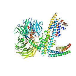
Cryo-EM structure of RCMV-E E27 bound to human DDB1 (deltaBPB) and rat STAT2 CCD
Descriptor: B27a, DNA damage-binding protein 1, Signal transducer and activator of transcription, ...
Authors:Lauer, S, Spahn, C.M.T, Schwefel, D.
Deposit date:2022-04-20
Release date:2022-11-09
Last modified:2024-07-24
Method:ELECTRON MICROSCOPY (3.78 Å)
Cite:Structural mechanism of CRL4-instructed STAT2 degradation via a novel cytomegaloviral DCAF receptor.
Embo J., 42, 2023
7ZNN
DownloadVisualize
BU of 7znn by Molmil
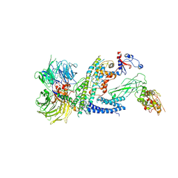
Cryo-EM structure of RCMV-E E27 bound to human DDB1 (deltaBPB) and full-length rat STAT2
Descriptor: B27a, DNA damage-binding protein 1, Signal transducer and activator of transcription, ...
Authors:Lauer, S, Spahn, C.M.T, Schwefel, D.
Deposit date:2022-04-21
Release date:2022-11-09
Last modified:2024-07-24
Method:ELECTRON MICROSCOPY (4.8 Å)
Cite:Structural mechanism of CRL4-instructed STAT2 degradation via a novel cytomegaloviral DCAF receptor.
Embo J., 42, 2023
1EG0
DownloadVisualize
BU of 1eg0 by Molmil
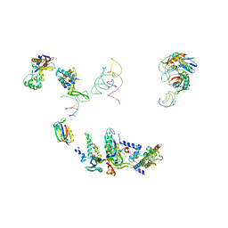
FITTING OF COMPONENTS WITH KNOWN STRUCTURE INTO AN 11.5 A CRYO-EM MAP OF THE E.COLI 70S RIBOSOME
Descriptor: FORMYL-METHIONYL-TRNA, FRAGMENT OF 16S RRNA HELIX 23, FRAGMENT OF 23S RRNA, ...
Authors:Gabashvili, I.S, Agrawal, R.K, Spahn, C.M.T, Grassucci, R.A, Svergun, D.I, Frank, J, Penczek, P.
Deposit date:2000-02-11
Release date:2000-03-06
Last modified:2024-02-07
Method:ELECTRON MICROSCOPY (11.5 Å)
Cite:Solution structure of the E. coli 70S ribosome at 11.5 A resolution.
Cell(Cambridge,Mass.), 100, 2000
6K2U
DownloadVisualize
BU of 6k2u by Molmil
Crystal structure of Thr66 ADP-ribosylated ubiquitin
Descriptor: ADENOSINE-5-DIPHOSPHORIBOSE, MAGNESIUM ION, Polyubiquitin-C, ...
Authors:Wang, X, Zhou, Y, Zhu, Y.
Deposit date:2019-05-15
Release date:2020-03-18
Last modified:2023-11-29
Method:X-RAY DIFFRACTION (2.554 Å)
Cite:Threonine ADP-Ribosylation of Ubiquitin by a Bacterial Effector Family Blocks Host Ubiquitination.
Mol.Cell, 78, 2020
7AH3
DownloadVisualize
BU of 7ah3 by Molmil
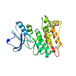
Kinase domain of cSrc in complex with a pyrazolopyrimidine
Descriptor: 1,2-ETHANEDIOL, DIMETHYL SULFOXIDE, GLYCEROL, ...
Authors:Dello Iacono, L, Kleinboelting, S, Fallacara, A.L, Rauh, D.
Deposit date:2020-09-24
Release date:2021-10-06
Last modified:2024-01-31
Method:X-RAY DIFFRACTION (1.95 Å)
Cite:Insights into the binding of pyrazolopyrimidines to Src kinase
To Be Published
6ZIP
DownloadVisualize
BU of 6zip by Molmil
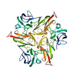
Crystal Structure of Two-Domain Laccase mutant R240A from Streptomyces griseoflavus
Descriptor: COPPER (II) ION, GLYCEROL, OXYGEN MOLECULE, ...
Authors:Gabdulkhakov, A.G, Tishchenko, T.V, Kolyadenko, I.A.
Deposit date:2020-06-26
Release date:2021-05-05
Last modified:2024-01-31
Method:X-RAY DIFFRACTION (2.05 Å)
Cite:The role of positive charged residue in the proton-transfer mechanism of two-domain laccase from Streptomyces griseoflavus Ac-993.
J.Biomol.Struct.Dyn., 40, 2022
6ZIJ
DownloadVisualize
BU of 6zij by Molmil
Crystal Structure of Two-Domain Laccase mutant R240H from Streptomyces griseoflavus
Descriptor: 1,2-ETHANEDIOL, COPPER (II) ION, GLYCEROL, ...
Authors:Gabdulkhakov, A.G, Tishchenko, T.V, Kolyadenko, I.A.
Deposit date:2020-06-26
Release date:2021-05-05
Last modified:2024-01-31
Method:X-RAY DIFFRACTION (1.6 Å)
Cite:The role of positive charged residue in the proton-transfer mechanism of two-domain laccase from Streptomyces griseoflavus Ac-993.
J.Biomol.Struct.Dyn., 40, 2022
1ICC
DownloadVisualize
BU of 1icc by Molmil
RAT OUTER MITOCHONDRIAL MEMBRANE CYTOCHROME B5
Descriptor: CYTOCHROME B5 OUTER MITOCHONDRIAL MEMBRANE ISOFORM, MAGNESIUM ION, PROTOPORPHYRIN IX CONTAINING FE
Authors:Terzyan, S, Zhang, X.
Deposit date:2001-03-30
Release date:2001-09-19
Last modified:2023-08-09
Method:X-RAY DIFFRACTION (2 Å)
Cite:Probing the differences between rat liver outer mitochondrial membrane cytochrome b5 and microsomal cytochromes b5.
Biochemistry, 40, 2001
1HT3
DownloadVisualize
BU of 1ht3 by Molmil
MERCURY INDUCED MODIFICATIONS IN THE STEREOCHEMISTRY OF THE ACTIVE SITE THROUGH CYS-73 IN A SERINE PROTEASE: CRYSTAL STRUCTURE OF THE COMPLEX OF A PARTIALLY MODIFIED PROTEINASE K WITH MERCURY AT 1.8 A RESOLUTION
Descriptor: CALCIUM ION, MERCURY (II) ION, PROTEINASE K
Authors:Gourinath, S.
Deposit date:2000-12-27
Release date:2001-06-27
Last modified:2018-01-24
Method:X-RAY DIFFRACTION (1.8 Å)
Cite:Mercury induced modifications in the stereochemistry of the active site through Cys-73 in a serine protease--crystal structure of the complex of a partially modified proteinase K with mercury at 1.8 A resolution
Indian J.Biochem.Biophys., 38, 2001
6FC7
DownloadVisualize
BU of 6fc7 by Molmil
Crystal Structure of Two-Domain Laccase mutant H165F from Streptomyces griseoflavus with high copper ions occupancy
Descriptor: COPPER (II) ION, GLYCEROL, OXYGEN MOLECULE, ...
Authors:Gabdulkhakov, A.G, Tishchenko, T.V.
Deposit date:2017-12-20
Release date:2019-01-30
Last modified:2024-01-17
Method:X-RAY DIFFRACTION (1.95 Å)
Cite:Investigations of Accessibility of T2/T3 Copper Center of Two-Domain Laccase fromStreptomyces griseoflavusAc-993.
Int J Mol Sci, 20, 2019
6FDJ
DownloadVisualize
BU of 6fdj by Molmil
Crystal Structure of Two-Domain Laccase mutant H165A from Streptomyces griseoflavus with high copper ions occupancy
Descriptor: COPPER (II) ION, GLYCEROL, OXYGEN MOLECULE, ...
Authors:Gabdulkhakov, A.G, Tishchenko, T.V.
Deposit date:2017-12-25
Release date:2019-01-30
Last modified:2024-01-17
Method:X-RAY DIFFRACTION (2.308 Å)
Cite:Investigations of Accessibility of T2/T3 Copper Center of Two-Domain Laccase fromStreptomyces griseoflavusAc-993.
Int J Mol Sci, 20, 2019
5AO1
DownloadVisualize
BU of 5ao1 by Molmil
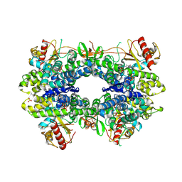
Crystal structure of human SAMHD1 (amino acid residues 115-583) bound to ddGTP
Descriptor: 2'-3'-DIDEOXYGUANOSINE-5'-TRIPHOSPHATE, DEOXYNUCLEOSIDE TRIPHOSPHATE TRIPHOSPHOHYDROLASE SAMHD1, FE (III) ION, ...
Authors:Schwefel, D, Taylor, I.A.
Deposit date:2015-09-09
Release date:2015-10-14
Last modified:2024-01-10
Method:X-RAY DIFFRACTION (2.545 Å)
Cite:Phospho-Dependent Regulation of Samhd1 Oligomerisation Couples Catalysis and Restriction.
Plos Pathog., 11, 2015
5AO2
DownloadVisualize
BU of 5ao2 by Molmil
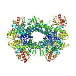
Crystal structure of human SAMHD1 (amino acid residues 115-583) R164A variant bound to dGTP
Descriptor: 2'-DEOXYGUANOSINE-5'-TRIPHOSPHATE, DEOXYNUCLEOSIDE TRIPHOSPHATE TRIPHOSPHOHYDROLASE SAMHD1, FE (III) ION
Authors:Schwefel, D, Taylor, I.A.
Deposit date:2015-09-09
Release date:2015-10-14
Last modified:2024-01-10
Method:X-RAY DIFFRACTION (2.966 Å)
Cite:Phospho-Dependent Regulation of Samhd1 Oligomerisation Couples Catalysis and Restriction.
Plos Pathog., 11, 2015
5AO0
DownloadVisualize
BU of 5ao0 by Molmil
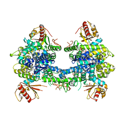
Crystal structure of human SAMHD1 (amino acid residues 41-583) bound to ddGTP
Descriptor: 2'-3'-DIDEOXYGUANOSINE-5'-TRIPHOSPHATE, DEOXYNUCLEOSIDE TRIPHOSPHATE TRIPHOSPHOHYDROLASE SAMHD1, FE (III) ION, ...
Authors:Schwefel, D, Taylor, I.A.
Deposit date:2015-09-09
Release date:2015-10-14
Last modified:2024-01-10
Method:X-RAY DIFFRACTION (3.731 Å)
Cite:Phospho-Dependent Regulation of Samhd1 Oligomerisation Couples Catalysis and Restriction.
Plos Pathog., 11, 2015
5AO3
DownloadVisualize
BU of 5ao3 by Molmil
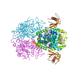
Crystal structure of human SAMHD1 (amino acid residues 115-626) bound to GTP
Descriptor: DEOXYNUCLEOSIDE TRIPHOSPHATE TRIPHOSPHOHYDROLASE SAMHD1, FE (III) ION, GUANOSINE-5'-TRIPHOSPHATE, ...
Authors:Schwefel, D, Taylor, I.A.
Deposit date:2015-09-09
Release date:2016-04-20
Last modified:2024-01-10
Method:X-RAY DIFFRACTION (3.004 Å)
Cite:Phospho-Dependent Regulation of Samhd1 Oligomerisation Couples Catalysis and Restriction.
Plos Pathog., 11, 2015

224931

数据于2024-09-11公开中

PDB statisticsPDBj update infoContact PDBjnumon