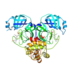
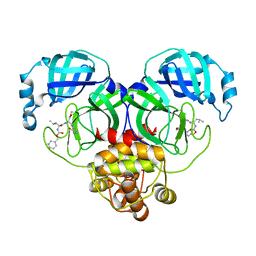
Loading
PDBj
MenuPDBj@FacebookPDBj@TwitterPDBj@YouTubewwPDB FoundationwwPDB
RCSB PDBPDBeBMRBAdv. SearchSearch help
Search by PDB author
6OAP
DownloadVisualize
BU of 6oap by Molmil
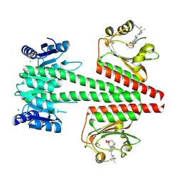
Crystal structure of a dual sensor histidine kinase in the green-light absorbing Pg state
Descriptor: Dual sensor histidine kinase, MAGNESIUM ION, PHYCOCYANOBILIN
Authors:Heewhan, S, Zhong, R, Xiaoli, Z, Sepalika, B, Xiaojing, Y.
Deposit date:2019-03-18
Release date:2019-09-18
Last modified:2023-10-11
Method:X-RAY DIFFRACTION (1.97 Å)
Cite:Structural basis of molecular logic OR in a dual-sensor histidine kinase.
Proc.Natl.Acad.Sci.USA, 116, 2019
6CF8
DownloadVisualize
BU of 6cf8 by Molmil
Crystal structure of Cj0843 lytic transglycosylase of Campylobacter jejuni at 1.87A resolution
Descriptor: Lytic transglycosylase, SULFATE ION
Authors:van den Akker, F, Kumar, V, Vijayaraghavan, J.
Deposit date:2018-02-13
Release date:2018-05-30
Method:X-RAY DIFFRACTION (1.87 Å)
Cite:Structural studies and molecular dynamics simulations suggest a processive mechanism of exolytic lytic transglycosylase from Campylobacter jejuni.
PLoS ONE, 13, 2018
6CFC
DownloadVisualize
BU of 6cfc by Molmil
Crystal structure of soluble lytic transglycosylase Cj0843 of Campylobacter jejuni in complex with Bulgecin A
Descriptor: 4-O-(4-O-SULFONYL-N-ACETYLGLUCOSAMININYL)-5-METHYLHYDROXY-L-PROLINE-TAURINE, Lytic transglycosylase, SULFATE ION
Authors:van den Akker, F, Kumar, V.
Deposit date:2018-02-14
Release date:2018-05-30
Last modified:2023-10-04
Method:X-RAY DIFFRACTION (2.04 Å)
Cite:Structural studies and molecular dynamics simulations suggest a processive mechanism of exolytic lytic transglycosylase from Campylobacter jejuni.
PLoS ONE, 13, 2018
8HVX
DownloadVisualize
BU of 8hvx by Molmil
Crystal structure of SARS-Cov-2 main protease Y54C mutant in complex with PF07304814
Descriptor: 3C-like proteinase nsp5, [(3~{S})-3-[[(2~{S})-2-[(4-methoxy-1~{H}-indol-2-yl)carbonylamino]-4-methyl-pentanoyl]amino]-2-oxidanylidene-4-[(3~{R})-2-oxidanylidene-3,4-dihydropyrrol-3-yl]butyl] dihydrogen phosphate
Authors:Zhou, X.L, Zhang, J, Li, J.
Deposit date:2022-12-28
Release date:2024-01-17
Last modified:2024-02-07
Method:X-RAY DIFFRACTION (1.75 Å)
Cite:Crystal structures of main protease (M pro ) mutants of SARS-CoV-2 variants bound to PF-07304814.
Mol Biomed, 4, 2023
8HVY
DownloadVisualize
BU of 8hvy by Molmil
Crystal structure of SARS-Cov-2 main protease K90R mutant in complex with PF07304814
Descriptor: 3C-like proteinase nsp5, [(3~{S})-3-[[(2~{S})-2-[(4-methoxy-1~{H}-indol-2-yl)carbonylamino]-4-methyl-pentanoyl]amino]-2-oxidanylidene-4-[(3~{R})-2-oxidanylidene-3,4-dihydropyrrol-3-yl]butyl] dihydrogen phosphate
Authors:Wang, J, Zhang, J, Li, J.
Deposit date:2022-12-28
Release date:2024-01-17
Last modified:2024-02-07
Method:X-RAY DIFFRACTION (1.97 Å)
Cite:Crystal structures of main protease (M pro ) mutants of SARS-CoV-2 variants bound to PF-07304814.
Mol Biomed, 4, 2023
8HVZ
DownloadVisualize
BU of 8hvz by Molmil
Crystal structure of SARS-Cov-2 main protease V186F mutant in complex with PF07304814
Descriptor: 3C-like proteinase nsp5, [(3~{S})-3-[[(2~{S})-2-[(4-methoxy-1~{H}-indol-2-yl)carbonylamino]-4-methyl-pentanoyl]amino]-2-oxidanylidene-4-[(3~{R})-2-oxidanylidene-3,4-dihydropyrrol-3-yl]butyl] dihydrogen phosphate
Authors:Zou, X.F, Zhang, J, Li, J.
Deposit date:2022-12-28
Release date:2024-01-17
Last modified:2024-02-07
Method:X-RAY DIFFRACTION (1.7 Å)
Cite:Crystal structures of main protease (M pro ) mutants of SARS-CoV-2 variants bound to PF-07304814.
Mol Biomed, 4, 2023
8HVW
DownloadVisualize
BU of 8hvw by Molmil
Crystal structure of SARS-Cov-2 main protease M49I mutant in complex with PF07304814
Descriptor: 3C-like proteinase nsp5, [(3~{S})-3-[[(2~{S})-2-[(4-methoxy-1~{H}-indol-2-yl)carbonylamino]-4-methyl-pentanoyl]amino]-2-oxidanylidene-4-[(3~{R})-2-oxidanylidene-3,4-dihydropyrrol-3-yl]butyl] dihydrogen phosphate
Authors:Zou, X.F, Zhang, J, Li, J.
Deposit date:2022-12-28
Release date:2024-01-17
Last modified:2024-02-07
Method:X-RAY DIFFRACTION (2.05 Å)
Cite:Crystal structures of main protease (M pro ) mutants of SARS-CoV-2 variants bound to PF-07304814.
Mol Biomed, 4, 2023
6CF9
DownloadVisualize
BU of 6cf9 by Molmil
Crystal structure of soluble lytic transglycosylase Cj0843 of Campylobacter jejuni at 2.3A resolution in I23 space group
Descriptor: 2-(2-METHOXYETHOXY)ETHANOL, ACETATE ION, Lytic transglycosylase
Authors:van den Akker, F, Kumar, V.
Deposit date:2018-02-13
Release date:2018-05-30
Last modified:2023-10-04
Method:X-RAY DIFFRACTION (2.29 Å)
Cite:Structural studies and molecular dynamics simulations suggest a processive mechanism of exolytic lytic transglycosylase from Campylobacter jejuni.
PLoS ONE, 13, 2018
8IG4
DownloadVisualize
BU of 8ig4 by Molmil
Crystal structure of SARS-Cov-2 main protease in complex with GC376
Descriptor: Non-structural protein 11, N~2~-[(benzyloxy)carbonyl]-N-{(2S)-1-hydroxy-3-[(3S)-2-oxopyrrolidin-3-yl]propan-2-yl}-L-leucinamide
Authors:Zhou, X.L, Zhang, J, Li, J.
Deposit date:2023-02-20
Release date:2024-03-06
Last modified:2024-05-08
Method:X-RAY DIFFRACTION (1.8 Å)
Cite:Structural Basis for Coronaviral Main Proteases Inhibition by the 3CLpro Inhibitor GC376.
J.Mol.Biol., 436, 2024
8IG6
DownloadVisualize
BU of 8ig6 by Molmil
Crystal structure of MERS main protease in complex with GC376
Descriptor: N~2~-[(benzyloxy)carbonyl]-N-{(2S)-1-hydroxy-3-[(3S)-2-oxopyrrolidin-3-yl]propan-2-yl}-L-leucinamide, ORF1a
Authors:Lin, C, Zhang, J, Li, J.
Deposit date:2023-02-20
Release date:2024-03-06
Last modified:2024-05-08
Method:X-RAY DIFFRACTION (2.07 Å)
Cite:Structural Basis for Coronaviral Main Proteases Inhibition by the 3CLpro Inhibitor GC376.
J.Mol.Biol., 436, 2024
8IGA
DownloadVisualize
BU of 8iga by Molmil
Crystal structure of SARS-Cov-2 main protease K90R mutant in complex with GC376
Descriptor: 3C-like proteinase nsp5, N~2~-[(benzyloxy)carbonyl]-N-{(2S)-1-hydroxy-3-[(3S)-2-oxopyrrolidin-3-yl]propan-2-yl}-L-leucinamide
Authors:Wang, J, Zhang, J, Li, J.
Deposit date:2023-02-20
Release date:2024-03-06
Last modified:2024-05-08
Method:X-RAY DIFFRACTION (1.63 Å)
Cite:Structural Basis for Coronaviral Main Proteases Inhibition by the 3CLpro Inhibitor GC376.
J.Mol.Biol., 436, 2024
8IG5
DownloadVisualize
BU of 8ig5 by Molmil
Crystal structure of SARS main protease in complex with GC376
Descriptor: 3C-like proteinase nsp5, N~2~-[(benzyloxy)carbonyl]-N-{(2S)-1-hydroxy-3-[(3S)-2-oxopyrrolidin-3-yl]propan-2-yl}-L-leucinamide
Authors:Lin, C, Zhang, J, Li, J.
Deposit date:2023-02-20
Release date:2024-03-06
Last modified:2024-05-08
Method:X-RAY DIFFRACTION (2.17 Å)
Cite:Structural Basis for Coronaviral Main Proteases Inhibition by the 3CLpro Inhibitor GC376.
J.Mol.Biol., 436, 2024
8IG9
DownloadVisualize
BU of 8ig9 by Molmil
Crystal structure of SARS-Cov-2 main protease M49I mutant in complex with GC376
Descriptor: 3C-like proteinase nsp5, N~2~-[(benzyloxy)carbonyl]-N-{(2S)-1-hydroxy-3-[(3S)-2-oxopyrrolidin-3-yl]propan-2-yl}-L-leucinamide
Authors:Zou, X.F, Zhang, J, Li, J.
Deposit date:2023-02-20
Release date:2024-03-06
Last modified:2024-10-09
Method:X-RAY DIFFRACTION (1.68 Å)
Cite:Structural Basis for Coronaviral Main Proteases Inhibition by the 3CLpro Inhibitor GC376.
J.Mol.Biol., 436, 2024
8IGB
DownloadVisualize
BU of 8igb by Molmil
Crystal structure of SARS-Cov-2 main protease P132H mutant in complex with GC376
Descriptor: 3C-like proteinase nsp5, N~2~-[(benzyloxy)carbonyl]-N-{(2S)-1-hydroxy-3-[(3S)-2-oxopyrrolidin-3-yl]propan-2-yl}-L-leucinamide
Authors:Li, W.W, Zhang, J, Li, J.
Deposit date:2023-02-20
Release date:2024-03-06
Last modified:2024-10-09
Method:X-RAY DIFFRACTION (1.72 Å)
Cite:Structural Basis for Coronaviral Main Proteases Inhibition by the 3CLpro Inhibitor GC376.
J.Mol.Biol., 436, 2024
8URN
DownloadVisualize
BU of 8urn by Molmil
Crystal structure of EscI(51-87)-linker-EtgA(18-152) fusion protein
Descriptor: EscI inner rod protein type III secretion system,EtgA protein, SULFATE ION
Authors:van den Akker, F.
Deposit date:2023-10-26
Release date:2024-02-14
Last modified:2024-03-20
Method:X-RAY DIFFRACTION (2.01 Å)
Cite:Structural insights into peptidoglycan glycosidase EtgA binding to the inner rod protein EscI of the type III secretion system via a designed EscI-EtgA fusion protein.
Protein Sci., 33, 2024
2I1T
DownloadVisualize
BU of 2i1t by Molmil
Solution structure of Jingzhaotoxin-III, a novel toxin inhibiting both Nav and Kv channels
Descriptor: Jingzhaotoxin-3
Authors:Liao, Z, Peng, K, Liang, S.
Deposit date:2006-08-15
Release date:2006-08-29
Last modified:2022-03-09
Method:SOLUTION NMR
Cite:Solution structure of Jingzhaotoxin-III, a novel toxin inhibiting both Nav and Kv channels
To be Published
5YKF
DownloadVisualize
BU of 5ykf by Molmil
Structure of pancreatic ATP-sensitive potassium channel bound with glibenclamide and ATPgammaS (3D class1 at 4.33A)
Descriptor: 5-chloro-N-(2-{4-[(cyclohexylcarbamoyl)sulfamoyl]phenyl}ethyl)-2-methoxybenzamide, ATP-binding cassette sub-family C member 8 isoform X2, ATP-sensitive inward rectifier potassium channel 11, ...
Authors:Chen, L, Wu, J.X.
Deposit date:2017-10-14
Release date:2018-04-18
Last modified:2019-11-06
Method:ELECTRON MICROSCOPY (4.33 Å)
Cite:Ligand binding and conformational changes of SUR1 subunit in pancreatic ATP-sensitive potassium channels.
Protein Cell, 9, 2018
5YKG
DownloadVisualize
BU of 5ykg by Molmil
Structure of pancreatic ATP-sensitive potassium channel bound with glibenclamide and ATPgammaS (Class2 at 4.57A)
Descriptor: 5-chloro-N-(2-{4-[(cyclohexylcarbamoyl)sulfamoyl]phenyl}ethyl)-2-methoxybenzamide, ATP-binding cassette sub-family C member 8 isoform X2, ATP-sensitive inward rectifier potassium channel 11, ...
Authors:Chen, L, Wu, J.X.
Deposit date:2017-10-14
Release date:2018-04-18
Last modified:2018-06-13
Method:ELECTRON MICROSCOPY (4.57 Å)
Cite:Ligand binding and conformational changes of SUR1 subunit in pancreatic ATP-sensitive potassium channels.
Protein Cell, 9, 2018
5YKE
DownloadVisualize
BU of 5yke by Molmil
Structure of pancreatic ATP-sensitive potassium channel bound with glibenclamide and ATPgammaS (focused refinement on TM at 4.11A)
Descriptor: 5-chloro-N-(2-{4-[(cyclohexylcarbamoyl)sulfamoyl]phenyl}ethyl)-2-methoxybenzamide, ATP-binding cassette sub-family C member 8 isoform X2, ATP-sensitive inward rectifier potassium channel 11
Authors:Chen, L, Wu, J.X.
Deposit date:2017-10-14
Release date:2018-04-18
Last modified:2018-06-13
Method:ELECTRON MICROSCOPY (4.11 Å)
Cite:Ligand binding and conformational changes of SUR1 subunit in pancreatic ATP-sensitive potassium channels.
Protein Cell, 9, 2018
5YW9
DownloadVisualize
BU of 5yw9 by Molmil
Structure of pancreatic ATP-sensitive potassium channel bound with ATPgammaS (class1 5.0A)
Descriptor: ATP-binding cassette sub-family C member 8 isoform X2, ATP-sensitive inward rectifier potassium channel 11, PHOSPHOTHIOPHOSPHORIC ACID-ADENYLATE ESTER
Authors:Chen, L, Wu, J.X.
Deposit date:2017-11-29
Release date:2018-05-02
Last modified:2018-06-13
Method:ELECTRON MICROSCOPY (5 Å)
Cite:Ligand binding and conformational changes of SUR1 subunit in pancreatic ATP-sensitive potassium channels.
Protein Cell, 9, 2018
5YWB
DownloadVisualize
BU of 5ywb by Molmil
Structure of pancreatic ATP-sensitive potassium channel bound with Mg-ADP (CTD class2 at 5.2A)
Descriptor: ADENOSINE-5'-DIPHOSPHATE, ATP-binding cassette sub-family C member 8 isoform X2, ATP-sensitive inward rectifier potassium channel 11, ...
Authors:Chen, L, Wu, J.X.
Deposit date:2017-11-29
Release date:2018-05-30
Last modified:2019-11-06
Method:ELECTRON MICROSCOPY (5.2 Å)
Cite:Ligand binding and conformational changes of SUR1 subunit in pancreatic ATP-sensitive potassium channels.
Protein Cell, 9, 2018
5YWC
DownloadVisualize
BU of 5ywc by Molmil
Structure of pancreatic ATP-sensitive potassium channel bound with Mg-ADP (CTD class1 at 4.3A)
Descriptor: ADENOSINE-5'-DIPHOSPHATE, ATP-binding cassette sub-family C member 8 isoform X2, ATP-sensitive inward rectifier potassium channel 11, ...
Authors:Chen, L, Wu, J.X.
Deposit date:2017-11-29
Release date:2018-05-02
Last modified:2018-06-13
Method:ELECTRON MICROSCOPY (4.3 Å)
Cite:Ligand binding and conformational changes of SUR1 subunit in pancreatic ATP-sensitive potassium channels.
Protein Cell, 9, 2018
5YW8
DownloadVisualize
BU of 5yw8 by Molmil
Structure of pancreatic ATP-sensitive potassium channel bound with ATPgammaS (all particles at 4.4A)
Descriptor: ATP-binding cassette sub-family C member 8 isoform X2, ATP-sensitive inward rectifier potassium channel 11, PHOSPHOTHIOPHOSPHORIC ACID-ADENYLATE ESTER
Authors:Chen, L, Wu, J.X.
Deposit date:2017-11-29
Release date:2018-05-02
Last modified:2018-06-13
Method:ELECTRON MICROSCOPY (4.4 Å)
Cite:Ligand binding and conformational changes of SUR1 subunit in pancreatic ATP-sensitive potassium channels.
Protein Cell, 9, 2018
5YWD
DownloadVisualize
BU of 5ywd by Molmil
Structure of pancreatic ATP-sensitive potassium channel bound with Mg-ADP (focused refinement of SUR1 ABC transporter module at 4.22A)
Descriptor: ADENOSINE-5'-DIPHOSPHATE, ATP-binding cassette sub-family C member 8 isoform X2, MAGNESIUM ION
Authors:Chen, L, Wu, J.X.
Deposit date:2017-11-29
Release date:2018-05-02
Last modified:2024-03-27
Method:ELECTRON MICROSCOPY (4.2 Å)
Cite:Ligand binding and conformational changes of SUR1 subunit in pancreatic ATP-sensitive potassium channels.
Protein Cell, 9, 2018
5YW7
DownloadVisualize
BU of 5yw7 by Molmil
Structure of pancreatic ATP-sensitive potassium channel bound with glibenclamide and ATPgammaS (focused refinement on SUR1 ABC transporter module at 4.4A)
Descriptor: 5-chloro-N-(2-{4-[(cyclohexylcarbamoyl)sulfamoyl]phenyl}ethyl)-2-methoxybenzamide, ATP-binding cassette sub-family C member 8 isoform X2, PHOSPHOTHIOPHOSPHORIC ACID-ADENYLATE ESTER
Authors:Chen, L, Wu, J.X.
Deposit date:2017-11-29
Release date:2018-05-02
Last modified:2024-03-27
Method:ELECTRON MICROSCOPY (4.4 Å)
Cite:Ligand binding and conformational changes of SUR1 subunit in pancreatic ATP-sensitive potassium channels.
Protein Cell, 9, 2018

225946

数据于2024-10-09公开中

PDB statisticsPDBj update infoContact PDBjnumon