7U0M
 
 | |
3HWK
 | |
3ICO
 | |
3H81
 | |
3H7F
 | |
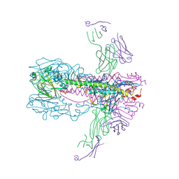
1K5G
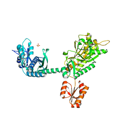
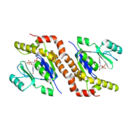
 | | Crystal structure of Ran-GDP-AlFx-RanBP1-RanGAP complex | | Descriptor: | ALUMINUM FLUORIDE, GTP-binding nuclear protein RAN, GUANOSINE-5'-DIPHOSPHATE, ... | | Authors: | Seewald, M.J, Koerner, C, Wittinghofer, A, Vetter, I.R. | | Deposit date: | 2001-10-10 | | Release date: | 2002-02-13 | | Last modified: | 2023-08-16 | | Method: | X-RAY DIFFRACTION (3.1 Å) | | Cite: | RanGAP mediates GTP hydrolysis without an arginine finger.
Nature, 415, 2002
|
|
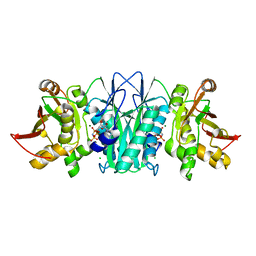
1K5D
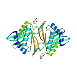
 | | Crystal structure of Ran-GPPNHP-RanBP1-RanGAP complex | | Descriptor: | GTP-binding nuclear protein RAN, MAGNESIUM ION, PHOSPHOAMINOPHOSPHONIC ACID-GUANYLATE ESTER, ... | | Authors: | Seewald, M.J, Koerner, C, Wittinghofer, A, Vetter, I.R. | | Deposit date: | 2001-10-10 | | Release date: | 2002-02-13 | | Last modified: | 2023-08-16 | | Method: | X-RAY DIFFRACTION (2.7 Å) | | Cite: | RanGAP mediates GTP hydrolysis without an arginine finger.
Nature, 415, 2002
|
|
5B8H
 | |
6UYN
 | |
5CC8
 | |
5CM7
 | |
6VEL
 | |
6UYH
 | |
6VU9
 | |
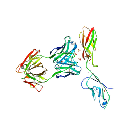
8SJ9
 | | Crystal structure of the H1 hemagglutinin COBRA X6 | | Descriptor: | 1,2-ETHANEDIOL, 2-acetamido-2-deoxy-beta-D-glucopyranose, 2-acetamido-2-deoxy-beta-D-glucopyranose-(1-4)-2-acetamido-2-deoxy-beta-D-glucopyranose, ... | | Authors: | Dzimianski, J.V, DuBois, R.M. | | Deposit date: | 2023-04-17 | | Release date: | 2024-05-01 | | Last modified: | 2024-08-28 | | Method: | X-RAY DIFFRACTION (3.25 Å) | | Cite: | Structural basis for the broad antigenicity of the computationally optimized influenza hemagglutinin X6.
Structure, 32, 2024
|
|
6WCI
 | |
6WNG
 | |
6VJU
 | |
6VUD
 | |
5DD7
 | |
6WCT
 | |
5DWM
 | |
3OA1
 | |
6WSA
 | |
6WSB
 | |


