Loading
PDBj
MenuPDBj@FacebookPDBj@TwitterPDBj@YouTubewwPDB FoundationwwPDB
RCSB PDBPDBeBMRBAdv. SearchSearch help
Search by PDB author
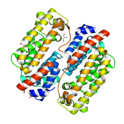
8HAR
DownloadVisualize
BU of 8har by Molmil
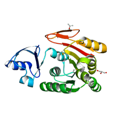
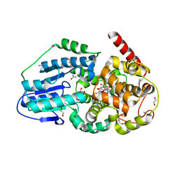
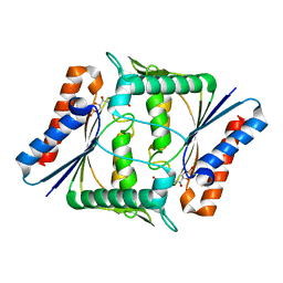
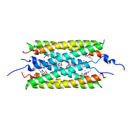
SAH-bound C-Methyltransferase Fur6 from Streptomyces sp. KO-3988
Descriptor: 2-AMINO-2-HYDROXYMETHYL-PROPANE-1,3-DIOL, Fur6, S-ADENOSYL-L-HOMOCYSTEINE
Authors:Noguchi, T, Nagata, R, Tomita, T, Kuzuyama, T.
Deposit date:2022-10-26
Release date:2023-11-01
Method:X-RAY DIFFRACTION (2.12 Å)
Cite:Reductive biosynthesis of meroterpenoids via transient diazotization
To Be Published
8I4J
DownloadVisualize
BU of 8i4j by Molmil
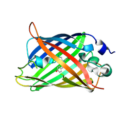
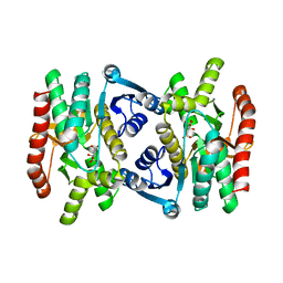
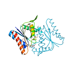
Structure of wild-type Azami Green from Galaxea fascicularis
Descriptor: Azami-Green
Authors:Otsubo, S, Takekawa, N, Imamura, H, Imada, K.
Deposit date:2023-01-19
Release date:2023-11-01
Last modified:2023-11-15
Method:X-RAY DIFFRACTION (1.62 Å)
Cite:Red fluorescent proteins engineered from green fluorescent proteins.
Proc.Natl.Acad.Sci.USA, 120, 2023
8I4K
DownloadVisualize
BU of 8i4k by Molmil
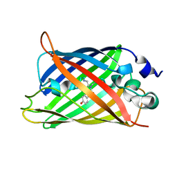
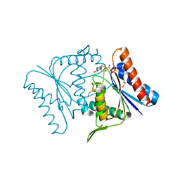
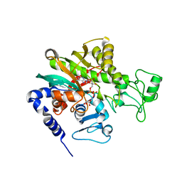
Structure of Azami Red1.0, a red fluorescent protein engineered from Azami Green
Descriptor: Azami Red1.0, CALCIUM ION
Authors:Otsubo, S, Takekawa, N, Imamura, H, Imada, K.
Deposit date:2023-01-19
Release date:2023-11-01
Last modified:2023-12-20
Method:X-RAY DIFFRACTION (1.84 Å)
Cite:Red fluorescent proteins engineered from green fluorescent proteins.
Proc.Natl.Acad.Sci.USA, 120, 2023
4NNO
DownloadVisualize
BU of 4nno by Molmil
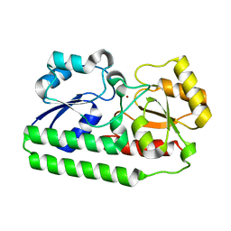
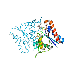
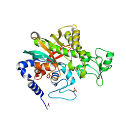
Crystal Structure of Manganese ABC transporter substrate-binding protein MntC from Staphylococcus Aureus bound to a Zinc ion
Descriptor: Lipoprotein, ZINC ION
Authors:Rouge, L, Ahuja, S, Sudhamsu, J.
Deposit date:2013-11-18
Release date:2014-11-26
Last modified:2023-09-20
Method:X-RAY DIFFRACTION (1.174 Å)
Cite:Structural analysis of bacterial ABC transporter inhibition by an antibody fragment.
Structure, 23, 2015
2YY8
DownloadVisualize
BU of 2yy8 by Molmil
Crystal structure of archaeal tRNA-methylase for position 56 (aTrm56) from Pyrococcus horikoshii, complexed with S-adenosyl-L-methionine
Descriptor: 5'-DEOXY-5'-METHYLTHIOADENOSINE, S-ADENOSYLMETHIONINE, UPF0106 protein PH0461
Authors:Kuratani, M, Yokoyama, S, RIKEN Structural Genomics/Proteomics Initiative (RSGI)
Deposit date:2007-04-27
Release date:2008-03-18
Last modified:2024-03-13
Method:X-RAY DIFFRACTION (2.48 Å)
Cite:Crystal structure and mutational study of a unique SpoU family archaeal methylase that forms 2'-O-methylcytidine at position 56 of tRNA
J.Mol.Biol., 375, 2008
2YTZ
DownloadVisualize
BU of 2ytz by Molmil
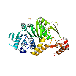
Complex structure of Trm1 from Pyrococcus horikoshii with S-adenosyl-L-Homocystein in the orthorhombic crystal-lattice
Descriptor: N(2),N(2)-dimethylguanosine tRNA methyltransferase, S-ADENOSYL-L-HOMOCYSTEINE, SULFATE ION
Authors:Ihsanawati, Shirouzu, M, Bessho, Y, Yokoyama, S, RIKEN Structural Genomics/Proteomics Initiative (RSGI)
Deposit date:2007-04-05
Release date:2007-10-09
Last modified:2023-10-25
Method:X-RAY DIFFRACTION (2.65 Å)
Cite:Crystal Structure of tRNA N(2),N(2)-Guanosine Dimethyltransferase Trm1 from Pyrococcus horikoshii
J.Mol.Biol., 383, 2008
4NNP
DownloadVisualize
BU of 4nnp by Molmil
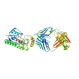
Crystal Structure of Apo Manganese ABC transporter MntC from Staphylococcus aureus bound to an antagonistic fab fragment
Descriptor: Heavy chain of antagonistic fab fragment, Light chain of antagonistic fab fragment, Lipoprotein
Authors:Rouge, L, Sudhamsu, J.
Deposit date:2013-11-18
Release date:2014-12-10
Last modified:2020-05-20
Method:X-RAY DIFFRACTION (2.69 Å)
Cite:Structural analysis of bacterial ABC transporter inhibition by an antibody fragment.
Structure, 23, 2015
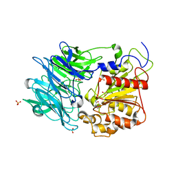
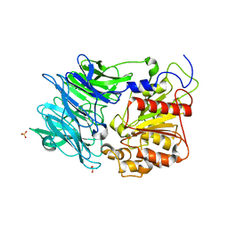
3VSM
DownloadVisualize
BU of 3vsm by Molmil
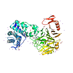
The crystal structure of novel chondroition lyase ODV-E66, baculovirus envelope protein
Descriptor: GLYCEROL, Occlusion-derived virus envelope protein E66
Authors:Kawaguchi, Y, Sugiura, N, Kimata, K, Kimura, M, Kakuta, Y.
Deposit date:2012-04-27
Release date:2013-05-22
Last modified:2024-03-20
Method:X-RAY DIFFRACTION (2 Å)
Cite:The crystal structure of novel chondroition lyase ODV-E66, baculovirus envelope protein
To be Published
2Z98
DownloadVisualize
BU of 2z98 by Molmil
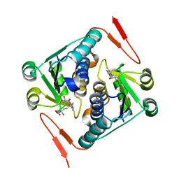
The crystal structure of AzoR (azoreductase) from Escherichia coli: Oxidized AzoR in tetragonal crystals (The resolution has improved from 1.8 (1v4b) to 1.4 angstrom)
Descriptor: 1,2-ETHANEDIOL, FLAVIN MONONUCLEOTIDE, FMN-dependent NADH-azoreductase
Authors:Ito, K.
Deposit date:2007-09-18
Release date:2008-03-11
Last modified:2023-11-01
Method:X-RAY DIFFRACTION (1.4 Å)
Cite:Expansion of Substrate Specificity and Catalytic Mechanism of Azoreductase by X-ray Crystallography and Site-directed Mutagenesis
J.Biol.Chem., 283, 2008
2Z9C
DownloadVisualize
BU of 2z9c by Molmil
The crystal structure of AzoR (azoreductase) from Escherichia coli: AzoR in complex with dicoumarol
Descriptor: BISHYDROXY[2H-1-BENZOPYRAN-2-ONE,1,2-BENZOPYRONE], FLAVIN MONONUCLEOTIDE, FMN-dependent NADH-azoreductase, ...
Authors:Ito, K.
Deposit date:2007-09-18
Release date:2008-03-11
Last modified:2023-11-01
Method:X-RAY DIFFRACTION (2.3 Å)
Cite:Expansion of Substrate Specificity and Catalytic Mechanism of Azoreductase by X-ray Crystallography and Site-directed Mutagenesis
J.Biol.Chem., 283, 2008
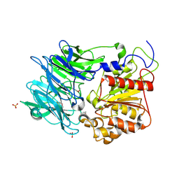
3VSN
DownloadVisualize
BU of 3vsn by Molmil
The crystal structure of novel chondroition lyase ODV-E66, baculovirus envelope protein
Descriptor: GLYCEROL, IODIDE ION, Occlusion-derived virus envelope protein E66
Authors:Kawaguchi, Y, Sugiura, N, Kimata, K, Kimura, M, Kakuta, Y.
Deposit date:2012-04-27
Release date:2013-05-22
Last modified:2024-03-20
Method:X-RAY DIFFRACTION (2 Å)
Cite:The crystal structure of novel chondroition lyase ODV-E66, baculovirus envelope protein
To be Published
3W1D
DownloadVisualize
BU of 3w1d by Molmil
Structure of a pressure sensitive YFP variant YFP-G3
Descriptor: Green Fluorescent protein
Authors:Imada, K, Yoshizawa, K, Kinoshita, M, Watanabe, T.M.
Deposit date:2012-11-14
Release date:2013-10-02
Last modified:2023-12-06
Method:X-RAY DIFFRACTION (1.5 Å)
Cite:Glycine insertion makes yellow fluorescent protein sensitive to hydrostatic pressure.
Plos One, 8, 2013
3W1C
DownloadVisualize
BU of 3w1c by Molmil
Structure of a pressure sensitive YFP variant YFP-G1
Descriptor: Green Fluorescent protein
Authors:Imada, K, Yoshizawa, K, Kinoshita, M, Watanabe, T.M.
Deposit date:2012-11-14
Release date:2013-10-02
Last modified:2023-12-06
Method:X-RAY DIFFRACTION (1.3 Å)
Cite:Glycine insertion makes yellow fluorescent protein sensitive to hydrostatic pressure.
Plos One, 8, 2013
2Z9D
DownloadVisualize
BU of 2z9d by Molmil
The crystal structure of AzoR (azoreductase) from Escherichia coli: Oxidized AzoR in orthorhombic crystals
Descriptor: FLAVIN MONONUCLEOTIDE, FMN-dependent NADH-azoreductase
Authors:Ito, K.
Deposit date:2007-09-18
Release date:2008-03-11
Last modified:2023-11-01
Method:X-RAY DIFFRACTION (2.1 Å)
Cite:Expansion of Substrate Specificity and Catalytic Mechanism of Azoreductase by X-ray Crystallography and Site-directed Mutagenesis
J.Biol.Chem., 283, 2008
2RSG
DownloadVisualize
BU of 2rsg by Molmil
Solution structure of the CERT PH domain
Descriptor: Collagen type IV alpha-3-binding protein
Authors:Sugiki, T, Takeuchi, K, Tokunaga, Y, Kumagai, K, Kawano, M, Nishijima, M, Hanada, K, Takahashi, H, Shimada, I.
Deposit date:2012-02-25
Release date:2012-08-15
Last modified:2024-05-15
Method:SOLUTION NMR
Cite:Structural basis for the Golgi association by the pleckstrin homology domain of the ceramide trafficking protein (CERT)
J.Biol.Chem., 287, 2012
2Z9B
DownloadVisualize
BU of 2z9b by Molmil
The crystal structure of AzoR (azoreductase) from Escherichia coli: Reduced AzoR in tetragonal crystals
Descriptor: 1,2-ETHANEDIOL, FLAVIN MONONUCLEOTIDE, FMN-dependent NADH-azoreductase
Authors:Ito, K.
Deposit date:2007-09-18
Release date:2008-03-11
Last modified:2023-11-01
Method:X-RAY DIFFRACTION (1.7 Å)
Cite:Expansion of Substrate Specificity and Catalytic Mechanism of Azoreductase by X-ray Crystallography and Site-directed Mutagenesis
J.Biol.Chem., 283, 2008
7WBK
DownloadVisualize
BU of 7wbk by Molmil
Crystal structure of Legionella pneumophila effector protein Lpg0081
Descriptor: Lpg0081, SULFATE ION
Authors:Lee, J, Kim, H, Oh, B.H.
Deposit date:2021-12-16
Release date:2022-06-15
Method:X-RAY DIFFRACTION (2.74 Å)
Cite:Reversible modification of mitochondrial ADP/ATP translocases by paired Legionella effector proteins.
Proc.Natl.Acad.Sci.USA, 119, 2022
7WBM
DownloadVisualize
BU of 7wbm by Molmil
Crystal structure of Legionella pneumophila effector protein Lpg0081
Descriptor: Lpg0081, [(2R,3S,4R,5R)-5-(6-AMINOPURIN-9-YL)-3,4-DIHYDROXY-OXOLAN-2-YL]METHYL [HYDROXY-[[(2R,3S,4R,5S)-3,4,5-TRIHYDROXYOXOLAN-2-YL]METHOXY]PHOSPHORYL] HYDROGEN PHOSPHATE
Authors:Lee, J, Kim, H, Oh, B.H.
Deposit date:2021-12-17
Release date:2022-06-15
Last modified:2023-11-29
Method:X-RAY DIFFRACTION (2.7 Å)
Cite:Reversible modification of mitochondrial ADP/ATP translocases by paired Legionella effector proteins.
Proc.Natl.Acad.Sci.USA, 119, 2022
7XGO
DownloadVisualize
BU of 7xgo by Molmil
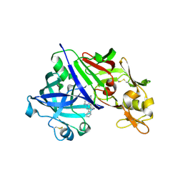
Human renin in complex with compound2
Descriptor: 2-acetamido-2-deoxy-beta-D-glucopyranose, Renin, UNKNOWN LIGAND
Authors:Kashima, A.
Deposit date:2022-04-05
Release date:2022-08-31
Method:X-RAY DIFFRACTION (2.1 Å)
Cite:Discovery of Novel 2-Carbamoyl Morpholine Derivatives as Highly Potent and Orally Active Direct Renin Inhibitors.
Acs Med.Chem.Lett., 13, 2022
7XGK
DownloadVisualize
BU of 7xgk by Molmil
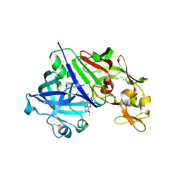
Human renin in complex with compound1
Descriptor: 2-acetamido-2-deoxy-beta-D-glucopyranose, Renin, UNKNOWN LIGAND
Authors:Kashima, A.
Deposit date:2022-04-05
Release date:2022-08-31
Method:X-RAY DIFFRACTION (2.4 Å)
Cite:Discovery of Novel 2-Carbamoyl Morpholine Derivatives as Highly Potent and Orally Active Direct Renin Inhibitors.
Acs Med.Chem.Lett., 13, 2022
3GFL
DownloadVisualize
BU of 3gfl by Molmil
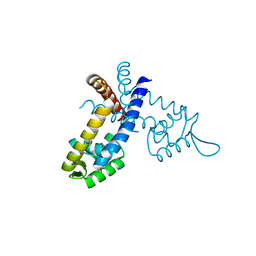
Crystal structure of the ST1710 mutant (R90A) protein
Descriptor: 146aa long hypothetical transcriptional regulator, CALCIUM ION
Authors:Kumarevel, T, Tanaka, T, Yokoyama, S, RIKEN Structural Genomics/Proteomics Initiative (RSGI)
Deposit date:2009-02-27
Release date:2009-08-25
Last modified:2023-11-01
Method:X-RAY DIFFRACTION (1.9 Å)
Cite:ST1710-DNA complex crystal structure reveals the DNA binding mechanism of the MarR family of regulators.
Nucleic Acids Res., 37, 2009
3GF2
DownloadVisualize
BU of 3gf2 by Molmil
Crystal structure of the hypothetical regulator ST1710 complexed with sodium salicylate
Descriptor: 146aa long hypothetical transcriptional regulator, 2-HYDROXYBENZOIC ACID
Authors:Kumarevel, T, Tanaka, T, Yokoyama, S, RIKEN Structural Genomics/Proteomics Initiative (RSGI)
Deposit date:2009-02-26
Release date:2009-08-25
Last modified:2023-11-01
Method:X-RAY DIFFRACTION (1.8 Å)
Cite:ST1710-DNA complex crystal structure reveals the DNA binding mechanism of the MarR family of regulators.
Nucleic Acids Res., 37, 2009
3GEZ
DownloadVisualize
BU of 3gez by Molmil
Crystal Structure of the hypothetical egulator from Sulfolobus tokodaii 7
Descriptor: 146aa long hypothetical transcriptional regulator, CALCIUM ION
Authors:Kumarevel, T, Tanaka, T, Yokoyama, S, RIKEN Structural Genomics/Proteomics Initiative (RSGI)
Deposit date:2009-02-26
Release date:2009-08-25
Last modified:2023-11-01
Method:X-RAY DIFFRACTION (2 Å)
Cite:ST1710-DNA complex crystal structure reveals the DNA binding mechanism of the MarR family of regulators.
Nucleic Acids Res., 37, 2009
3GFJ
DownloadVisualize
BU of 3gfj by Molmil
Crystal structure of the ST1710 mutant (R89A) protein
Descriptor: 146aa long hypothetical transcriptional regulator, CALCIUM ION
Authors:Kumarevel, T, Tanaka, T, Yokoyama, S, RIKEN Structural Genomics/Proteomics Initiative (RSGI)
Deposit date:2009-02-27
Release date:2009-08-25
Last modified:2023-11-01
Method:X-RAY DIFFRACTION (2.2 Å)
Cite:ST1710-DNA complex crystal structure reveals the DNA binding mechanism of the MarR family of regulators.
Nucleic Acids Res., 37, 2009
3GFI
DownloadVisualize
BU of 3gfi by Molmil
Crystal structure of ST1710 complexed with its promoter DNA
Descriptor: 146aa long hypothetical transcriptional regulator, 5'-D(*TP*AP*AP*CP*AP*AP*TP*AP*GP*CP*AP*AP*A)-3', 5'-D(*TP*TP*GP*CP*TP*AP*TP*TP*GP*T)-3'
Authors:Kumarevel, T, Tanaka, T, Yokoyama, S, RIKEN Structural Genomics/Proteomics Initiative (RSGI)
Deposit date:2009-02-26
Release date:2009-08-25
Last modified:2024-03-20
Method:X-RAY DIFFRACTION (2.1 Å)
Cite:ST1710-DNA complex crystal structure reveals the DNA binding mechanism of the MarR family of regulators.
Nucleic Acids Res., 37, 2009

221716

数据于2024-06-26公开中

PDB statisticsPDBj update infoContact PDBjnumon