Loading
PDBj
MenuPDBj@FacebookPDBj@TwitterPDBj@YouTubewwPDB FoundationwwPDB
RCSB PDBPDBeBMRBAdv. SearchSearch help
Search by PDB author
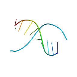
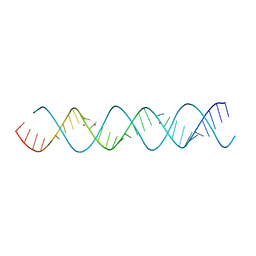
4RBQ
DownloadVisualize
BU of 4rbq by Molmil
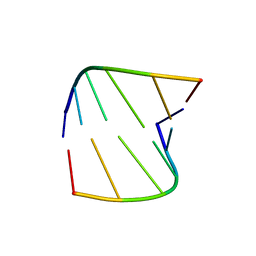
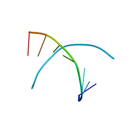
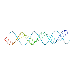
32 base pair oligo(U) RNA
Descriptor: POTASSIUM ION, U-Helix RNA from Trypanosome editing
Authors:Mooers, B.H.M.
Deposit date:2014-09-12
Release date:2015-11-11
Last modified:2024-04-03
Method:X-RAY DIFFRACTION (1.05 Å)
Cite:Structure of the Trypanosome RNA Editing U-Helix with 16 Contiguous Us
To be Published
253D
DownloadVisualize
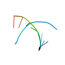
BU of 253d by Molmil
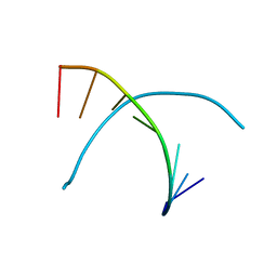
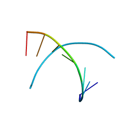
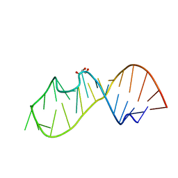
CRYSTAL STRUCTURE OF THE B-DNA NONAMER D(GCGTACGCG) WITH A NOVEL D[G*(G.C)] BASE-TRIPLET INVOLVING THE MINOR GROOVE
Descriptor: DNA (5'-D(*GP*CP*GP*TP*AP*CP*GP*CP*G)-3'), MAGNESIUM ION
Authors:Mooers, B.H.M, Ho, P.S.
Deposit date:1996-03-20
Release date:1996-04-15
Last modified:2024-02-14
Method:X-RAY DIFFRACTION (2.2 Å)
Cite:Crystal Structure of the B-DNA Nonamer d(GCGTACGCG) with a Novel d[G*(G.C)] Base-triplet Involving the Minor Groove
To be Published, 1996
339D
DownloadVisualize
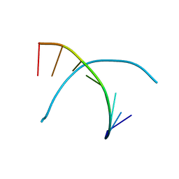
BU of 339d by Molmil
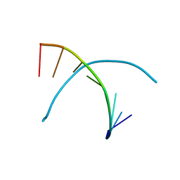
STRUCTURAL PARAMETERS FROM SINGLE-CRYSTAL STRUCTURES FOR ACCURATE MODELS OF A-DNA
Descriptor: DNA (5'-D(*GP*(5CM)P*GP*CP*GP*(5CM)P*GP*C)-3')
Authors:Mooers, B.H.M, Eichman, B.F, Ho, P.S.
Deposit date:1997-06-26
Release date:1997-07-21
Last modified:2024-02-21
Method:X-RAY DIFFRACTION (2.2 Å)
Cite:Structural Parameters from Single-Crystal Structures for Accurate Models of A-DNA
To be Published
338D
DownloadVisualize
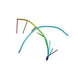
BU of 338d by Molmil
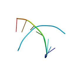
STRUCTURAL PARAMETERS FROM SINGLE-CRYSTAL STRUCTURES FOR ACCURATE MODELS OF A-DNA
Descriptor: DNA (5'-D(*GP*(5CM)P*GP*CP*GP*CP*GP*C)-3')
Authors:Mooers, B.H.M, Eichman, B.F, Ho, P.S.
Deposit date:1997-06-26
Release date:1997-07-21
Last modified:2024-02-21
Method:X-RAY DIFFRACTION (1.85 Å)
Cite:Structural Parameters from Single-Crystal Structures for Accurate Models of A-DNA
To be Published
340D
DownloadVisualize
BU of 340d by Molmil
STRUCTURAL PARAMETERS FROM SINGLE-CRYSTAL STRUCTURES FOR ACCURATE MODELS OF A-DNA
Descriptor: DNA (5'-D(*GP*(5CM)P*GP*(5CM)P*GP*CP*GP*C)-3')
Authors:Mooers, B.H.M, Eichman, B.F, Ho, P.S.
Deposit date:1997-06-26
Release date:1997-07-21
Last modified:2024-02-21
Method:X-RAY DIFFRACTION (1.6 Å)
Cite:Structural Parameters from Single-Crystal Structures for Accurate Models of A-DNA
To be Published
343D
DownloadVisualize
BU of 343d by Molmil
STRUCTURAL PARAMETERS FROM SINGLE-CRYSTAL STRUCTURES FOR ACCURATE MODELS OF A-DNA
Descriptor: DNA (5'-D(*GP*(5CM)P*TP*AP*GP*C)-3')
Authors:Mooers, B.H.M, Eichman, B.F, Ho, P.S.
Deposit date:1997-06-26
Release date:1997-07-21
Last modified:2024-02-21
Method:X-RAY DIFFRACTION (2.1 Å)
Cite:Structural Parameters from Single-Crystal Structures for Accurate Models of A-DNA
To be Published
345D
DownloadVisualize
BU of 345d by Molmil
STRUCTURAL PARAMETERS FROM SINGLE-CRYSTAL STRUCTURES FOR ACCURATE MODELS OF A-DNA
Descriptor: DNA (5'-D(*GP*(5CM)P*GP*CP*GP*CP*GP*C)-3')
Authors:Mooers, B.H.M, Eichman, B.F, Ho, P.S.
Deposit date:1997-08-15
Release date:1997-08-29
Last modified:2024-02-21
Method:X-RAY DIFFRACTION (1.85 Å)
Cite:Structural Parameters from Single-Crystal Structures for Accurate Models of A-DNA
To be Published
341D
DownloadVisualize
BU of 341d by Molmil
STRUCTURAL PARAMETERS FROM SINGLE-CRYSTAL STRUCTURES FOR ACCURATE MODELS OF A-DNA
Descriptor: DNA (5'-D(*GP*(5CM)P*GP*(5CM)P*GP*CP*GP*C)-3')
Authors:Mooers, B.H.M, Eichman, B.F, Ho, P.S.
Deposit date:1997-06-26
Release date:1997-07-21
Last modified:2024-02-21
Method:X-RAY DIFFRACTION (1.75 Å)
Cite:Structural Parameters from Single-Crystal Structures For Accurate Models of A-DNA
To be Published
342D
DownloadVisualize
BU of 342d by Molmil
STRUCTURAL PARAMETERS FROM SINGLE-CRYSTAL STRUCTURES FOR ACCURATE MODELS OF A-DNA
Descriptor: DNA (5'-D(*GP*(5CM)P*GP*(5CM)P*GP*(5CM)P*GP*C)-3')
Authors:Mooers, B.H.M, Eichman, B.F, Ho, P.S.
Deposit date:1997-06-26
Release date:1997-07-21
Last modified:2024-02-21
Method:X-RAY DIFFRACTION (2.1 Å)
Cite:Structural Parameters from Single-Crystal Structures for Accurate Models of A-DNA
To be Published
346D
DownloadVisualize
BU of 346d by Molmil
STRUCTURAL PARAMETERS FROM SINGLE-CRYSTAL STRUCTURES FOR ACCURATE MODELS OF A-DNA
Descriptor: DNA (5'-D(*GP*(5CM)P*GP*(5CM)P*GP*(5CM)P*GP*C)-3')
Authors:Mooers, B.H.M, Eichman, B.F, Ho, P.S.
Deposit date:1997-08-26
Release date:1997-08-29
Last modified:2024-02-21
Method:X-RAY DIFFRACTION (2.1 Å)
Cite:Structural Parameters from Single-Crystal Structures for Accurate Models of A-DNA
To be Published
337D
DownloadVisualize
BU of 337d by Molmil
STRUCTURAL PARAMETERS FROM SINGLE-CRYSTAL STRUCTURES FOR ACCURATE MODELS OF A-DNA
Descriptor: DNA (5'-D(*GP*CP*GP*(5CM)P*GP*(5CM)P*GP*C)-3')
Authors:Mooers, B.H.M, Eichman, B.F, Ho, P.S.
Deposit date:1997-06-26
Release date:1997-07-21
Last modified:2024-02-21
Method:X-RAY DIFFRACTION (1.85 Å)
Cite:Structural Parameters from Single-Crystal Structures for Accurate Models of A-DNA
To be Published
8G2K
DownloadVisualize
BU of 8g2k by Molmil
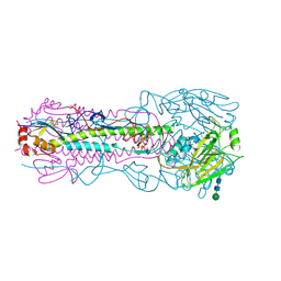
Structure of the H3 hemagglutinin of A/California/7/2004
Descriptor: 2-acetamido-2-deoxy-beta-D-glucopyranose, 2-acetamido-2-deoxy-beta-D-glucopyranose-(1-4)-2-acetamido-2-deoxy-beta-D-glucopyranose, Hemagglutinin, ...
Authors:Venkatramani, L, Mooers, B.H.M, Air, G.M.
Deposit date:2023-02-05
Release date:2024-05-15
Last modified:2024-10-16
Method:X-RAY DIFFRACTION (2.35 Å)
Cite:Structure of the H3 hemagglutinin of A/California/7/2004
To Be Published
7JU5
DownloadVisualize
BU of 7ju5 by Molmil
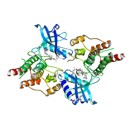
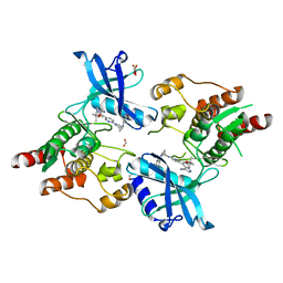
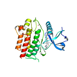
Structure of RET protein tyrosine kinase in complex with pralsetinib
Descriptor: FORMIC ACID, Pralsetinib, Proto-oncogene tyrosine-protein kinase receptor Ret
Authors:Terzyan, S.S, Shen, T, Wu, J, Mooers, B.H.M.
Deposit date:2020-08-19
Release date:2020-11-11
Last modified:2023-10-18
Method:X-RAY DIFFRACTION (1.9 Å)
Cite:Structural basis of acquired resistance to selpercatinib and pralsetinib mediated by non-gatekeeper RET mutations.
Ann Oncol, 32, 2021
7JU6
DownloadVisualize
BU of 7ju6 by Molmil
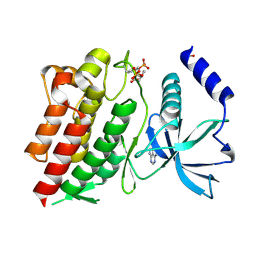
Structure of RET protein tyrosine kinase in complex with selpercatinib
Descriptor: FORMIC ACID, Proto-oncogene tyrosine-protein kinase receptor Ret, Selpercatinib
Authors:Terzyan, S.S, Shen, T, Wu, J, Mooers, B.H.M.
Deposit date:2020-08-19
Release date:2020-11-11
Last modified:2023-10-18
Method:X-RAY DIFFRACTION (2.06 Å)
Cite:Structural basis of acquired resistance to selpercatinib and pralsetinib mediated by non-gatekeeper RET mutations.
Ann Oncol, 32, 2021
1FHZ
DownloadVisualize
BU of 1fhz by Molmil
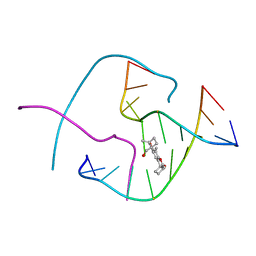
PSORALEN CROSS-LINKED D(CCGGTACCGG) FORMS HOLLIDAY JUNCTION
Descriptor: 4'-HYDROXYMETHYL-4,5',8-TRIMETHYLPSORALEN, DNA (5'-D(*CP*CP*GP*GP*TP*AP*CP*CP*GP*G)-3')
Authors:Eichman, B.F, Mooers, B.H.M, Alberti, M, Hearst, J.E, Ho, P.S.
Deposit date:2000-08-02
Release date:2001-04-21
Last modified:2024-02-07
Method:X-RAY DIFFRACTION (2.2 Å)
Cite:The crystal structures of psoralen cross-linked DNAs: drug-dependent formation of Holliday junctions.
J.Mol.Biol., 308, 2001
1FHY
DownloadVisualize
BU of 1fhy by Molmil
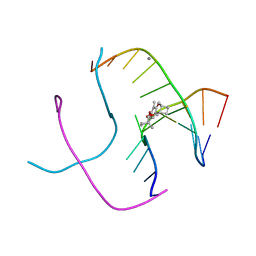
PSORALEN CROSS-LINKED D(CCGCTAGCGG) FORMS HOLLIDAY JUNCTION
Descriptor: 4'-HYDROXYMETHYL-4,5',8-TRIMETHYLPSORALEN, CALCIUM ION, DNA (5'-D(*CP*CP*GP*CP*TP*AP*GP*CP*GP*G)-3')
Authors:Eichman, B.F, Mooers, B.H.M, Alberti, M, Hearst, J.E, Ho, P.S.
Deposit date:2000-08-02
Release date:2001-04-21
Last modified:2024-02-07
Method:X-RAY DIFFRACTION (2.2 Å)
Cite:The crystal structures of psoralen cross-linked DNAs: drug-dependent formation of Holliday junctions.
J.Mol.Biol., 308, 2001
6NE7
DownloadVisualize
BU of 6ne7 by Molmil
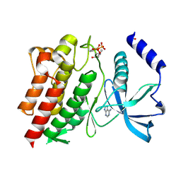
Structure of G810A mutant of RET protein tyrosine kinase domain.
Descriptor: ADENOSINE MONOPHOSPHATE, FORMIC ACID, Proto-oncogene tyrosine-protein kinase receptor Ret
Authors:Terzyan, S.S, Shen, T, Wu, J, Mooers, B.H.M.
Deposit date:2018-12-17
Release date:2019-06-05
Last modified:2024-10-16
Method:X-RAY DIFFRACTION (1.99 Å)
Cite:Structural basis of resistance of mutant RET protein-tyrosine kinase to its inhibitors nintedanib and vandetanib.
J.Biol.Chem., 294, 2019
6NEC
DownloadVisualize
BU of 6nec by Molmil
STRUCTURE OF RET PROTEIN TYROSINE KINASE DOMAIN IN COMPLEX WITH NINTEDANIB
Descriptor: FORMIC ACID, Proto-oncogene tyrosine-protein kinase receptor Ret, methyl (3Z)-3-{[(4-{methyl[(4-methylpiperazin-1-yl)acetyl]amino}phenyl)amino](phenyl)methylidene}-2-oxo-2,3-dihydro-1H-indole-6-carboxylate
Authors:Terzyan, S.S, Shen, T, Wu, J, Mooers, B.H.M.
Deposit date:2018-12-17
Release date:2019-06-05
Last modified:2023-10-11
Method:X-RAY DIFFRACTION (1.87 Å)
Cite:Structural basis of resistance of mutant RET protein-tyrosine kinase to its inhibitors nintedanib and vandetanib.
J.Biol.Chem., 294, 2019
6NJA
DownloadVisualize
BU of 6nja by Molmil
Structure of WT RET protein tyrosine kinase domain at 1.92A resolution.
Descriptor: ADENINE, FORMIC ACID, Proto-oncogene tyrosine-protein kinase receptor Ret
Authors:Terzyan, S.S, Shen, T, Wu, J, Mooers, B.H.M.
Deposit date:2019-01-02
Release date:2019-06-05
Last modified:2024-10-23
Method:X-RAY DIFFRACTION (1.92 Å)
Cite:Structural basis of resistance of mutant RET protein-tyrosine kinase to its inhibitors nintedanib and vandetanib.
J.Biol.Chem., 294, 2019
3C8R
DownloadVisualize
BU of 3c8r by Molmil
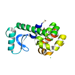
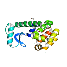
Contributions of all 20 amino acids at site 96 to stability and structure of T4 lysozyme
Descriptor: BETA-MERCAPTOETHANOL, CHLORIDE ION, Lysozyme
Authors:Mooers, B.H.M.
Deposit date:2008-02-13
Release date:2009-02-17
Last modified:2023-08-30
Method:X-RAY DIFFRACTION (1.8 Å)
Cite:Contributions of all 20 amino acids at site 96 to the stability and structure of T4 lysozyme.
Protein Sci., 18, 2009
3C7W
DownloadVisualize
BU of 3c7w by Molmil
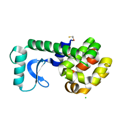
Contributions of all 20 amino acids at site 96 to the stability and structure of T4 lysozyme
Descriptor: BETA-MERCAPTOETHANOL, CHLORIDE ION, Lysozyme
Authors:Mooers, B.H.M.
Deposit date:2008-02-08
Release date:2009-02-17
Last modified:2023-08-30
Method:X-RAY DIFFRACTION (1.77 Å)
Cite:Contributions of all 20 amino acids at site 96 to the stability and structure of T4 lysozyme.
Protein Sci., 18, 2009
3C7Z
DownloadVisualize
BU of 3c7z by Molmil
T4 lysozyme mutant D89A/R96H at room temperature
Descriptor: BETA-MERCAPTOETHANOL, CHLORIDE ION, Lysozyme
Authors:Mooers, B.H.M.
Deposit date:2008-02-08
Release date:2009-02-17
Last modified:2023-08-30
Method:X-RAY DIFFRACTION (1.67 Å)
Cite:Contributions of all 20 amino acids at site 96 to the stability and structure of T4 lysozyme.
Protein Sci., 18, 2009
5DA6
DownloadVisualize
BU of 5da6 by Molmil
Atomic resolution crystal structure of double-stranded RNA 32 base pairs long determined from random starting phases angles in the presence of pseudo translational symmetry using the direct methods program SIR2014.
Descriptor: POTASSIUM ION, RNA (32-MER)
Authors:Mooers, B.H.M.
Deposit date:2015-08-19
Release date:2016-04-13
Last modified:2024-03-06
Method:X-RAY DIFFRACTION (1.05 Å)
Cite:Direct-methods structure determination of a trypanosome RNA-editing substrate fragment with translational pseudosymmetry.
Acta Crystallogr D Struct Biol, 72, 2016
5D99
DownloadVisualize
BU of 5d99 by Molmil
3DW4 redetermined by direct methods starting from random phase angles
Descriptor: GLYCEROL, RNA (27-MER) hairpin from sarcin-ricin domain of E. coli 23S rRNA
Authors:Mooers, B.H.M.
Deposit date:2015-08-18
Release date:2016-04-13
Last modified:2024-03-06
Method:X-RAY DIFFRACTION (0.97 Å)
Cite:Direct-methods structure determination of a trypanosome RNA-editing substrate fragment with translational pseudosymmetry.
Acta Crystallogr D Struct Biol, 72, 2016
4PCO
DownloadVisualize
BU of 4pco by Molmil
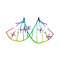
Crystal structure of double-stranded RNA with four terminal GU wobble base pairs
Descriptor: CHLORIDE ION, COBALT HEXAMMINE(III), RNA (5'-D(*GP*GP*UP*GP*GP*CP*UP*GP*UP*U)-3')
Authors:Mooers, B.H.M.
Deposit date:2014-04-15
Release date:2015-10-14
Last modified:2023-12-27
Method:X-RAY DIFFRACTION (1.32 Å)
Cite:Structures and Energetics of Four Adjacent GU Pairs That Stabilize an RNA Helix.
J.Phys.Chem.B, 119, 2015


123>

226707

건을2024-10-30부터공개중

PDB statisticsPDBj update infoContact PDBjnumon