Loading
PDBj
MenuPDBj@FacebookPDBj@TwitterPDBj@YouTubewwPDB FoundationwwPDB
RCSB PDBPDBeBMRBAdv. SearchSearch help
Search by PDB author
1CQN
DownloadVisualize
BU of 1cqn by Molmil
PROTEIN AGGREGATION AND ALZHEIMER'S DISEASE: CRYSTALLOGRAPHIC ANALYSIS OF THE PHENOMENON. ENGINEERED VERSION OF THE RIBOSOMAL PROTEIN S6 USED AS A STABLE SCAFFOLD TO STUDY OLIGOMERIZATION.
Descriptor: RIBOSOMAL PROTEIN S6
Authors:Kristensen, O, Otzen, D.E, Oliveberg, M.
Deposit date:1999-08-08
Release date:2000-09-08
Last modified:2024-02-07
Method:X-RAY DIFFRACTION (2.1 Å)
Cite:Designed protein tetramer zipped together with a hydrophobic Alzheimer homology: a structural clue to amyloid assembly.
Proc.Natl.Acad.Sci.USA, 97, 2000
1CQM
DownloadVisualize
BU of 1cqm by Molmil
PROTEIN AGGREGATION AND ALZHEIMER'S DISEASE: CRYSTALLOGRAPHIC ANALYSIS OF THE PHENOMENON. ENGINEERED VERSION OF THE RIBOSOMAL PROTEIN S6 USED AS A STABLE SCAFFOLD TO STUDY OLIGOMERIZATION.
Descriptor: RIBOSOMAL PROTEIN S6
Authors:Kristensen, O, Otzen, D.E, Oliveberg, M.
Deposit date:1999-08-08
Release date:2000-09-08
Last modified:2024-02-07
Method:X-RAY DIFFRACTION (1.65 Å)
Cite:Designed protein tetramer zipped together with a hydrophobic Alzheimer homology: a structural clue to amyloid assembly.
Proc.Natl.Acad.Sci.USA, 97, 2000
1CRM
DownloadVisualize
BU of 1crm by Molmil
STRUCTURE AND FUNCTION OF CARBONIC ANHYDRASES
Descriptor: CARBONIC ANHYDRASE I, CHLORIDE ION, HYDROSULFURIC ACID, ...
Authors:Yadava, V.S, Kannan, K.K.
Deposit date:1994-03-04
Release date:1995-02-07
Last modified:2024-02-07
Method:X-RAY DIFFRACTION (2 Å)
Cite:Structure and Function of Carbonic Anhydrases
Biomolecular Structure, Conformation, Function and Evolution, 1, 1981
1I6U
DownloadVisualize
BU of 1i6u by Molmil
RNA-PROTEIN INTERACTIONS: THE CRYSTAL STRUCTURE OF RIBOSOMAL PROTEIN S8/RRNA COMPLEX FROM METHANOCOCCUS JANNASCHII
Descriptor: 16S RRNA FRAGMENT, 30S RIBOSOMAL PROTEIN S8P, SULFATE ION
Authors:Tishchenko, S, Nikulin, A, Fomenkova, N, Nevskaya, N, Nikonov, O, Dumas, P, Moine, H, Ehresmann, B, Ehresmann, C, Piendl, W, Lamzin, V, Garber, M, Nikonov, S.
Deposit date:2001-03-05
Release date:2001-08-03
Last modified:2024-10-09
Method:X-RAY DIFFRACTION (2.6 Å)
Cite:Detailed analysis of RNA-protein interactions within the ribosomal protein S8-rRNA complex from the archaeon Methanococcus jannaschii.
J.Mol.Biol., 311, 2001
1QJH
DownloadVisualize
BU of 1qjh by Molmil
Protein Aggregation and Alzheimer's Disease: Crystallographic Analysis of the Phenomenon. Engineered version of the ribosomal protein S6 used as a stable scaffold to study oligomerization.
Descriptor: 30S ribosomal protein S6, MAGNESIUM ION
Authors:Kristensen, O, Otzen, D.E, Oliveberg, M.
Deposit date:1999-06-24
Release date:2000-06-29
Last modified:2023-12-13
Method:X-RAY DIFFRACTION (2.2 Å)
Cite:Designed protein tetramer zipped together with a hydrophobic Alzheimer homology: a structural clue to amyloid assembly.
Proc. Natl. Acad. Sci. U.S.A., 97, 2000
2J4R
DownloadVisualize
BU of 2j4r by Molmil
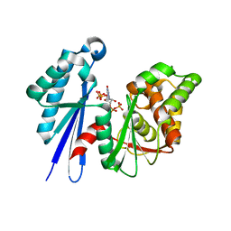
Structural Study of the Aquifex aeolicus PPX-GPPA enzyme
Descriptor: EXOPOLYPHOSPHATASE, GUANOSINE-5',3'-TETRAPHOSPHATE
Authors:Kristensen, O.
Deposit date:2006-09-05
Release date:2007-10-16
Last modified:2023-12-13
Method:X-RAY DIFFRACTION (2.71 Å)
Cite:Structure of the Ppx/Gppa Phosphatase from Aquifex Aeolicus in Complex with the Alarmone Ppgpp
J.Mol.Biol., 375, 2008
2J7K
DownloadVisualize
BU of 2j7k by Molmil
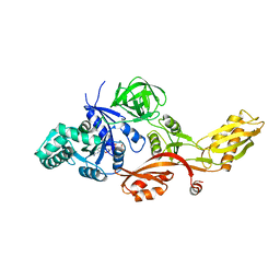
Crystal structure of the T84A mutant EF-G:GDPCP complex
Descriptor: ELONGATION FACTOR G, MAGNESIUM ION, PHOSPHOMETHYLPHOSPHONIC ACID GUANYLATE ESTER
Authors:Hansson, S, Logan, D.T.
Deposit date:2006-10-12
Release date:2007-10-23
Last modified:2023-12-13
Method:X-RAY DIFFRACTION (2.9 Å)
Cite:New Insights Into the Role of the P-Loop Lysine: Implications from the Crystal Structure of a Mutant EF-G:Gdpcp Complex
To be Published
1I2A
DownloadVisualize
BU of 1i2a by Molmil
CRYSTAL STRUCTURE OF L1 RIBOSOMAL PROTEIN FROM METHANOCOCCUS JANNASCHII WITH 1.85A RESOLUTION.
Descriptor: 50S RIBOSOMAL PROTEIN L1P, PENTANAL
Authors:Smolinskaya, Y, Nikonov, S.V.
Deposit date:2001-02-07
Release date:2003-12-09
Last modified:2023-08-09
Method:X-RAY DIFFRACTION (1.85 Å)
Cite:

2BXJ
DownloadVisualize
BU of 2bxj by Molmil
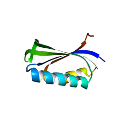
Double Mutant of the Ribosomal Protein S6
Descriptor: 30S RIBOSOMAL PROTEIN S6
Authors:Otzen, D.E.
Deposit date:2005-07-26
Release date:2005-10-26
Last modified:2023-12-13
Method:X-RAY DIFFRACTION (2.4 Å)
Cite:Antagonism, Non-Native Interactions and Non-Two-State Folding in S6 Revealed by Double-Mutant Cycle Analysis.
Protein Eng.Des.Sel., 18, 2005
1FY2
DownloadVisualize
BU of 1fy2 by Molmil
Aspartyl Dipeptidase
Descriptor: ASPARTYL DIPEPTIDASE, CADMIUM ION
Authors:Hakansson, K, Wang, A.H.-J, Miller, C.G.
Deposit date:2000-09-28
Release date:2001-01-10
Last modified:2024-02-07
Method:X-RAY DIFFRACTION (1.2 Å)
Cite:The structure of aspartyl dipeptidase reveals a unique fold with a Ser-His-Glu catalytic triad.
Proc.Natl.Acad.Sci.USA, 97, 2000
1FYE
DownloadVisualize
BU of 1fye by Molmil
Aspartyl Dipeptidase (Anisotropic B-Factor Refinement)
Descriptor: ASPARTYL DIPEPTIDASE, CADMIUM ION
Authors:Hakansson, K, Wang, A.H.-J, Miller, C.G.
Deposit date:2000-09-29
Release date:2001-01-10
Last modified:2024-02-07
Method:X-RAY DIFFRACTION (1.2 Å)
Cite:The structure of aspartyl dipeptidase reveals a unique fold with a Ser-His-Glu catalytic triad.
Proc.Natl.Acad.Sci.USA, 97, 2000
1G0F
DownloadVisualize
BU of 1g0f by Molmil
SITE-SPECIFIC MUTANT (HIS64 REPLACED WITH ALA) OF HUMAN CARBONIC ANHYDRASE II
Descriptor: CARBONIC ANHYDRASE II, MERCURY (II) ION, ZINC ION
Authors:Duda, D, McKenna, R.
Deposit date:2000-10-06
Release date:2000-10-18
Last modified:2024-02-07
Method:X-RAY DIFFRACTION (1.6 Å)
Cite:Structural and kinetic analysis of the chemical rescue of the proton transfer function of carbonic anhydrase II.
Biochemistry, 40, 2001
1G0E
DownloadVisualize
BU of 1g0e by Molmil
SITE-SPECIFIC MUTANT (HIS64 REPLACED WITH ALA) OF HUMAN CARBONIC ANHYDRASE II COMPLEXED WITH 4-METHYLIMIDAZOLE
Descriptor: 4-METHYLIMIDAZOLE, CARBONIC ANHYDRASE II, MERCURY (II) ION, ...
Authors:Duda, D, McKenna, R.
Deposit date:2000-10-06
Release date:2000-10-18
Last modified:2024-02-07
Method:X-RAY DIFFRACTION (1.6 Å)
Cite:Structural and kinetic analysis of the chemical rescue of the proton transfer function of carbonic anhydrase II.
Biochemistry, 40, 2001
<1234


226262

건을2024-10-16부터공개중

PDB statisticsPDBj update infoContact PDBjnumon