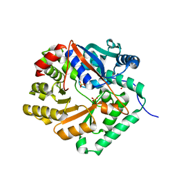
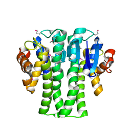
Loading
PDBj
MenuPDBj@FacebookPDBj@TwitterPDBj@YouTubewwPDB FoundationwwPDB
RCSB PDBPDBeBMRBAdv. SearchSearch help
Search by PDB author
3EXQ
DownloadVisualize
BU of 3exq by Molmil
Crystal structure of a NUDIX family hydrolase from Lactobacillus brevis
Descriptor: NUDIX family hydrolase, SULFATE ION
Authors:Palani, K, Burley, S.K, Swaminathan, S, New York SGX Research Center for Structural Genomics (NYSGXRC)
Deposit date:2008-10-16
Release date:2008-11-04
Last modified:2023-12-27
Method:X-RAY DIFFRACTION (2 Å)
Cite:Crystal structure of a NUDIX family hydrolase from Lactobacillus brevis
To be Published
4M1V
DownloadVisualize
BU of 4m1v by Molmil
Crystal structure of the ancestral soluble variant of the Human Phosphate Binding Protein (HPBP)
Descriptor: 1,2-ETHANEDIOL, ACETATE ION, GLYCEROL, ...
Authors:Gonzalez, D, Hiblot, J, Darbinian, N, Miller, J.S, Gotthard, G, Amini, S, Chabriere, E, Elias, M.
Deposit date:2013-08-04
Release date:2014-01-01
Last modified:2023-09-20
Method:X-RAY DIFFRACTION (1.3 Å)
Cite:Ancestral mutations as a tool for solubilizing proteins: The case of a hydrophobic phosphate-binding protein.
FEBS Open Bio, 4, 2014
1Y9Q
DownloadVisualize
BU of 1y9q by Molmil
Crystal Structure of HTH_3 family Transcriptional Regulator from Vibrio cholerae
Descriptor: D-METHIONINE, ZINC ION, transcriptional regulator, ...
Authors:Kumaran, D, Swaminathan, S, Burley, S.K, New York SGX Research Center for Structural Genomics (NYSGXRC)
Deposit date:2004-12-16
Release date:2005-01-04
Last modified:2021-02-03
Method:X-RAY DIFFRACTION (1.9 Å)
Cite:Crystal Structure of HTH_3 family Transcriptional Regulator from Vibrio cholerae
To be Published
2IMO
DownloadVisualize
BU of 2imo by Molmil
Crystal structure of allantoate amidohydrolase from Escherichia coli at pH 4.6
Descriptor: Allantoate amidohydrolase
Authors:Agarwal, R, Burley, S.K, Swaminathan, S, New York SGX Research Center for Structural Genomics (NYSGXRC)
Deposit date:2006-10-04
Release date:2006-10-17
Last modified:2021-02-03
Method:X-RAY DIFFRACTION (2.8 Å)
Cite:Structural Analysis of a Ternary Complex of Allantoate Amidohydrolase from Escherichia coli Reveals its Mechanics.
J.Mol.Biol., 368, 2007
1YDF
DownloadVisualize
BU of 1ydf by Molmil
Crystal structure of a HAD-like phosphatase from Streptococcus pneumoniae
Descriptor: MAGNESIUM ION, hydrolase, haloacid dehalogenase-like family
Authors:Seetharaman, J, Swaminathan, S, Burley, S.K, New York SGX Research Center for Structural Genomics (NYSGXRC)
Deposit date:2004-12-23
Release date:2005-01-18
Last modified:2024-02-14
Method:X-RAY DIFFRACTION (2.6 Å)
Cite:Crystal Structure of a Hypothetical protein, hydrolase haloacid dehalogenase-like family
To be Published
1YBF
DownloadVisualize
BU of 1ybf by Molmil
Crystal structure of AMP nucleosidase from Bacteroides thetaiotaomicron VPI-5482
Descriptor: AMP nucleosidase
Authors:Krishnamurthy, N.R, Swaminathan, S, Burley, S.K, New York SGX Research Center for Structural Genomics (NYSGXRC)
Deposit date:2004-12-20
Release date:2005-01-04
Last modified:2021-02-03
Method:X-RAY DIFFRACTION (2.9 Å)
Cite:Crystal structure of AMP nucleosidase from Bacteroides thetaiotaomicron
To be published
2IMG
DownloadVisualize
BU of 2img by Molmil
Crystal structure of dual specificity protein phosphatase 23 from Homo sapiens in complex with ligand malate ion
Descriptor: D-MALATE, Dual specificity protein phosphatase 23
Authors:Agarwal, R, Burley, S.K, Swaminathan, S, New York SGX Research Center for Structural Genomics (NYSGXRC)
Deposit date:2006-10-04
Release date:2006-10-17
Last modified:2021-02-03
Method:X-RAY DIFFRACTION (1.93 Å)
Cite:Structure of human dual specificity protein phosphatase 23, VHZ, enzyme-substrate/product complex.
J.Biol.Chem., 283, 2008
2ICS
DownloadVisualize
BU of 2ics by Molmil
Crystal structure of an adenine deaminase
Descriptor: ADENINE, Adenine Deaminase, ZINC ION
Authors:Sugadev, R, Kumaran, D, Burley, S.K, Swaminathan, S, New York SGX Research Center for Structural Genomics (NYSGXRC)
Deposit date:2006-09-13
Release date:2006-10-17
Last modified:2021-02-03
Method:X-RAY DIFFRACTION (2.3 Å)
Cite:Crystal structure of an adenine deaminase
TO BE PUBLISHED
1YBD
DownloadVisualize
BU of 1ybd by Molmil
Crystal structure analysis of uridylate kinase from Neisseria meningitidis
Descriptor: FORMIC ACID, GLYCEROL, Uridylate kinase
Authors:Kumaran, D, Swaminathan, S, Burley, S.K, New York SGX Research Center for Structural Genomics (NYSGXRC)
Deposit date:2004-12-20
Release date:2005-02-15
Last modified:2021-02-03
Method:X-RAY DIFFRACTION (2.6 Å)
Cite:Crystal structure analysis of uridylate kinase from Neisseria meningitidis
To be Published
1YAV
DownloadVisualize
BU of 1yav by Molmil
Crystal structure of CBS domain-containing protein ykuL from Bacillus subtilis
Descriptor: SULFATE ION, hypothetical protein BSU14130
Authors:Kumaran, D, Swaminathan, S, Burley, S.K, New York SGX Research Center for Structural Genomics (NYSGXRC)
Deposit date:2004-12-17
Release date:2004-12-28
Last modified:2024-02-14
Method:X-RAY DIFFRACTION (2.1 Å)
Cite:Crystal Structure of a hypothetical protein (ykul) containing CBS domains from Bacillus subtilis.
To be Published
1YBE
DownloadVisualize
BU of 1ybe by Molmil
Crystal Structure of a Nicotinate phosphoribosyltransferase
Descriptor: Nicotinate phosphoribosyltransferase
Authors:Seetharaman, J, Swaminathan, S, Burley, S.K, New York SGX Research Center for Structural Genomics (NYSGXRC)
Deposit date:2004-12-20
Release date:2005-01-11
Last modified:2024-02-14
Method:X-RAY DIFFRACTION (2.5 Å)
Cite:Crystal Structure of a Nicotinate phosphoribosyltransferase
To be Published
1Y9I
DownloadVisualize
BU of 1y9i by Molmil
Crystal structure of low temperature requirement C protein from Listeria monocytogenes
Descriptor: CALCIUM ION, GLYCEROL, MAGNESIUM ION, ...
Authors:Kumaran, D, Swaminathan, S, Burley, S.K, New York SGX Research Center for Structural Genomics (NYSGXRC)
Deposit date:2004-12-15
Release date:2004-12-28
Last modified:2021-02-03
Method:X-RAY DIFFRACTION (1.8 Å)
Cite:Crystal structure of phosphatidylglycerophosphatase (PGPase), a putative membrane-bound lipid phosphatase, reveals a novel binuclear metal binding site and two "proton wires".
Proteins, 64, 2006
1YIR
DownloadVisualize
BU of 1yir by Molmil
Crystal Structure of a Nicotinate Phosphoribosyltransferase
Descriptor: Nicotinate phosphoribosyltransferase 2, SULFATE ION
Authors:Seetharaman, J, Swaminathan, S, Burley, S.K, New York SGX Research Center for Structural Genomics (NYSGXRC)
Deposit date:2005-01-12
Release date:2005-02-01
Last modified:2024-02-14
Method:X-RAY DIFFRACTION (2.1 Å)
Cite:Crystal Structure of a Nicotinate Phosphoribosyltransferase
To be Published
2IOJ
DownloadVisualize
BU of 2ioj by Molmil
Crystal structure of protein AF1212 from Archaeoglobus fulgidus, Pfam DRTGG
Descriptor: Hypothetical protein AF_1212
Authors:Tyagi, R, Burley, S.K, Swaminathan, S, New York SGX Research Center for Structural Genomics (NYSGXRC)
Deposit date:2006-10-10
Release date:2006-10-31
Last modified:2021-02-03
Method:X-RAY DIFFRACTION (2.15 Å)
Cite:The crystal structure of a hypothetical protein (Y1212_ARCFU) from Archeoglobus fulgidus
To be Published
4DLL
DownloadVisualize
BU of 4dll by Molmil
Crystal structure of a 2-hydroxy-3-oxopropionate reductase from Polaromonas sp. JS666
Descriptor: 2-hydroxy-3-oxopropionate reductase, SULFATE ION
Authors:Agarwal, R, Chamala, S, Evans, B, Foti, R, Gizzi, A, Hillerich, B, Kar, A, Lafleur, J, Seidel, R, Villigas, G, Zencheck, W, Almo, S.C, Swaminathan, S, New York Structural Genomics Research Consortium (NYSGRC)
Deposit date:2012-02-06
Release date:2012-02-15
Last modified:2023-12-06
Method:X-RAY DIFFRACTION (2.11 Å)
Cite:Crystal structure of a 2-hydroxy-3-oxopropionate reductase from Polaromonas sp. JS666
To be Published
3NHM
DownloadVisualize
BU of 3nhm by Molmil
Crystal structure of a response regulator from Myxococcus xanthus
Descriptor: Response regulator
Authors:Palani, K, Burley, S.K, Swaminathan, S, New York SGX Research Center for Structural Genomics (NYSGXRC)
Deposit date:2010-06-14
Release date:2010-07-21
Last modified:2023-11-22
Method:X-RAY DIFFRACTION (2.19 Å)
Cite:Crystal structure of a response regulator from Myxococcus xanthus
To be Published
3NAS
DownloadVisualize
BU of 3nas by Molmil
The crystal structure of beta-phosphoglucomutase from Bacillus subtilis
Descriptor: beta-phosphoglucomutase
Authors:Zhang, Z, Burley, S.K, Swaminathan, S, New York SGX Research Center for Structural Genomics (NYSGXRC)
Deposit date:2010-06-02
Release date:2010-07-28
Last modified:2021-02-10
Method:X-RAY DIFFRACTION (3 Å)
Cite:The crystal structure of beta-phosphoglucomutase from Bacillus subtilis
To be Published
3NIV
DownloadVisualize
BU of 3niv by Molmil
The crystal structure of Glutathione S-transferase from Legionella pneumophila
Descriptor: Glutathione S-transferase
Authors:Zhang, Z, Burley, S.K, Swaminathan, S, New York SGX Research Center for Structural Genomics (NYSGXRC)
Deposit date:2010-06-16
Release date:2010-07-14
Last modified:2021-02-10
Method:X-RAY DIFFRACTION (2.3 Å)
Cite:The crystal structure of Glutathione S-transferase from Legionella pneumophila
To be Published
3NND
DownloadVisualize
BU of 3nnd by Molmil
The crystal structure of ABC transporter from Rhodopseudomonas palustris
Descriptor: Possible substrate binding protein of ABC transporter system
Authors:Zhang, Z, Burley, S.K, Swaminathan, S, New York SGX Research Center for Structural Genomics (NYSGXRC)
Deposit date:2010-06-23
Release date:2010-10-13
Last modified:2023-12-27
Method:X-RAY DIFFRACTION (2.8 Å)
Cite:The crystal structure of ABC transporter from Rhodopseudomonas palustris
To be Published
3NO1
DownloadVisualize
BU of 3no1 by Molmil
Crystal Structure of Mandelate racemase/muconate lactonizing enzyme from a Marine actinobacterium in complex with magnesium
Descriptor: MAGNESIUM ION, Mandelate racemase/muconate lactonizing enzyme
Authors:Satyanarayana, L, Burley, S.K, Swaminathan, S, New York SGX Research Center for Structural Genomics (NYSGXRC)
Deposit date:2010-06-24
Release date:2010-07-07
Last modified:2023-11-22
Method:X-RAY DIFFRACTION (2.16 Å)
Cite:Crystal Structure of Mandelate racemase/muconate lactonizing enzyme from a Marine actinobacterium in complex with magnesium
To be Published
1NJR
DownloadVisualize
BU of 1njr by Molmil
Crystal structure of yeast ymx7, an ADP-ribose-1''-monophosphatase
Descriptor: 32.1 kDa protein in ADH3-RCA1 intergenic region, Xylitol
Authors:Kumaran, D, Eswaramoorthy, S, Studier, F.W, Swaminathan, S, Burley, S.K, New York SGX Research Center for Structural Genomics (NYSGXRC)
Deposit date:2003-01-02
Release date:2004-08-17
Last modified:2021-02-03
Method:X-RAY DIFFRACTION (1.9 Å)
Cite:Structure and mechanism of ADP-ribose-1''-monophosphatase (Appr-1''-pase), a ubiquitous cellular processing enzyme
Protein Sci., 14, 2005
1NKQ
DownloadVisualize
BU of 1nkq by Molmil
Crystal structure of yeast ynq8, a fumarylacetoacetate hydrolase family protein
Descriptor: ACETIC ACID, CALCIUM ION, Hypothetical 28.8 kDa protein in PSD1-SKO1 intergenic region, ...
Authors:Eswaramoorthy, S, Kumaran, D, Daniels, B, Studier, F.W, Swaminathan, S, Burley, S.K, New York SGX Research Center for Structural Genomics (NYSGXRC)
Deposit date:2003-01-03
Release date:2004-06-15
Last modified:2021-02-03
Method:X-RAY DIFFRACTION (2.2 Å)
Cite:Crtystal Structure of Yeast Hypothetical Protein YNQ8_YEAST
To be Published
2NN4
DownloadVisualize
BU of 2nn4 by Molmil
Crystal structure of Bacillus subtilis yqgQ, Pfam DUF910
Descriptor: Hypothetical protein yqgQ
Authors:Damodharan, L, Eswaramoorthy, S, Burley, S.K, Swaminathan, S, New York SGX Research Center for Structural Genomics (NYSGXRC)
Deposit date:2006-10-23
Release date:2006-10-31
Last modified:2023-12-27
Method:X-RAY DIFFRACTION (2.1 Å)
Cite:Structure of YqgQ protein from Bacillus subtilis, a conserved hypothetical protein.
Acta Crystallogr.,Sect.F, 66, 2010
2NXO
DownloadVisualize
BU of 2nxo by Molmil
Crystal structure of protein SCO4506 from Streptomyces coelicolor, Pfam DUF178
Descriptor: Hypothetical protein SCO4506
Authors:Tyagi, R, Eswaramoorthy, S, Burley, S.K, Swaminathan, S, New York SGX Research Center for Structural Genomics (NYSGXRC)
Deposit date:2006-11-17
Release date:2006-12-19
Last modified:2023-12-27
Method:X-RAY DIFFRACTION (2.04 Å)
Cite:The crystal structure of a hypothetical protein SCO4506 (gene ID: Q9L0T8) from Streptomyces coelicolor to 2.04 Angstrom resolution
To be Published
2NYG
DownloadVisualize
BU of 2nyg by Molmil
Crystal structure of YokD protein from Bacillus subtilis
Descriptor: COENZYME A, YokD protein
Authors:Madegowda, M, Eswaramoorthy, S, Burley, S.K, Swaminathan, S, New York SGX Research Center for Structural Genomics (NYSGXRC)
Deposit date:2006-11-20
Release date:2006-12-12
Last modified:2023-12-27
Method:X-RAY DIFFRACTION (2.6 Å)
Cite:Crystal structure of YokD protein from Bacillus subtilis
To be Published

224931

건을2024-09-11부터공개중

PDB statisticsPDBj update infoContact PDBjnumon