Loading
PDBj
MenuPDBj@FacebookPDBj@TwitterPDBj@YouTubewwPDB FoundationwwPDB
RCSB PDBPDBeBMRBAdv. SearchSearch help
Search by PDB author
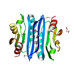
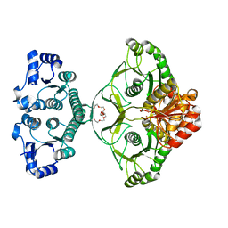
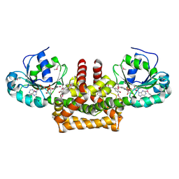
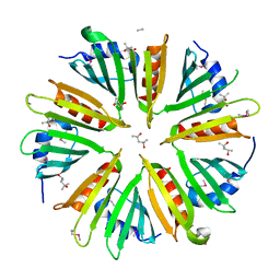
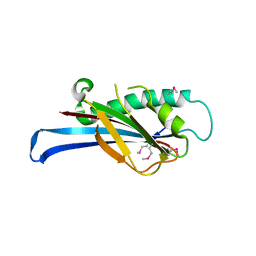
3L5O
DownloadVisualize
BU of 3l5o by Molmil
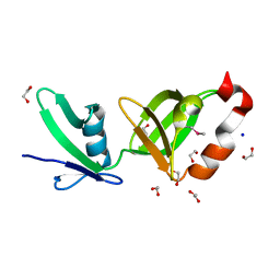
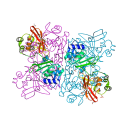
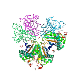
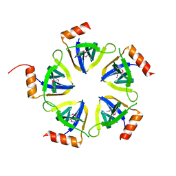
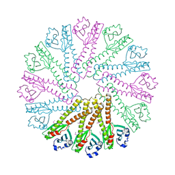
Crystal structure of protein with unknown function from DUF364 family (ZP_00559375.1) from Desulfitobacterium hafniense DCB-2 at 2.01 A resolution
Descriptor: 1,2-ETHANEDIOL, CHLORIDE ION, IMIDAZOLE, ...
Authors:Joint Center for Structural Genomics, Joint Center for Structural Genomics (JCSG)
Deposit date:2009-12-22
Release date:2010-02-02
Last modified:2023-02-01
Method:X-RAY DIFFRACTION (2.01 Å)
Cite:Structure of the first representative of Pfam family PF04016 (DUF364) reveals enolase and Rossmann-like folds that combine to form a unique active site with a possible role in heavy-metal chelation.
Acta Crystallogr.,Sect.F, 66, 2010
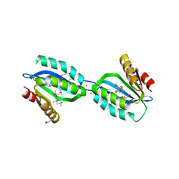
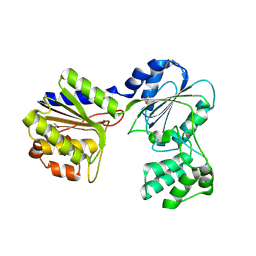
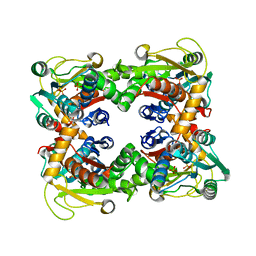
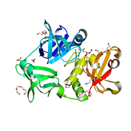
2QTP
DownloadVisualize
BU of 2qtp by Molmil
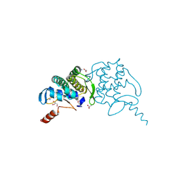
Crystal structure of a duf1185 family protein (spo0826) from silicibacter pomeroyi dss-3 at 2.10 A resolution
Descriptor: Uncharacterized protein
Authors:Joint Center for Structural Genomics (JCSG)
Deposit date:2007-08-02
Release date:2007-08-21
Last modified:2023-01-25
Method:X-RAY DIFFRACTION (2.1 Å)
Cite:Structures of the first representatives of Pfam family PF06684 (DUF1185) reveal a novel variant of the Bacillus chorismate mutase fold and suggest a role in amino-acid metabolism.
Acta Crystallogr.,Sect.F, 66, 2010
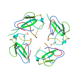
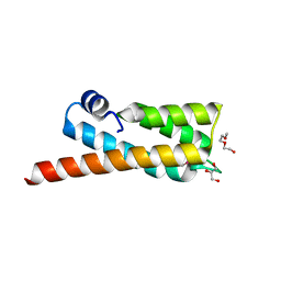
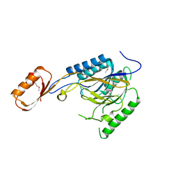
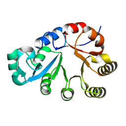
2OOK
DownloadVisualize
BU of 2ook by Molmil
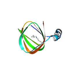
Crystal structure of a protein with unknown function (YP_749275.1) from Shewanella Frigidimarina NCIMB 400 at 1.80 A resolution
Descriptor: 1,2-ETHANEDIOL, Hypothetical protein
Authors:Joint Center for Structural Genomics (JCSG)
Deposit date:2007-01-25
Release date:2007-02-06
Last modified:2023-01-25
Method:X-RAY DIFFRACTION (1.8 Å)
Cite:Open and closed conformations of two SpoIIAA-like proteins (YP_749275.1 and YP_001095227.1) provide insights into membrane association and ligand binding.
Acta Crystallogr.,Sect.F, 66, 2010
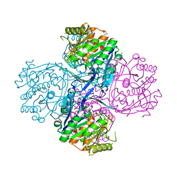
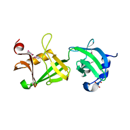
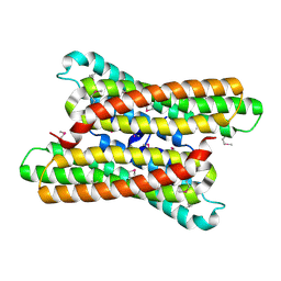
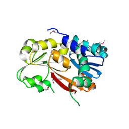
2Q3L
DownloadVisualize
BU of 2q3l by Molmil
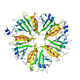
CRYSTAL STRUCTURE OF AN UNCHARACTERIZED PROTEIN FROM DUF3478 FAMILY WITH A SPOIIAA-LIKE FOLD (SHEW_3102) FROM SHEWANELLA LOIHICA PV-4 AT 2.25 A RESOLUTION
Descriptor: (4S)-2-METHYL-2,4-PENTANEDIOL, CHLORIDE ION, SODIUM ION, ...
Authors:Joint Center for Structural Genomics (JCSG)
Deposit date:2007-05-30
Release date:2007-06-12
Last modified:2023-01-25
Method:X-RAY DIFFRACTION (2.25 Å)
Cite:Open and closed conformations of two SpoIIAA-like proteins (YP_749275.1 and YP_001095227.1) provide insights into membrane association and ligand binding.
Acta Crystallogr.,Sect.F, 66, 2010
6WKP
DownloadVisualize
BU of 6wkp by Molmil
Crystal structure of RNA-binding domain of nucleocapsid phosphoprotein from SARS CoV-2, monoclinic crystal form
Descriptor: 2-(N-MORPHOLINO)-ETHANESULFONIC ACID, Nucleoprotein, ZINC ION
Authors:Chang, C, Michalska, K, Jedrzejczak, R, Maltseva, N, Endres, M, Godzik, A, Kim, Y, Joachimiak, A, Center for Structural Genomics of Infectious Diseases (CSGID)
Deposit date:2020-04-16
Release date:2020-04-29
Last modified:2024-02-28
Method:X-RAY DIFFRACTION (2.67 Å)
Cite:Epitopes recognition of SARS-CoV-2 nucleocapsid RNA binding domain by human monoclonal antibodies.
Iscience, 27, 2024
2AFB
DownloadVisualize
BU of 2afb by Molmil
Crystal structure of 2-dehydro-3- deoxygluconokinase (EC 2.7.1.45) (tm0067) from THERMOTOGA MARITIMA at 2.05 A resolution
Descriptor: 2-keto-3-deoxygluconate kinase, CALCIUM ION, NICKEL (II) ION
Authors:Joint Center for Structural Genomics (JCSG)
Deposit date:2005-07-25
Release date:2006-02-14
Last modified:2023-01-25
Method:X-RAY DIFFRACTION (2.05 Å)
Cite:Crystal structure of 2-keto-3-deoxygluconate kinase (TM0067) from Thermotoga maritima at 2.05 A resolution.
Proteins, 70, 2007
1ZCZ
DownloadVisualize
BU of 1zcz by Molmil
Crystal structure of Phosphoribosylaminoimidazolecarboxamide formyltransferase / IMP cyclohydrolase (TM1249) from THERMOTOGA MARITIMA at 1.88 A resolution
Descriptor: Bifunctional purine biosynthesis protein purH, POTASSIUM ION, TETRAETHYLENE GLYCOL
Authors:Joint Center for Structural Genomics (JCSG)
Deposit date:2005-04-13
Release date:2005-04-26
Last modified:2023-09-20
Method:X-RAY DIFFRACTION (1.88 Å)
Cite:Crystal structure of AICAR transformylase IMP cyclohydrolase (TM1249) from Thermotoga maritima at 1.88 A resolution.
Proteins, 71, 2008
2B8N
DownloadVisualize
BU of 2b8n by Molmil
Crystal structure of Glycerate kinase (EC 2.7.1.31) (tm1585) from THERMOTOGA MARITIMA at 2.70 A resolution
Descriptor: glycerate kinase, putative
Authors:Joint Center for Structural Genomics (JCSG)
Deposit date:2005-10-07
Release date:2005-11-08
Last modified:2023-01-25
Method:X-RAY DIFFRACTION (2.53 Å)
Cite:Crystal structure of a glycerate kinase (TM1585) from Thermotoga maritima at 2.70 A resolution reveals a new fold
Proteins, 65, 2006
2OOC
DownloadVisualize
BU of 2ooc by Molmil
Crystal structure of Histidine Phosphotransferase ShpA (NP_419930.1) from Caulobacter crescentus at 1.52 A resolution
Descriptor: GLYCEROL, Histidine phosphotransferase, TETRAETHYLENE GLYCOL
Authors:Joint Center for Structural Genomics (JCSG)
Deposit date:2007-01-25
Release date:2007-02-06
Last modified:2023-01-25
Method:X-RAY DIFFRACTION (1.52 Å)
Cite:Crystal structure of histidine phosphotransfer protein ShpA, an essential regulator of stalk biogenesis in Caulobacter crescentus.
J.Mol.Biol., 390, 2009
2RE3
DownloadVisualize
BU of 2re3 by Molmil
CRYSTAL STRUCTURE OF a DUF1285 family protein (SPO_0140) FROM SILICIBACTER POMEROYI DSS-3 AT 2.50 A RESOLUTION
Descriptor: GLYCEROL, Uncharacterized protein
Authors:Joint Center for Structural Genomics (JCSG)
Deposit date:2007-09-25
Release date:2007-10-09
Last modified:2023-01-25
Method:X-RAY DIFFRACTION (2.5 Å)
Cite:Structures of the first representatives of Pfam family PF06938 (DUF1285) reveal a new fold with repeated structural motifs and possible involvement in signal transduction.
Acta Crystallogr.,Sect.F, 66, 2010
2PV7
DownloadVisualize
BU of 2pv7 by Molmil
Crystal structure of chorismate mutase / prephenate dehydrogenase (tyrA) (1574749) from Haemophilus influenzae RD at 2.00 A resolution
Descriptor: NICOTINAMIDE-ADENINE-DINUCLEOTIDE, T-protein [Includes: Chorismate mutase (EC 5.4.99.5) (CM) and Prephenate dehydrogenase (EC 1.3.1.12) (PDH)], TYROSINE
Authors:Joint Center for Structural Genomics (JCSG)
Deposit date:2007-05-09
Release date:2007-05-22
Last modified:2023-01-25
Method:X-RAY DIFFRACTION (2 Å)
Cite:The structure of Haemophilus influenzae prephenate dehydrogenase suggests unique features of bifunctional TyrA enzymes.
Acta Crystallogr.,Sect.F, 66, 2010
1O58
DownloadVisualize
BU of 1o58 by Molmil
Crystal structure of O-acetylserine sulfhydrylase (TM0665) from Thermotoga maritima at 1.80 A resolution
Descriptor: O-acetylserine sulfhydrylase, PHOSPHATE ION
Authors:Joint Center for Structural Genomics (JCSG)
Deposit date:2003-08-20
Release date:2003-09-02
Last modified:2024-05-22
Method:X-RAY DIFFRACTION (1.8 Å)
Cite:Crystal structure of O-acetylserine sulfhydrylase (TM0665) from Thermotoga maritima at 1.8 A resolution
Proteins, 56, 2004
2Q8U
DownloadVisualize
BU of 2q8u by Molmil
CRYSTAL STRUCTURE OF MRE11 FROM THERMOTOGA MARITIMA MSB8 (TM1635) AT 2.20 A RESOLUTION
Descriptor: Exonuclease, putative
Authors:Joint Center for Structural Genomics (JCSG)
Deposit date:2007-06-11
Release date:2007-06-26
Last modified:2023-01-25
Method:X-RAY DIFFRACTION (2.2 Å)
Cite:Crystal structure of the first eubacterial Mre11 nuclease reveals novel features that may discriminate substrates during DNA repair.
J.Mol.Biol., 397, 2010
1O5H
DownloadVisualize
BU of 1o5h by Molmil
Crystal structure of formiminotetrahydrofolate cyclodeaminase (TM1560) from Thermotoga maritima at 2.80 A resolution
Descriptor: Formiminotetrahydrofolate cyclodeaminase
Authors:Joint Center for Structural Genomics (JCSG)
Deposit date:2003-09-17
Release date:2003-09-30
Last modified:2023-01-25
Method:X-RAY DIFFRACTION (2.8 Å)
Cite:Crystal structure of a formiminotetrahydrofolate cyclodeaminase (TM1560) from Thermotoga maritima at 2.80 A resolution reveals a new fold
Proteins, 58, 2005
2RA9
DownloadVisualize
BU of 2ra9 by Molmil
Crystal structure of a duf1285 family protein (sbal_2486) from shewanella baltica os155 at 1.40 A resolution
Descriptor: 1,2-ETHANEDIOL, SODIUM ION, Uncharacterized protein DUF1285
Authors:Joint Center for Structural Genomics (JCSG)
Deposit date:2007-09-14
Release date:2007-10-16
Last modified:2023-01-25
Method:X-RAY DIFFRACTION (1.4 Å)
Cite:Structures of the first representatives of Pfam family PF06938 (DUF1285) reveal a new fold with repeated structural motifs and possible involvement in signal transduction.
Acta Crystallogr.,Sect.F, 66, 2010
6WLC
DownloadVisualize
BU of 6wlc by Molmil
Crystal Structure of NSP15 Endoribonuclease from SARS CoV-2 in the Complex with Uridine-5'-Monophosphate
Descriptor: 1,2-ETHANEDIOL, 2-AMINO-2-HYDROXYMETHYL-PROPANE-1,3-DIOL, ACETATE ION, ...
Authors:Kim, Y, Maltseva, N, Jedrzejczak, R, Endres, M, Chang, C, Godzik, A, Michalska, K, Joachimiak, A, Center for Structural Genomics of Infectious Diseases (CSGID)
Deposit date:2020-04-19
Release date:2020-04-29
Last modified:2023-10-18
Method:X-RAY DIFFRACTION (1.82 Å)
Cite:Tipiracil binds to uridine site and inhibits Nsp15 endoribonuclease NendoU from SARS-CoV-2.
Commun Biol, 4, 2021
3BYQ
DownloadVisualize
BU of 3byq by Molmil
Crystal structure of a duf1185 family protein (bb2672) from bordetella bronchiseptica rb50 at 1.70 A resolution
Descriptor: 1,2-ETHANEDIOL, CHLORIDE ION, TETRAETHYLENE GLYCOL, ...
Authors:Joint Center for Structural Genomics (JCSG)
Deposit date:2008-01-16
Release date:2008-01-29
Last modified:2023-02-01
Method:X-RAY DIFFRACTION (1.7 Å)
Cite:Structures of the first representatives of Pfam family PF06684 (DUF1185) reveal a novel variant of the Bacillus chorismate mutase fold and suggest a role in amino-acid metabolism.
Acta Crystallogr.,Sect.F, 66, 2010
3BY7
DownloadVisualize
BU of 3by7 by Molmil
CRYSTAL STRUCTURE OF A PROTEIN STRUCTURALLY SIMILAR TO SM/LSM-LIKE RNA-BINDING PROTEINS (JCVI_PEP_1096686650277) FROM UNCULTURED MARINE ORGANISM AT 2.60 A RESOLUTION
Descriptor: uncharacterized protein
Authors:Joint Center for Structural Genomics (JCSG)
Deposit date:2008-01-15
Release date:2008-01-29
Last modified:2023-02-01
Method:X-RAY DIFFRACTION (2.6 Å)
Cite:Crystal structure of a novel Sm-like protein of putative cyanophage origin at 2.60 A resolution.
Proteins, 75, 2009
3B77
DownloadVisualize
BU of 3b77 by Molmil
Crystal structure of a ph domain containing bacterial protein (exig_2160) from exiguobacterium sibiricum 255-15 at 2.42 A resolution
Descriptor: Uncharacterized protein
Authors:Joint Center for Structural Genomics (JCSG)
Deposit date:2007-10-30
Release date:2007-11-13
Last modified:2023-01-25
Method:X-RAY DIFFRACTION (2.42 Å)
Cite:Bacterial pleckstrin homology domains: a prokaryotic origin for the PH domain.
J.Mol.Biol., 396, 2010
3H0N
DownloadVisualize
BU of 3h0n by Molmil
Crystal structure of a duf1470 family protein (jann_2411) from jannaschia sp. ccs1 at 1.45 A resolution
Descriptor: ACETATE ION, GLYCEROL, NICKEL (II) ION, ...
Authors:Joint Center for Structural Genomics (JCSG)
Deposit date:2009-04-09
Release date:2009-04-21
Last modified:2023-02-01
Method:X-RAY DIFFRACTION (1.45 Å)
Cite:The structure of Jann_2411 (DUF1470) from Jannaschia sp. at 1.45  resolution reveals a new fold (the ABATE domain) and suggests its possible role as a transcription regulator.
Acta Crystallogr.,Sect.F, 66, 2010
1O5U
DownloadVisualize
BU of 1o5u by Molmil
Crystal structure of a duf861 family protein (tm1112) from thermotoga maritima at 1.83 A resolution
Descriptor: UNKNOWN LIGAND, novel Thermotoga maritima enzyme TM1112
Authors:Joint Center for Structural Genomics (JCSG)
Deposit date:2003-10-06
Release date:2003-11-11
Last modified:2023-09-20
Method:X-RAY DIFFRACTION (1.83 Å)
Cite:Crystal structure of a novel Thermotoga maritima enzyme (TM1112) from the cupin family at 1.83 A resolution
Proteins, 56, 2004
3HSA
DownloadVisualize
BU of 3hsa by Molmil
Crystal structure of pleckstrin homology domain (YP_926556.1) from SHEWANELLA AMAZONENSIS SB2B at 1.99 A resolution
Descriptor: DI(HYDROXYETHYL)ETHER, GLYCEROL, pleckstrin homology domain
Authors:Joint Center for Structural Genomics (JCSG)
Deposit date:2009-06-10
Release date:2009-06-23
Last modified:2023-02-01
Method:X-RAY DIFFRACTION (1.99 Å)
Cite:Bacterial pleckstrin homology domains: a prokaryotic origin for the PH domain.
J.Mol.Biol., 396, 2010
3DCX
DownloadVisualize
BU of 3dcx by Molmil
Crystal structure of a duf1696 family protein with a pleckstrin-homology domain (shew_0819) from shewanella loihica pv-4 at 2.00 A resolution
Descriptor: (4S)-2-METHYL-2,4-PENTANEDIOL, CHLORIDE ION, Protein of Unknown Function (DUF1696) with Pleckstrin-homology Domains
Authors:Joint Center for Structural Genomics (JCSG)
Deposit date:2008-06-04
Release date:2008-07-29
Last modified:2023-02-01
Method:X-RAY DIFFRACTION (2 Å)
Cite:Bacterial pleckstrin homology domains: a prokaryotic origin for the PH domain.
J.Mol.Biol., 396, 2010
3H41
DownloadVisualize
BU of 3h41 by Molmil
CRYSTAL STRUCTURE OF A NLPC/P60 FAMILY PROTEIN (BCE_2878) FROM BACILLUS CEREUS ATCC 10987 AT 1.79 A RESOLUTION
Descriptor: ALANINE, D-GLUTAMIC ACID, NLP/P60 family protein, ...
Authors:Joint Center for Structural Genomics (JCSG)
Deposit date:2009-04-17
Release date:2009-05-05
Last modified:2023-02-01
Method:X-RAY DIFFRACTION (1.79 Å)
Cite:Structure of the gamma-D-glutamyl-L-diamino acid endopeptidase YkfC from Bacillus cereus in complex with L-Ala-[gamma]-D-Glu: insights into substrate recognition by NlpC/P60 cysteine peptidases
Acta Crystallogr.,Sect.F, 66, 2010
1O1Z
DownloadVisualize
BU of 1o1z by Molmil
Crystal structure of glycerophosphodiester phosphodiesterase (GDPD) (TM1621) from Thermotoga maritima at 1.60 A resolution
Descriptor: SODIUM ION, glycerophosphodiester phosphodiesterase
Authors:Joint Center for Structural Genomics (JCSG)
Deposit date:2003-02-12
Release date:2003-04-01
Last modified:2024-05-22
Method:X-RAY DIFFRACTION (1.6 Å)
Cite:Crystal structure of a glycerophosphodiester phosphodiesterase (GDPD) from Thermotoga maritima (TM1621) at 1.60 A resolution.
Proteins, 56, 2004

222926

건을2024-07-24부터공개중

PDB statisticsPDBj update infoContact PDBjnumon