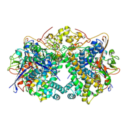
9EQL
 
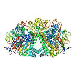
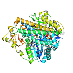
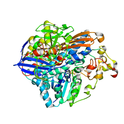
 | | Hydrogenase-1 Ni-B state poised at +300mV | | Descriptor: | CARBONMONOXIDE-(DICYANO) IRON, CHLORIDE ION, FE3-S4 CLUSTER, ... | | Authors: | Carr, S.B, Li, W, Wong, K.l, Ash, P.A, Vincent, K.A. | | Deposit date: | 2024-03-21 | | Release date: | 2025-04-02 | | Method: | X-RAY DIFFRACTION (1.51 Å) | | Cite: | Glutamate flick enables proton tunnelling during fast redox biocatalysis
To Be Published
|
|
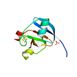
9ERA
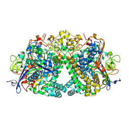
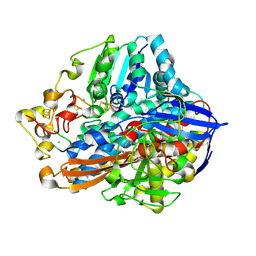
 | | Hydrogenase-1 Ni-Lii state | | Descriptor: | CARBONMONOXIDE-(DICYANO) IRON, CHLORIDE ION, FE3-S4 CLUSTER, ... | | Authors: | Wong, K.L, Carr, S.B, Ash, P.A, Vincent, K.A. | | Deposit date: | 2024-03-22 | | Release date: | 2025-04-02 | | Method: | X-RAY DIFFRACTION (1.65 Å) | | Cite: | Glutamate flick enables proton tunnelling during fast redox biocatalysis
To Be Published
|
|
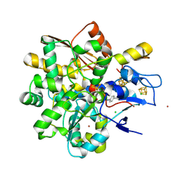
9ERB
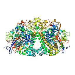
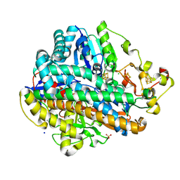
 | | Hydrogenase-2 Ni-B state | | Descriptor: | CARBONMONOXIDE-(DICYANO) IRON, CHLORIDE ION, FE3-S4 CLUSTER, ... | | Authors: | Carr, S.B, Li, W, Wong, K.l, Ash, P.A, Vincent, K.A. | | Deposit date: | 2024-03-22 | | Release date: | 2025-04-02 | | Method: | X-RAY DIFFRACTION (1.3 Å) | | Cite: | Glutamate flick enables proton tunnelling during fast redox biocatalysis
To Be Published
|
|
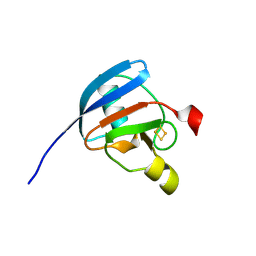
9ER7
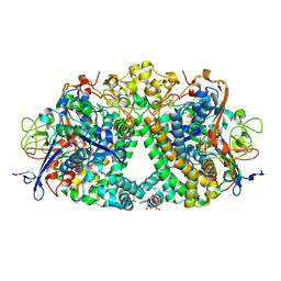
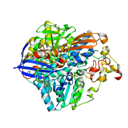
 | | Hydrogenase-1 Ni-SCO state | | Descriptor: | 2-AMINO-2-HYDROXYMETHYL-PROPANE-1,3-DIOL, CARBON MONOXIDE, CARBONMONOXIDE-(DICYANO) IRON, ... | | Authors: | Carr, S.B, Li, W, Wong, K.l, Ash, P.A, Vincent, K.A. | | Deposit date: | 2024-03-22 | | Release date: | 2025-04-02 | | Method: | X-RAY DIFFRACTION (1.4 Å) | | Cite: | Glutamate flick enables proton tunnelling during fast redox biocatalysis
To Be Published
|
|
9ER6
 | | Hydrogenase-1 Ni-SI state | | Descriptor: | 2-AMINO-2-HYDROXYMETHYL-PROPANE-1,3-DIOL, CARBONMONOXIDE-(DICYANO) IRON, CHLORIDE ION, ... | | Authors: | Carr, S.B, Li, W, Wong, K.L, Ash, P.A, Vincent, K.A. | | Deposit date: | 2024-03-22 | | Release date: | 2025-04-02 | | Method: | X-RAY DIFFRACTION (1.45 Å) | | Cite: | Glutamate flick enables proton tunnelling during fast redox biocatalysis
To Be Published
|
|
9ER5
 | | Hydrogenase-1 Ni-B state poised at +100mV | | Descriptor: | CARBONMONOXIDE-(DICYANO) IRON, CHLORIDE ION, FE3-S4 CLUSTER, ... | | Authors: | Carr, S.B, Li, W, Wong, K.l, Ash, P.A, Vincent, K.A. | | Deposit date: | 2024-03-22 | | Release date: | 2025-04-02 | | Method: | X-RAY DIFFRACTION (1.4 Å) | | Cite: | Glutamate flick enables proton tunnelling during fast redox biocatalysis
To Be Published
|
|
9ER9
 | | Hydrogenase-1 Ni-R state | | Descriptor: | 2-[[2-[2-[2-[bis(2-hydroxy-2-oxoethyl)amino]phenoxy]ethoxy]phenyl]-(2-hydroxy-2-oxoethyl)amino]ethanoic acid, CARBONMONOXIDE-(DICYANO) IRON, CHLORIDE ION, ... | | Authors: | Carr, S.B, Li, W, Wong, K.l, Ash, P.A, Vincent, K.A. | | Deposit date: | 2024-03-22 | | Release date: | 2025-04-02 | | Method: | X-RAY DIFFRACTION (1.4 Å) | | Cite: | Glutamate flick enables proton tunnelling during fast redox biocatalysis
To Be Published
|
|
9ERR
 | | Hydrogenase-2 Ni-SCO state | | Descriptor: | CARBON MONOXIDE, CARBONMONOXIDE-(DICYANO) IRON, FE3-S4 CLUSTER, ... | | Authors: | Carr, S.B, Li, W, Wong, K.l, Ash, P.A, Vincent, K.A. | | Deposit date: | 2024-03-25 | | Release date: | 2025-04-09 | | Method: | X-RAY DIFFRACTION (1.4 Å) | | Cite: | Glutamate flick enables proton tunnelling during fast redox biocatalysis
To Be Published
|
|
9GYL
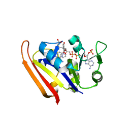
 | | Ferredoxin Wild-type -Oxidised state | | Descriptor: | FE2/S2 (INORGANIC) CLUSTER, Ferredoxin-1, chloroplastic, ... | | Authors: | Carr, S.B, Wei, J, Vincent, K.A. | | Deposit date: | 2024-10-02 | | Release date: | 2025-05-28 | | Last modified: | 2025-06-11 | | Method: | X-RAY DIFFRACTION (1.19 Å) | | Cite: | Cyanophenylalanine as an Infrared Probe for Iron-Sulfur Cluster Redox State in Multicenter Metalloenzymes.
Chembiochem, 2025
|
|
9GZL
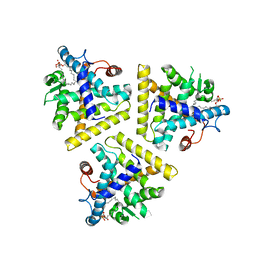
 | | Apo FeFe Hydrogenase from Desulfovibrio desulfuricans labelled with cyanophenylalanine | | Descriptor: | CHLORIDE ION, IRON/SULFUR CLUSTER, LITHIUM ION, ... | | Authors: | Carr, S.B, Duan, Z, Rodriguez-Macia, P, Vincent, K.A. | | Deposit date: | 2024-10-04 | | Release date: | 2025-05-28 | | Last modified: | 2025-06-11 | | Method: | X-RAY DIFFRACTION (1.02 Å) | | Cite: | Cyanophenylalanine as an Infrared Probe for Iron-Sulfur Cluster Redox State in Multicenter Metalloenzymes.
Chembiochem, 2025
|
|
9GYU
 | | Ferredoxin CNF-labelled reduced state | | Descriptor: | FE2/S2 (INORGANIC) CLUSTER, Ferredoxin-1, chloroplastic | | Authors: | Carr, S.B, Wei, J, Vincent, K.A. | | Deposit date: | 2024-10-03 | | Release date: | 2025-05-28 | | Last modified: | 2025-06-11 | | Method: | X-RAY DIFFRACTION (1.1 Å) | | Cite: | Cyanophenylalanine as an Infrared Probe for Iron-Sulfur Cluster Redox State in Multicenter Metalloenzymes.
Chembiochem, 2025
|
|
9GYD
 | | Ferredoxin Wild-type -As-isolated state | | Descriptor: | FE2/S2 (INORGANIC) CLUSTER, Ferredoxin-1, chloroplastic | | Authors: | Carr, S.B, Wei, J, Vincent, K.A. | | Deposit date: | 2024-10-01 | | Release date: | 2025-05-28 | | Last modified: | 2025-06-11 | | Method: | X-RAY DIFFRACTION (0.92 Å) | | Cite: | Cyanophenylalanine as an Infrared Probe for Iron-Sulfur Cluster Redox State in Multicenter Metalloenzymes.
Chembiochem, 2025
|
|
9GYR
 | | Ferredoxin CNF labelled, oxidised state | | Descriptor: | FE2/S2 (INORGANIC) CLUSTER, Ferredoxin-1, chloroplastic | | Authors: | Carr, S.B, Wei, J, Vincent, K.A. | | Deposit date: | 2024-10-02 | | Release date: | 2025-05-28 | | Last modified: | 2025-06-11 | | Method: | X-RAY DIFFRACTION (1.1 Å) | | Cite: | Cyanophenylalanine as an Infrared Probe for Iron-Sulfur Cluster Redox State in Multicenter Metalloenzymes.
Chembiochem, 2025
|
|
9GZ4
 | | FeFe Hydrogenase from Desulfovibrio desulfuricans labelled with cyanophenylalanine - reduced state | | Descriptor: | CHLORIDE ION, IRON/SULFUR CLUSTER, LITHIUM ION, ... | | Authors: | Carr, S.B, Duan, Z, Rodroguez-Macia, P, Vincent, K.A. | | Deposit date: | 2024-10-03 | | Release date: | 2025-05-28 | | Last modified: | 2025-06-11 | | Method: | X-RAY DIFFRACTION (0.96 Å) | | Cite: | Cyanophenylalanine as an Infrared Probe for Iron-Sulfur Cluster Redox State in Multicenter Metalloenzymes.
Chembiochem, 2025
|
|
9GYN
 | | Ferredoxin Wild-type - Reduced state | | Descriptor: | FE2/S2 (INORGANIC) CLUSTER, Ferredoxin-1, chloroplastic | | Authors: | Carr, S.B, Wei, J, Vincent, K.A. | | Deposit date: | 2024-10-02 | | Release date: | 2025-05-28 | | Last modified: | 2025-06-11 | | Method: | X-RAY DIFFRACTION (1 Å) | | Cite: | Cyanophenylalanine as an Infrared Probe for Iron-Sulfur Cluster Redox State in Multicenter Metalloenzymes.
Chembiochem, 2025
|
|
9GZ0
 | | FeFe Hydrogenase from Desulfovibrio desulfuricans labelled with cyanophenylalanine - oxidised state | | Descriptor: | CHLORIDE ION, IRON/SULFUR CLUSTER, LITHIUM ION, ... | | Authors: | Carr, S.B, Duan, Z, Rodroguez-Macia, P, Vincent, K.A. | | Deposit date: | 2024-10-03 | | Release date: | 2025-05-28 | | Last modified: | 2025-06-11 | | Method: | X-RAY DIFFRACTION (1.02 Å) | | Cite: | Cyanophenylalanine as an Infrared Probe for Iron-Sulfur Cluster Redox State in Multicenter Metalloenzymes.
Chembiochem, 2025
|
|
8POZ
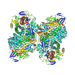
 | | Crystal Structure of the C120G variant of the membrane-bound [NiFe]-Hydrogenase from Cupriavidus necator in the H2-reduced state at 1.65 A Resolution. | | Descriptor: | CHLORIDE ION, FE3-S4 CLUSTER, FE4-S3 CLUSTER, ... | | Authors: | Schmidt, A, Kalms, J, Scheerer, P. | | Deposit date: | 2023-07-05 | | Release date: | 2024-09-18 | | Last modified: | 2025-02-26 | | Method: | X-RAY DIFFRACTION (1.65 Å) | | Cite: | Stepwise conversion of the Cys 6 [4Fe-3S] to a Cys 4 [4Fe-4S] cluster and its impact on the oxygen tolerance of [NiFe]-hydrogenase.
Chem Sci, 14, 2023
|
|
8POY
 | | Crystal Structure of the C120G variant of the membrane-bound [NiFe]-Hydrogenase from Cupriavidus necator in the air-oxidized state at 1.93 A Resolution. | | Descriptor: | CHLORIDE ION, FE3-S4 CLUSTER, FE4-S3 CLUSTER, ... | | Authors: | Schmidt, A, Kalms, J, Scheerer, P. | | Deposit date: | 2023-07-05 | | Release date: | 2025-02-26 | | Method: | X-RAY DIFFRACTION (1.93 Å) | | Cite: | Stepwise conversion of the Cys 6 [4Fe-3S] to a Cys 4 [4Fe-4S] cluster and its impact on the oxygen tolerance of [NiFe]-hydrogenase.
Chem Sci, 14, 2023
|
|
8POW
 | | Crystal Structure of the C19G variant of the membrane-bound [NiFe]-Hydrogenase from Cupriavidus necator in the air-oxidized state at 1.61 A Resolution. | | Descriptor: | CHLORIDE ION, FE3-S4 CLUSTER, Fe4S4, ... | | Authors: | Kalms, J, Schmidt, A, Scheerer, P. | | Deposit date: | 2023-07-05 | | Release date: | 2023-11-15 | | Last modified: | 2024-11-20 | | Method: | X-RAY DIFFRACTION (1.61 Å) | | Cite: | Stepwise conversion of the Cys 6 [4Fe-3S] to a Cys 4 [4Fe-4S] cluster and its impact on the oxygen tolerance of [NiFe]-hydrogenase.
Chem Sci, 14, 2023
|
|
8POX
 | | Crystal Structure of the C19G variant of the membrane-bound [NiFe]-Hydrogenase from Cupriavidus necator in the H2-reduced state at 1.6 A Resolution. | | Descriptor: | CHLORIDE ION, FE3-S4 CLUSTER, Fe4S4, ... | | Authors: | Kalms, J, Schmidt, A, Scheerer, P. | | Deposit date: | 2023-07-05 | | Release date: | 2024-09-18 | | Method: | X-RAY DIFFRACTION (1.6 Å) | | Cite: | Stepwise conversion of the Cys 6 [4Fe-3S] to a Cys 4 [4Fe-4S] cluster and its impact on the oxygen tolerance of [NiFe]-hydrogenase.
Chem Sci, 14, 2023
|
|
8POU
 | | Crystal Structure of the C19G/C120G variant of the membrane-bound [NiFe]-Hydrogenase from Cupriavidus necator in the air-oxidized state at 1.65 A Resolution. | | Descriptor: | CHLORIDE ION, FE3-S4 CLUSTER, IRON/SULFUR CLUSTER, ... | | Authors: | Schmidt, A, Kalms, J, Scheerer, P. | | Deposit date: | 2023-07-05 | | Release date: | 2023-11-01 | | Last modified: | 2024-02-07 | | Method: | X-RAY DIFFRACTION (1.65 Å) | | Cite: | Stepwise conversion of the Cys 6 [4Fe-3S] to a Cys 4 [4Fe-4S] cluster and its impact on the oxygen tolerance of [NiFe]-hydrogenase.
Chem Sci, 14, 2023
|
|
8POV
 | | Crystal Structure of the C19G/C120G variant of the membrane-bound [NiFe]-Hydrogenase from Cupriavidus necator in the H2-reduced state at 1.92 A Resolution. | | Descriptor: | FE3-S4 CLUSTER, IRON/SULFUR CLUSTER, MAGNESIUM ION, ... | | Authors: | Schmidt, A, Kalms, J, Scheerer, P. | | Deposit date: | 2023-07-05 | | Release date: | 2023-11-01 | | Method: | X-RAY DIFFRACTION (1.92 Å) | | Cite: | Stepwise conversion of the Cys 6 [4Fe-3S] to a Cys 4 [4Fe-4S] cluster and its impact on the oxygen tolerance of [NiFe]-hydrogenase.
Chem Sci, 14, 2023
|
|
3IA4
 | |
6G94
 | |
1HG4
 | |


