Loading
PDBj
MenuPDBj@FacebookPDBj@TwitterPDBj@YouTubewwPDB FoundationwwPDB
RCSB PDBPDBeBMRBAdv. SearchSearch help
Search by PDB author
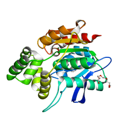
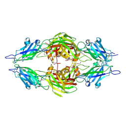
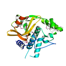
4H40
DownloadVisualize
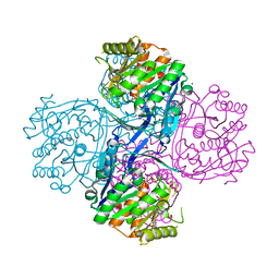
BU of 4h40 by Molmil
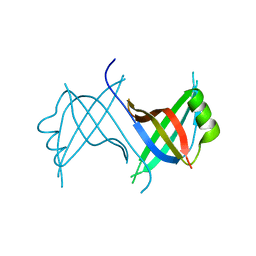
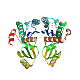
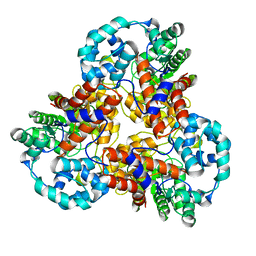
Crystal structure of a putative cell adhesion protein (BF2867) from Bacteroides fragilis NCTC 9343 at 2.57 A resolution
Descriptor: CHLORIDE ION, putative cell adhesion protein
Authors:Joint Center for Structural Genomics (JCSG)
Deposit date:2012-09-14
Release date:2012-10-03
Last modified:2023-02-01
Method:X-RAY DIFFRACTION (2.57 Å)
Cite:A Distinct Type of Pilus from the Human Microbiome.
Cell, 165, 2016
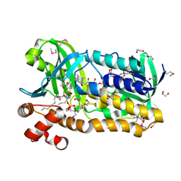
4H4J
DownloadVisualize
BU of 4h4j by Molmil
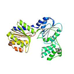
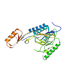
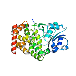
Crystal structure of a N-acetylmuramoyl-L-alanine amidase (BACUNI_02947) from Bacteroides uniformis ATCC 8492 at 1.15 A resolution
Descriptor: hypothetical protein
Authors:Joint Center for Structural Genomics (JCSG)
Deposit date:2012-09-17
Release date:2012-10-17
Last modified:2023-02-01
Method:X-RAY DIFFRACTION (1.15 Å)
Cite:Structure-guided functional characterization of DUF1460 reveals a highly specific NlpC/P60 amidase family.
Structure, 22, 2014
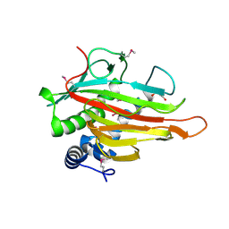
2QMQ
DownloadVisualize
BU of 2qmq by Molmil
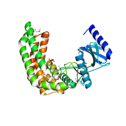
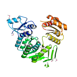
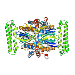
Crystal structure of a n-myc downstream regulated 2 protein (ndrg2, syld, ndr2, ai182517, au040374) from mus musculus at 1.70 A resolution
Descriptor: BENZOIC ACID, MAGNESIUM ION, NONAETHYLENE GLYCOL, ...
Authors:Joint Center for Structural Genomics (JCSG)
Deposit date:2007-07-16
Release date:2007-09-04
Last modified:2023-09-20
Method:X-RAY DIFFRACTION (1.7 Å)
Cite:Crystal structure of the human N-Myc downstream-regulated gene 2 protein provides insight into its role as a tumor suppressor.
J.Biol.Chem., 286, 2011
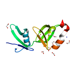
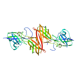
2RA9
DownloadVisualize
BU of 2ra9 by Molmil
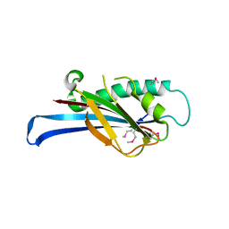
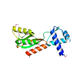
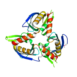
Crystal structure of a duf1285 family protein (sbal_2486) from shewanella baltica os155 at 1.40 A resolution
Descriptor: 1,2-ETHANEDIOL, SODIUM ION, Uncharacterized protein DUF1285
Authors:Joint Center for Structural Genomics (JCSG)
Deposit date:2007-09-14
Release date:2007-10-16
Last modified:2023-01-25
Method:X-RAY DIFFRACTION (1.4 Å)
Cite:Structures of the first representatives of Pfam family PF06938 (DUF1285) reveal a new fold with repeated structural motifs and possible involvement in signal transduction.
Acta Crystallogr.,Sect.F, 66, 2010
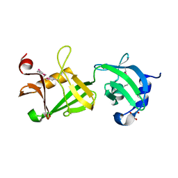
2RE3
DownloadVisualize
BU of 2re3 by Molmil
CRYSTAL STRUCTURE OF a DUF1285 family protein (SPO_0140) FROM SILICIBACTER POMEROYI DSS-3 AT 2.50 A RESOLUTION
Descriptor: GLYCEROL, Uncharacterized protein
Authors:Joint Center for Structural Genomics (JCSG)
Deposit date:2007-09-25
Release date:2007-10-09
Last modified:2023-01-25
Method:X-RAY DIFFRACTION (2.5 Å)
Cite:Structures of the first representatives of Pfam family PF06938 (DUF1285) reveal a new fold with repeated structural motifs and possible involvement in signal transduction.
Acta Crystallogr.,Sect.F, 66, 2010
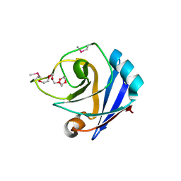
1ZX8
DownloadVisualize
BU of 1zx8 by Molmil
CRYSTAL STRUCTURE OF an atypical cyclophilin (peptidylprolyl cis-trans isomerase) (TM1367) FROM THERMOTOGA MARITIMA AT 1.90 A RESOLUTION
Descriptor: NICKEL (II) ION, PENTAETHYLENE GLYCOL, hypothetical protein TM1367
Authors:Joint Center for Structural Genomics (JCSG)
Deposit date:2005-06-07
Release date:2005-07-26
Last modified:2023-01-25
Method:X-RAY DIFFRACTION (1.9 Å)
Cite:Crystal structure of TM1367 from Thermotoga maritima at 1.90 A resolution reveals an atypical member of the cyclophilin (peptidylprolyl isomerase) fold.
Proteins, 63, 2006
1Z9F
DownloadVisualize
BU of 1z9f by Molmil
Crystal structure of single stranded DNA-binding protein (TM0604) from Thermotoga maritima at 2.60 A resolution
Descriptor: Single-strand binding protein
Authors:Joint Center for Structural Genomics (JCSG)
Deposit date:2005-04-01
Release date:2005-04-12
Last modified:2023-09-20
Method:X-RAY DIFFRACTION (2.3 Å)
Cite:Crystal structure of a single-stranded DNA-binding protein (TM0604) from Thermotoga maritima at 2.60 A resolution.
Proteins, 63, 2006
2B8N
DownloadVisualize
BU of 2b8n by Molmil
Crystal structure of Glycerate kinase (EC 2.7.1.31) (tm1585) from THERMOTOGA MARITIMA at 2.70 A resolution
Descriptor: glycerate kinase, putative
Authors:Joint Center for Structural Genomics (JCSG)
Deposit date:2005-10-07
Release date:2005-11-08
Last modified:2023-01-25
Method:X-RAY DIFFRACTION (2.53 Å)
Cite:Crystal structure of a glycerate kinase (TM1585) from Thermotoga maritima at 2.70 A resolution reveals a new fold
Proteins, 65, 2006
1ZKG
DownloadVisualize
BU of 1zkg by Molmil
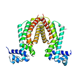
Crystal structure of Transcriptional regulator, TETR family (tm1030) from Thermotoga maritima at 2.30 A resolution
Descriptor: UNKNOWN LIGAND, transcriptional regulator, TetR family
Authors:Joint Center for Structural Genomics (JCSG)
Deposit date:2005-05-02
Release date:2005-07-26
Last modified:2023-01-25
Method:X-RAY DIFFRACTION (2.3 Å)
Cite:Crystal structure of TM1030 from Thermotoga maritima at 2.3 A resolution reveals molecular details of its transcription repressor function.
Proteins, 68, 2007
2QTP
DownloadVisualize
BU of 2qtp by Molmil
Crystal structure of a duf1185 family protein (spo0826) from silicibacter pomeroyi dss-3 at 2.10 A resolution
Descriptor: Uncharacterized protein
Authors:Joint Center for Structural Genomics (JCSG)
Deposit date:2007-08-02
Release date:2007-08-21
Last modified:2023-01-25
Method:X-RAY DIFFRACTION (2.1 Å)
Cite:Structures of the first representatives of Pfam family PF06684 (DUF1185) reveal a novel variant of the Bacillus chorismate mutase fold and suggest a role in amino-acid metabolism.
Acta Crystallogr.,Sect.F, 66, 2010
2A6A
DownloadVisualize
BU of 2a6a by Molmil
Crystal structure of Glycoprotein endopeptidase (tm0874) from THERMOTOGA MARITIMA at 2.50 A resolution
Descriptor: UNKNOWN LIGAND, hypothetical protein TM0874
Authors:Joint Center for Structural Genomics (JCSG)
Deposit date:2005-07-02
Release date:2005-07-19
Last modified:2023-01-25
Method:X-RAY DIFFRACTION (2.5 Å)
Cite:Structure of an essential bacterial protein YeaZ (TM0874) from Thermotoga maritima at 2.5 A resolution.
Acta Crystallogr.,Sect.F, 66, 2010
2Q8U
DownloadVisualize
BU of 2q8u by Molmil
CRYSTAL STRUCTURE OF MRE11 FROM THERMOTOGA MARITIMA MSB8 (TM1635) AT 2.20 A RESOLUTION
Descriptor: Exonuclease, putative
Authors:Joint Center for Structural Genomics (JCSG)
Deposit date:2007-06-11
Release date:2007-06-26
Last modified:2023-01-25
Method:X-RAY DIFFRACTION (2.2 Å)
Cite:Crystal structure of the first eubacterial Mre11 nuclease reveals novel features that may discriminate substrates during DNA repair.
J.Mol.Biol., 397, 2010
2Q83
DownloadVisualize
BU of 2q83 by Molmil
Crystal structure of ytaA (2635576) from Bacillus subtilis at 2.50 A resolution
Descriptor: 1,2-ETHANEDIOL, ADENOSINE, CITRIC ACID, ...
Authors:Joint Center for Structural Genomics (JCSG)
Deposit date:2007-06-08
Release date:2007-06-26
Last modified:2023-01-25
Method:X-RAY DIFFRACTION (2.5 Å)
Cite:Genomics, evolution, and crystal structure of a new family of bacterial spore kinases.
Proteins, 78, 2010
1IJC
DownloadVisualize
BU of 1ijc by Molmil
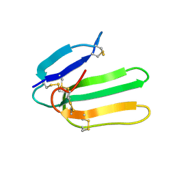
Solution Structure of Bucandin, a Neurotoxin from the Venom of the Malayan Krait
Descriptor: bucandin
Authors:Torres, A.M, Kini, R.M, Nirthanan, S, Kuchel, P.W.
Deposit date:2001-04-25
Release date:2001-12-21
Last modified:2022-02-23
Method:SOLUTION NMR
Cite:NMR structure of bucandin, a neurotoxin from the venom of the Malayan krait (Bungarus candidus).
Biochem.J., 360, 2001
2AFB
DownloadVisualize
BU of 2afb by Molmil
Crystal structure of 2-dehydro-3- deoxygluconokinase (EC 2.7.1.45) (tm0067) from THERMOTOGA MARITIMA at 2.05 A resolution
Descriptor: 2-keto-3-deoxygluconate kinase, CALCIUM ION, NICKEL (II) ION
Authors:Joint Center for Structural Genomics (JCSG)
Deposit date:2005-07-25
Release date:2006-02-14
Last modified:2023-01-25
Method:X-RAY DIFFRACTION (2.05 Å)
Cite:Crystal structure of 2-keto-3-deoxygluconate kinase (TM0067) from Thermotoga maritima at 2.05 A resolution.
Proteins, 70, 2007
7SUT
DownloadVisualize
BU of 7sut by Molmil
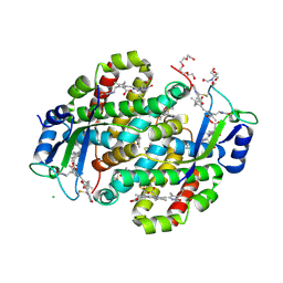
Light harvesting phycobiliprotein HaPE645 from the cryptophyte Hemiselmis andersenii CCMP644
Descriptor: (15,16)-DIHYDROBILIVERDIN (SINGLY LINKED), 2-[BIS-(2-HYDROXY-ETHYL)-AMINO]-2-HYDROXYMETHYL-PROPANE-1,3-DIOL, CHLORIDE ION, ...
Authors:Rathbone, H.W, Michie, K.A, Laos, A.L, Curmi, P.M.G.
Deposit date:2021-11-18
Release date:2023-10-25
Last modified:2023-11-29
Method:X-RAY DIFFRACTION (1.49 Å)
Cite:Molecular dissection of the soluble photosynthetic antenna from the cryptophyte alga Hemiselmis andersenii.
Commun Biol, 6, 2023
1J6U
DownloadVisualize
BU of 1j6u by Molmil
Crystal structure of UDP-N-acetylmuramate-alanine ligase MurC (TM0231) from Thermotoga maritima at 2.3 A resolution
Descriptor: UDP-N-acetylmuramate-alanine ligase MurC
Authors:Joint Center for Structural Genomics (JCSG)
Deposit date:2002-08-29
Release date:2002-11-06
Last modified:2023-01-25
Method:X-RAY DIFFRACTION (2.3 Å)
Cite:Crystal structure of an Udp-n-acetylmuramate-alanine ligase MurC (TM0231) from Thermotoga maritima at 2.3 A resolution.
Proteins, 55, 2004
1J5Y
DownloadVisualize
BU of 1j5y by Molmil
Crystal structure of transcriptional regulator (TM1602) from Thermotoga maritima at 2.3 A resolution
Descriptor: NICKEL (II) ION, POTASSIUM ION, TRANSCRIPTIONAL REGULATOR, ...
Authors:Joint Center for Structural Genomics (JCSG)
Deposit date:2002-07-05
Release date:2002-07-31
Last modified:2023-01-25
Method:X-RAY DIFFRACTION (2.3 Å)
Cite:Crystal structure of a transcription regulator (TM1602) from Thermotoga maritima at 2.3 A resolution.
Proteins, 67, 2007
1J5S
DownloadVisualize
BU of 1j5s by Molmil
Crystal structure of uronate isomerase (TM0064) from Thermotoga maritima at 2.85 A resolution
Descriptor: URONATE ISOMERASE
Authors:Joint Center for Structural Genomics (JCSG)
Deposit date:2002-07-02
Release date:2002-07-17
Last modified:2024-05-22
Method:X-RAY DIFFRACTION (2.85 Å)
Cite:Crystal structure of uronate isomerase (TM0064) from Thermotoga maritima at 2.85 A resolution.
Proteins, 53, 2003
1KQ3
DownloadVisualize
BU of 1kq3 by Molmil
CRYSTAL STRUCTURE OF A GLYCEROL DEHYDROGENASE (TM0423) FROM THERMOTOGA MARITIMA AT 1.5 A RESOLUTION
Descriptor: 2-AMINO-2-HYDROXYMETHYL-PROPANE-1,3-DIOL, CHLORIDE ION, ZINC ION, ...
Authors:Wilson, I.A, Miller, M.D, Joint Center for Structural Genomics (JCSG)
Deposit date:2002-01-03
Release date:2002-02-27
Last modified:2024-02-14
Method:X-RAY DIFFRACTION (1.5 Å)
Cite:Structural genomics of the Thermotoga maritima proteome implemented in a high-throughput structure determination pipeline
Proc.Natl.Acad.Sci.USA, 99, 2002
1KQ4
DownloadVisualize
BU of 1kq4 by Molmil
CRYSTAL STRUCTURE OF A THY1-COMPLEMENTING PROTEIN (TM0449) FROM THERMOTOGA MARITIMA AT 2.25 A RESOLUTION
Descriptor: FLAVIN-ADENINE DINUCLEOTIDE, HYPOTHETICAL PROTEIN TM0449
Authors:Wilson, I.A, Miller, M.D, Joint Center for Structural Genomics (JCSG)
Deposit date:2002-01-03
Release date:2002-02-27
Last modified:2017-10-11
Method:X-RAY DIFFRACTION (2.25 Å)
Cite:Structural genomics of the Thermotoga maritima proteome implemented in a high-throughput structure determination pipeline
Proc.Natl.Acad.Sci.USA, 99, 2002
3CM1
DownloadVisualize
BU of 3cm1 by Molmil
Crystal structure of SsgA-like sporulation-specific cell division protein (YP_290167.1) from Thermobifida fusca YX-ER1 at 2.60 A resolution
Descriptor: SsgA-like sporulation-specific cell division protein
Authors:Joint Center for Structural Genomics (JCSG), Chruszcz, M, Minor, W, Wang, S.
Deposit date:2008-03-20
Release date:2008-04-01
Last modified:2023-02-01
Method:X-RAY DIFFRACTION (2.6 Å)
Cite:Structural and functional characterizations of SsgB, a conserved activator of developmental cell division in morphologically complex actinomycetes.
J.Biol.Chem., 284, 2009
3PAY
DownloadVisualize
BU of 3pay by Molmil
Crystal structure of a putative adhesin (BACOVA_04077) from Bacteroides ovatus at 2.50 A resolution
Descriptor: DI(HYDROXYETHYL)ETHER, putative adhesin
Authors:Joint Center for Structural Genomics (JCSG)
Deposit date:2010-10-19
Release date:2010-11-10
Last modified:2020-04-22
Method:X-RAY DIFFRACTION (2.5 Å)
Cite:A Distinct Type of Pilus from the Human Microbiome.
Cell, 165, 2016
3OZ2
DownloadVisualize
BU of 3oz2 by Molmil
Crystal structure of a geranylgeranyl bacteriochlorophyll reductase-like (Ta0516) from Thermoplasma acidophilum at 1.60 A resolution
Descriptor: (2R)-3-{[(R)-{[(2S)-2,3-dihydroxypropyl]oxy}(hydroxy)phosphoryl]oxy}-2-[(6Z)-tridec-6-enoyloxy]propyl (9Z)-octadec-9-enoate, 1,2-ETHANEDIOL, Digeranylgeranylglycerophospholipid reductase, ...
Authors:Joint Center for Structural Genomics (JCSG)
Deposit date:2010-09-24
Release date:2010-10-27
Last modified:2023-02-01
Method:X-RAY DIFFRACTION (1.6 Å)
Cite:Insights into substrate specificity of geranylgeranyl reductases revealed by the structure of digeranylgeranylglycerophospholipid reductase, an essential enzyme in the biosynthesis of archaeal membrane lipids.
J.Mol.Biol., 404, 2010
3PL0
DownloadVisualize
BU of 3pl0 by Molmil
Crystal structure of a bsmA homolog (Mpe_A2762) from Methylobium petroleophilum PM1 at 1.91 A resolution
Descriptor: CHLORIDE ION, GLYCEROL, Uncharacterized protein
Authors:Joint Center for Structural Genomics (JCSG)
Deposit date:2010-11-12
Release date:2010-12-08
Last modified:2023-02-01
Method:X-RAY DIFFRACTION (1.91 Å)
Cite:Crystal structure of a member of a novel family of dioxygenases (PF10014) reveals a conserved cupin fold and active site.
Proteins, 82, 2014

223166

건을2024-07-31부터공개중

PDB statisticsPDBj update infoContact PDBjnumon