Loading
PDBj
MenuPDBj@FacebookPDBj@X(formerly Twitter)PDBj@BlueSkyPDBj@YouTubewwPDB FoundationwwPDB
RCSB PDBPDBeBMRBAdv. SearchSearch help
Search by PDB author
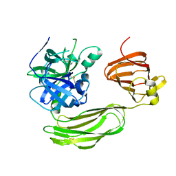
6BQM
DownloadVisualize
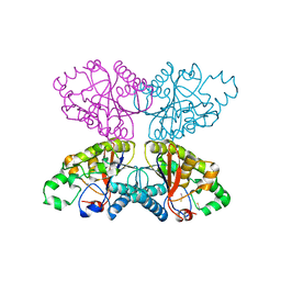
BU of 6bqm by Molmil
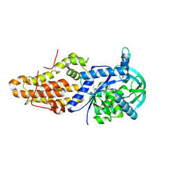
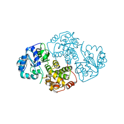
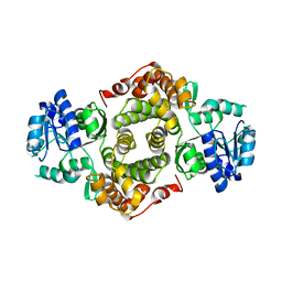
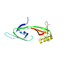
Secreted serine protease VesC from Vibrio cholerae
Descriptor: serine protease VesC
Authors:Park, Y.J, Korotkov, K.V, Delarosa, J.R, Turley, S, DiMaio, F, Hol, W.G.J.
Deposit date:2017-11-28
Release date:2018-11-28
Last modified:2023-10-04
Method:X-RAY DIFFRACTION (2.2 Å)
Cite:Suppressor Mutations in Type II Secretion Mutants of Vibrio cholerae: Inactivation of the VesC Protease.
Msphere, 5, 2020
6CML
DownloadVisualize
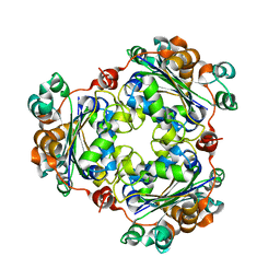
BU of 6cml by Molmil
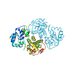
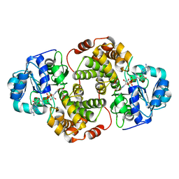
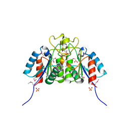
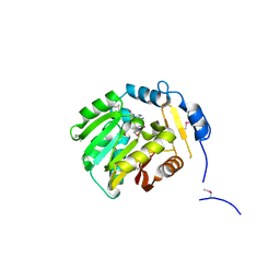
Trypanosoma brucei methionyl-tRNA synthetase in complex with inhibitor (Chem 2093)
Descriptor: 1-[(5-chloro-1H-imidazo[4,5-b]pyridin-2-yl)methyl]-4-[(2-chloro-4-methoxyphenyl)methyl]-3-ethyl-1,3-dihydro-2H-imidazol-2-one, GLYCEROL, METHIONINE, ...
Authors:Barros-Alvarez, X, Hol, W.G.J.
Deposit date:2018-03-05
Release date:2019-03-13
Last modified:2024-03-13
Method:X-RAY DIFFRACTION (2.7 Å)
Cite:Structure-guided discovery of selective methionyl-tRNA synthetase inhibitors with potent activity against Trypanosoma brucei
Rsc Med Chem, 11, 2020
1UV7
DownloadVisualize
BU of 1uv7 by Molmil
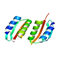
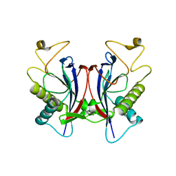
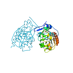
periplasmic domain of EpsM from Vibrio cholerae
Descriptor: GENERAL SECRETION PATHWAY PROTEIN M
Authors:Abendroth, J, Hol, W.G.J.
Deposit date:2004-01-15
Release date:2004-04-23
Last modified:2024-11-06
Method:X-RAY DIFFRACTION (1.7 Å)
Cite:The Crystal Structure of the Periplasmic Domain of the Type II Secretion System Protein Epsm from Vibrio Cholerae: The Simplest Version of the Ferredoxin Fold
J.Mol.Biol., 338, 2004
1W97
DownloadVisualize
BU of 1w97 by Molmil
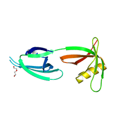
cyto-EpsL: the cytoplasmic domain of EpsL, an inner membrane component of the type II secretion system of Vibrio cholerae
Descriptor: TYPE II SECRETION SYSTEM PROTEIN L
Authors:Abendroth, J, Bagdasarian, M, Sansdkvist, M, Hol, W.G.J.
Deposit date:2004-10-06
Release date:2004-11-30
Last modified:2019-05-08
Method:X-RAY DIFFRACTION (2.7 Å)
Cite:The Structure of the Cytoplasmic Domain of Epsl, an Inner Membrane Component of the Type II Secretion System of Vibrio Cholerae: An Unusual Member of the Actin-Like ATPase Superfamily
J.Mol.Biol., 344, 2004
1SYR
DownloadVisualize
BU of 1syr by Molmil
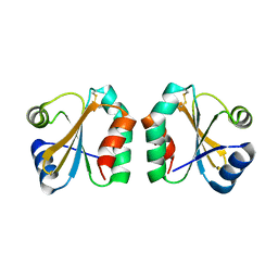
Initial Structural Analysis of Plasmodium falciparum thioredoxin
Descriptor: thioredoxin
Authors:Robien, M.A, Hol, W.G.J, Structural Genomics of Pathogenic Protozoa Consortium (SGPP)
Deposit date:2004-04-01
Release date:2004-04-13
Last modified:2024-10-16
Method:X-RAY DIFFRACTION (2.95 Å)
Cite:Initial Structural Analysis of Plasmodium falciparum thioredoxin
To be Published
1U8R
DownloadVisualize
BU of 1u8r by Molmil
Crystal Structure of an IdeR-DNA Complex Reveals a Conformational Change in Activated IdeR for Base-specific Interactions
Descriptor: COBALT (II) ION, Iron-dependent repressor ideR, SODIUM ION, ...
Authors:Wisedchaisri, G, Holmes, R.K, Hol, W.G.J.
Deposit date:2004-08-06
Release date:2004-10-05
Last modified:2023-08-23
Method:X-RAY DIFFRACTION (2.75 Å)
Cite:Crystal Structure of an IdeR-DNA Complex Reveals a Conformational Change in Activated IdeR for Base-specific Interactions.
J.Mol.Biol., 342, 2004
1SVV
DownloadVisualize
BU of 1svv by Molmil
Initial Stuctural Analysis of Leishmania major Threonine Aldolase
Descriptor: THREONINE ALDOLASE, UNKNOWN LIGAND
Authors:Hol, W.G.J, Robien, M.A, Structural Genomics of Pathogenic Protozoa Consortium (SGPP)
Deposit date:2004-03-30
Release date:2004-08-17
Last modified:2024-11-13
Method:X-RAY DIFFRACTION (2.1 Å)
Cite:Initial Structural Analysis of Leishmania major Threonine Aldolase
To be Published
1TC5
DownloadVisualize
BU of 1tc5 by Molmil
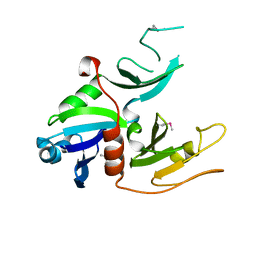
Structural Analysis of a probable eukaryotic D-amino acid tRNA deacylase
Descriptor: Probable eukaryotic D-amino acid tRNA deacylase, LMAJ005534AAA
Authors:Robien, M.A, Hol, W.G.J, Structural Genomics of Pathogenic Protozoa Consortium (SGPP)
Deposit date:2004-05-20
Release date:2004-06-08
Last modified:2024-04-03
Method:X-RAY DIFFRACTION (1.93 Å)
Cite:Structural analysis of LMAJ005534AAA, a probable eukaryotic D-amino acid tRNA deacylase from Leishmania major
To be Published
1M66
DownloadVisualize
BU of 1m66 by Molmil
Crystal Structure of Leishmania mexicana GPDH Complexed with Inhibitor 2-bromo-6-chloro-purine
Descriptor: 2-BROMO-6-CHLORO-PURINE, Glycerol-3-phosphate dehydrogenase, PALMITIC ACID
Authors:Choe, J, Suresh, S, Wisedchaisri, G, Kennedy, K.J, Gelb, M.H, Hol, W.G.J.
Deposit date:2002-07-12
Release date:2002-12-11
Last modified:2024-02-14
Method:X-RAY DIFFRACTION (1.9 Å)
Cite:Anomalous differences of light elements in determining precise binding modes of ligands to glycerol-3-phosphate dehydrogenase
Chem.Biol., 9, 2002
1M67
DownloadVisualize
BU of 1m67 by Molmil
Crystal Structure of Leishmania mexicana GPDH Complexed with Inhibitor 2-bromo-6-hydroxy-purine
Descriptor: 2-BROMO-6-HYDROXY-PURINE, Glycerol-3-phosphate dehydrogenase, PALMITIC ACID
Authors:Choe, J, Suresh, S, Wisedchaisri, G, Kennedy, K.J, Gelb, M.H, Hol, W.G.J.
Deposit date:2002-07-12
Release date:2002-12-11
Last modified:2024-02-14
Method:X-RAY DIFFRACTION (2.5 Å)
Cite:Anomalous differences of light elements in determining precise binding modes of ligands to glycerol-3-phosphate dehydrogenase
Chem.Biol., 9, 2002
1N1E
DownloadVisualize
BU of 1n1e by Molmil
Crystal structure of Leishmania mexicana Glycerol-3-phosphate dehydrogenase complexed with DHAP and NAD
Descriptor: ADENOSINE 5'-(TRIHYDROGEN DIPHOSPHATE) P'-5'-ESTER WITH 3-(AMINOCARBONYL)-4-(1-HYDROXYL-2-OXO-3-PHOSPHONOOXY-PROPYL)-1-BETA-D-RIBOFURANOSYLPYRIDINIUM INNER SALT, glycerol-3-phosphate dehydrogenase
Authors:Choe, J, Hol, W.G.J.
Deposit date:2002-10-17
Release date:2003-05-27
Last modified:2024-02-14
Method:X-RAY DIFFRACTION (1.9 Å)
Cite:Leishmania mexicana Glycerol-3-phosphate Dehydrogenase Showed Conformational Changes Upon Binding a Bi-substrate Adduct
J.Mol.Biol., 320, 2003
1N57
DownloadVisualize
BU of 1n57 by Molmil
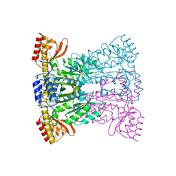
Crystal Structure of Chaperone Hsp31
Descriptor: Chaperone Hsp31, MAGNESIUM ION
Authors:Quigley, P.M, Korotkov, K, Baneyx, F, Hol, W.G.J.
Deposit date:2002-11-04
Release date:2003-03-18
Last modified:2024-11-13
Method:X-RAY DIFFRACTION (1.6 Å)
Cite:The 1.6A Crystal Structure of the Class of Chaperone Represented by Escherichia coli Hsp31 Reveals a Putative Catalytic Triad
Proc.Natl.Acad.Sci.USA, 100, 2003
1X6O
DownloadVisualize
BU of 1x6o by Molmil
Structural Analysis of Leishmania braziliensis eukaryotic initiation factor 5a
Descriptor: DI(HYDROXYETHYL)ETHER, eukaryotic initiation factor 5a
Authors:Bosch, J, Robien, M.A, Hol, W.G.J, Structural Genomics of Pathogenic Protozoa Consortium (SGPP)
Deposit date:2004-08-11
Release date:2004-08-24
Last modified:2024-02-14
Method:X-RAY DIFFRACTION (1.6 Å)
Cite:Structural Analysis of Leishmania braziliensis eukaryotic initiation factor 5a
To be Published
1YJ8
DownloadVisualize
BU of 1yj8 by Molmil
Initial structural analysis of Plasmodium falciparum Glycerol-3-phosphate dehydrogenase
Descriptor: glycerol-3-phosphate dehydrogenase
Authors:Robien, M.A, Hol, W.G.J, Structural Genomics of Pathogenic Protozoa Consortium (SGPP)
Deposit date:2005-01-13
Release date:2005-02-01
Last modified:2024-02-14
Method:X-RAY DIFFRACTION (2.85 Å)
Cite:Initial structural analysis of Plasmodium falciparum Glycerol-3-phosphate dehydrogenase
To be Published
2A0K
DownloadVisualize
BU of 2a0k by Molmil
Crystal structure of Nucleoside 2-deoxyribosyltransferase from Trypanosoma brucei at 1.8 A resolution
Descriptor: GLYCEROL, Nucleoside 2-deoxyribosyltransferase, SULFATE ION
Authors:Bosch, J, Robien, M.A, Hol, W.G.J, Structural Genomics of Pathogenic Protozoa Consortium (SGPP)
Deposit date:2005-06-16
Release date:2005-07-26
Last modified:2024-10-09
Method:X-RAY DIFFRACTION (1.8 Å)
Cite:Using fragment cocktail crystallography to assist inhibitor design of Trypanosoma brucei nucleoside 2-deoxyribosyltransferase.
J.Med.Chem., 49, 2006
2A0U
DownloadVisualize
BU of 2a0u by Molmil
Crystal structure of the eukaryotic initiation factor 2B from Leishmania major at 2.1 A resolution
Descriptor: SULFATE ION, initiation factor 2B
Authors:Bosch, J, Hol, W.G.J, Structural Genomics of Pathogenic Protozoa Consortium (SGPP)
Deposit date:2005-06-17
Release date:2005-08-02
Last modified:2024-11-06
Method:X-RAY DIFFRACTION (2.1 Å)
Cite:Crystal structure of the eukaryotic initiation factor 2B from Leishmania major at 2.1 A resolution
To be Published
1Y1X
DownloadVisualize
BU of 1y1x by Molmil
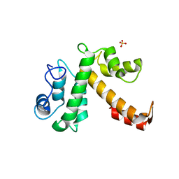
Structural analysis of a homolog of programmed cell death 6 protein from Leishmania major Friedlin
Descriptor: CALCIUM ION, Leishmania Major Homolog of Programmed Cell Death 6 Protein, SULFATE ION
Authors:Robien, M.A, Hol, W.G.J, Structural Genomics of Pathogenic Protozoa Consortium (SGPP)
Deposit date:2004-11-19
Release date:2004-12-07
Last modified:2024-04-03
Method:X-RAY DIFFRACTION (1.95 Å)
Cite:Structural analysis of a homolog of programmed cell death 6 protein from Leishmania major Friedlin
To be Published
1XTD
DownloadVisualize
BU of 1xtd by Molmil
Structural Analysis of Leishmania mexicana eukaryotic initiation factor 5a
Descriptor: eukaryotic initiation factor 5a
Authors:Bosch, J, Hol, W.G.J, Structural Genomics of Pathogenic Protozoa Consortium (SGPP)
Deposit date:2004-10-21
Release date:2004-10-26
Last modified:2024-10-30
Method:X-RAY DIFFRACTION (2.7 Å)
Cite:Structural Analysis of Leishmania mexicana eukaryotic initiation factor 5a
To be Published
1XTP
DownloadVisualize
BU of 1xtp by Molmil
Structural Analysis of Leishmania major LMAJ004091AAA, a SAM-dependent methyltransferase of the DUF858/Pfam05891 family
Descriptor: LMAJ004091AAA, S-ADENOSYL-L-HOMOSELENOCYSTEINE
Authors:Robien, M.A, Hol, W.G.J, Structural Genomics of Pathogenic Protozoa Consortium (SGPP)
Deposit date:2004-10-22
Release date:2004-11-09
Last modified:2011-07-13
Method:X-RAY DIFFRACTION (1.94 Å)
Cite:Structural Analysis of Leishmania major LMAJ004091AAA
To be Published
1XUP
DownloadVisualize
BU of 1xup by Molmil
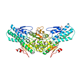
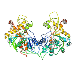
ENTEROCOCCUS CASSELIFLAVUS GLYCEROL KINASE COMPLEXED WITH GLYCEROL
Descriptor: GLYCEROL, Glycerol kinase
Authors:Yeh, J.I, Charrier, V, Paulo, J, Hou, L, Darbon, E, Hol, W.G.J, Deutscher, J.
Deposit date:2004-10-26
Release date:2004-12-14
Last modified:2024-02-14
Method:X-RAY DIFFRACTION (2.75 Å)
Cite:Structures of Enterococcal Glycerol Kinase in the Absence and Presence of Glycerol: Correlation of Conformation to Substrate Binding and a Mechanism of Activation by Phosphorylation
Biochemistry, 43, 2004
1ZLK
DownloadVisualize
BU of 1zlk by Molmil
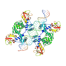
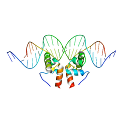
Crystal Structure of the Mycobacterium tuberculosis Hypoxic Response Regulator DosR C-terminal Domain-DNA Complex
Descriptor: 5'-D(*CP*GP*TP*GP*GP*CP*CP*AP*GP*GP*GP*TP*TP*AP*GP*GP*GP*AP*CP*TP*TP*TP*AP*GP*TP*CP*CP*CP*CP*AP*AP*AP*GP*CP*GP*CP*GP*GP*GP*CP*CP*AP*T)-3', 5'-D(*GP*GP*CP*CP*CP*GP*CP*GP*CP*TP*TP*TP*GP*GP*GP*GP*AP*CP*TP*AP*AP*AP*GP*TP*CP*CP*CP*TP*AP*AP*CP*CP*CP*TP*GP*GP*CP*CP*AP*CP*GP*AP*T)-3', Dormancy Survival Regulator
Authors:Wisedchaisri, G, Wu, M, Rice, A.E, Roberts, D.M, Sherman, D.R, Hol, W.G.J.
Deposit date:2005-05-06
Release date:2006-01-31
Last modified:2023-08-23
Method:X-RAY DIFFRACTION (3.1 Å)
Cite:Structures of Mycobacterium tuberculosis DosR and DosR-DNA complex involved in gene activation during adaptation to hypoxic latency.
J.Mol.Biol., 354, 2005
1XQ9
DownloadVisualize
BU of 1xq9 by Molmil
Structure of Phosphoglycerate Mutase from Plasmodium falciparum at 2.6 Resolution
Descriptor: THIOCYANATE ION, phosphoglycerate mutase
Authors:Caruthers, J.M, Hol, W.G.J, Structural Genomics of Pathogenic Protozoa Consortium (SGPP)
Deposit date:2004-10-11
Release date:2004-12-21
Last modified:2024-02-14
Method:X-RAY DIFFRACTION (2.58 Å)
Cite:Structure of Phosphoglycerate Mutase from Plasmodium falciparum at 2.6 Resolution
TO BE PUBLISHED
1XIQ
DownloadVisualize
BU of 1xiq by Molmil
Plasmodium falciparum Nucleoside diphosphate kinase B
Descriptor: Nucleoside diphosphate kinase B
Authors:Robien, M.A, Bosch, J, Hol, W.G.J, Structural Genomics of Pathogenic Protozoa Consortium (SGPP)
Deposit date:2004-09-21
Release date:2004-10-12
Last modified:2024-02-14
Method:X-RAY DIFFRACTION (3.05 Å)
Cite:Crystal structure of Nucleoside diphosphate kinase B from Plasmodium falciparum
To be Published
1XO7
DownloadVisualize
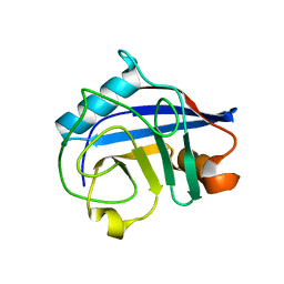
BU of 1xo7 by Molmil
Crystal structure of cyclophilin from Trypanosoma cruzi
Descriptor: cyclophilin
Authors:Caruthers, J.M, Hol, W.G.J, Structural Genomics of Pathogenic Protozoa Consortium (SGPP)
Deposit date:2004-10-05
Release date:2004-12-21
Last modified:2023-08-23
Method:X-RAY DIFFRACTION (1.61 Å)
Cite:Crystal structure of cyclophilin from Trypanosoma cruzi
TO BE PUBLISHED
1XQ7
DownloadVisualize
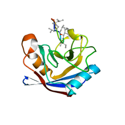
BU of 1xq7 by Molmil
Cyclophilin from Trypanosoma cruzi bound to cyclosporin A
Descriptor: CYCLOSPORIN A, PEPTIDYL-PROLYL CIS-TRANS ISOMERASE
Authors:Caruthers, J.M, Hol, W.G.J, Structural Genomics of Pathogenic Protozoa Consortium (SGPP)
Deposit date:2004-10-11
Release date:2004-12-21
Last modified:2023-11-15
Method:X-RAY DIFFRACTION (2.07 Å)
Cite:Cyclophilin from Trypanosoma Cruzi Bound to Cyclosporin A
To be Published

227344

건을2024-11-13부터공개중

PDB statisticsPDBj update infoContact PDBjnumon