Loading
PDBj
MenuPDBj@FacebookPDBj@TwitterPDBj@YouTubewwPDB FoundationwwPDB
RCSB PDBPDBeBMRBAdv. SearchSearch help
Search by PDB author
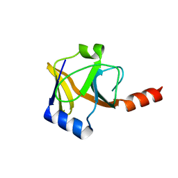
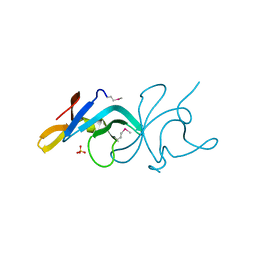
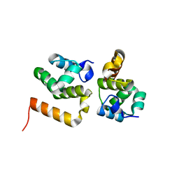
3MAB
DownloadVisualize
BU of 3mab by Molmil
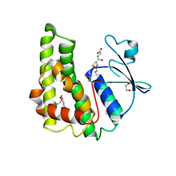
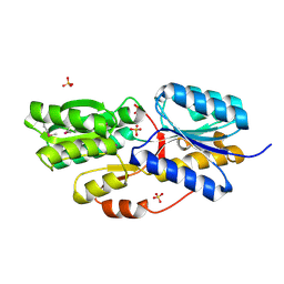
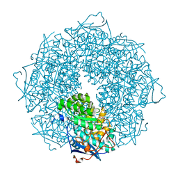
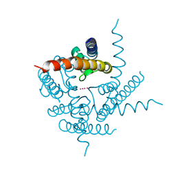
CRYSTAL STRUCTURE OF AN UNCHARACTERIZED PROTEIN FROM LISTERIA MONOCYTOGENES, Triclinic FORM
Descriptor: uncharacterized protein
Authors:Madegowda, M, Chruszcz, M, Minor, W, Burley, S.K, Swaminathan, S, New York SGX Research Center for Structural Genomics (NYSGXRC)
Deposit date:2010-03-23
Release date:2010-04-14
Last modified:2023-09-06
Method:X-RAY DIFFRACTION (1.42 Å)
Cite:Crystal structure of an uncharacterized protein from listeria monocytogenes
To be Published
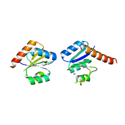
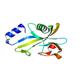
2QGO
DownloadVisualize
BU of 2qgo by Molmil
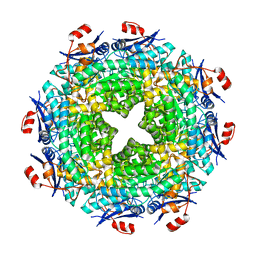
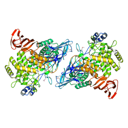
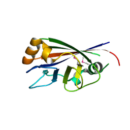
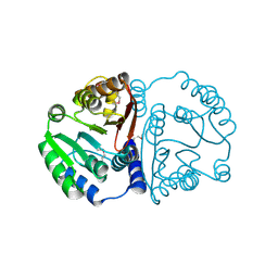
Crystal structure of a putative Fe-S biosynthesis protein from Lactobacillus acidophilus
Descriptor: Putative Fe-S biosynthesis protein
Authors:Agarwal, R, Burley, S.K, Swaminathan, S, New York SGX Research Center for Structural Genomics (NYSGXRC)
Deposit date:2007-06-29
Release date:2007-07-31
Last modified:2024-02-21
Method:X-RAY DIFFRACTION (2.04 Å)
Cite:Structural analysis of Fe-S cluster proteins.
To be Published
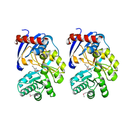
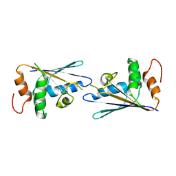
3N53
DownloadVisualize
BU of 3n53 by Molmil
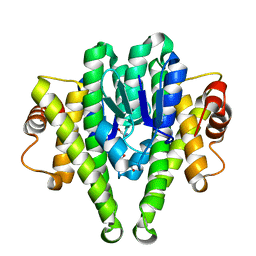
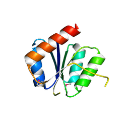
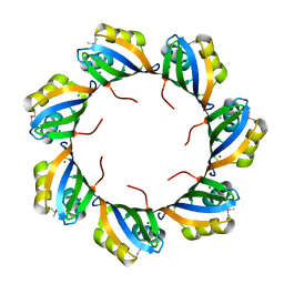
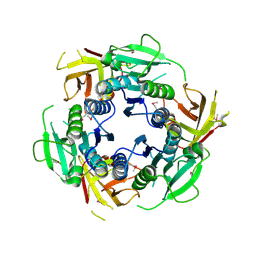
Crystal structure of a response regulator receiver modulated diguanylate cyclase from Pelobacter carbinolicus
Descriptor: Response regulator receiver modulated diguanylate cyclase
Authors:Palani, K, Zhang, Z, Burley, S.K, Swaminathan, S, New York SGX Research Center for Structural Genomics (NYSGXRC)
Deposit date:2010-05-24
Release date:2010-07-28
Last modified:2023-11-22
Method:X-RAY DIFFRACTION (2.2 Å)
Cite:Crystal structure of a response regulator receiver modulated diguanylate cyclase from Pelobacter carbinolicus
To be Published
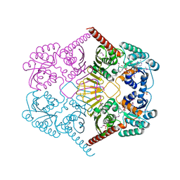
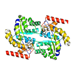
2QVH
DownloadVisualize
BU of 2qvh by Molmil
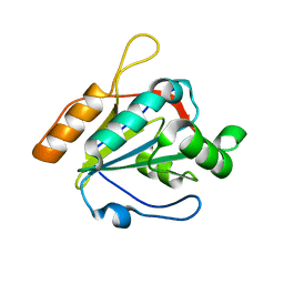
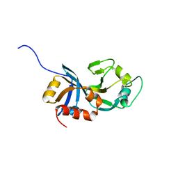
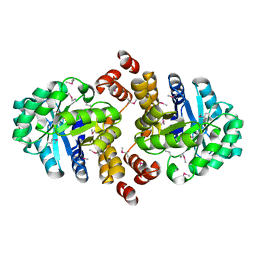
Crystal structure of O-succinylbenzoate synthase complexed with O-succinyl benzoate (OSB)
Descriptor: 2-SUCCINYLBENZOATE, MAGNESIUM ION, O-succinylbenzoate-CoA synthase
Authors:Sugadev, R, Burley, S.K, Swaminathan, S, New York SGX Research Center for Structural Genomics (NYSGXRC)
Deposit date:2007-08-08
Release date:2007-08-28
Last modified:2024-11-06
Method:X-RAY DIFFRACTION (1.76 Å)
Cite:Crystal structure of O-succinylbenzoate synthase complexed with O-succinyl benzoate.
To be Published
3M2T
DownloadVisualize
BU of 3m2t by Molmil
The crystal structure of dehydrogenase from Chromobacterium violaceum
Descriptor: NICOTINAMIDE-ADENINE-DINUCLEOTIDE, Probable dehydrogenase, SULFATE ION
Authors:Zhang, Z, Burley, S.K, Swaminathan, S, New York SGX Research Center for Structural Genomics (NYSGXRC)
Deposit date:2010-03-08
Release date:2010-04-07
Last modified:2024-10-16
Method:X-RAY DIFFRACTION (2.3 Å)
Cite:The crystal structure of dehydrogenase from Chromobacterium violaceum
To be Published
3M3M
DownloadVisualize
BU of 3m3m by Molmil
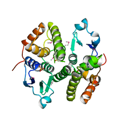
Crystal structure of glutathione S-transferase from Pseudomonas fluorescens [Pf-5]
Descriptor: 1,2-ETHANEDIOL, GLUTATHIONE, Glutathione S-transferase, ...
Authors:Bagaria, A, Burley, S.K, Swaminathan, S, New York SGX Research Center for Structural Genomics (NYSGXRC)
Deposit date:2010-03-09
Release date:2010-03-16
Last modified:2021-02-10
Method:X-RAY DIFFRACTION (1.75 Å)
Cite:Crystal structure of glutathione S-transferase from Pseudomonas fluorescens [Pf-5]
To be Published
3MQT
DownloadVisualize
BU of 3mqt by Molmil
Crystal structure of a mandelate racemase/muconate lactonizing enzyme from Shewanella pealeana
Descriptor: MAGNESIUM ION, Mandelate racemase/muconate lactonizing protein
Authors:Agarwal, R, Burley, S.K, Swaminathan, S, New York SGX Research Center for Structural Genomics (NYSGXRC)
Deposit date:2010-04-28
Release date:2010-05-12
Last modified:2023-09-06
Method:X-RAY DIFFRACTION (2.1 Å)
Cite:Crystal STRUCTURE OF A MANDELATE RACEMASE/MUCONATE LACTONIZING ENZYME FROM SHEWANELLA PEALEANA
To be Published
3M0F
DownloadVisualize
BU of 3m0f by Molmil
Crystal structure of Glutathione S Transferase in complex with glutathione from Pseudomonas fluorescens
Descriptor: GLUTATHIONE, uncharacterized protein GST_N
Authors:Agarwal, R, Burley, S.K, Swaminathan, S, New York SGX Research Center for Structural Genomics (NYSGXRC)
Deposit date:2010-03-03
Release date:2010-03-16
Last modified:2024-02-21
Method:X-RAY DIFFRACTION (1.6 Å)
Cite:Crystal structure of Glutathione S Transferase in complex with glutathione from Pseudomonas fluorescens
To be Published
3M8N
DownloadVisualize
BU of 3m8n by Molmil
Crystal structure of a possible gutathione S-tranferase from Rhodopseudomonas palustris
Descriptor: Possible glutathione S-transferase, SULFATE ION
Authors:Damodharan, L, Burley, S.K, Swaminathan, S, New York SGX Research Center for Structural Genomics (NYSGXRC)
Deposit date:2010-03-18
Release date:2010-04-07
Last modified:2021-02-10
Method:X-RAY DIFFRACTION (2.04 Å)
Cite:Crystal structure of a possible gutathione S-tranferase from Rhodopseudomonas palustris
To be Published
2RJO
DownloadVisualize
BU of 2rjo by Molmil
Crystal structure of Twin-arginine translocation pathway signal protein from Burkholderia phytofirmans
Descriptor: SULFATE ION, Twin-arginine translocation pathway signal protein, beta-D-galactopyranose
Authors:Agarwal, R, Burley, S.K, Swaminathan, S, New York SGX Research Center for Structural Genomics (NYSGXRC)
Deposit date:2007-10-15
Release date:2007-10-23
Last modified:2024-11-06
Method:X-RAY DIFFRACTION (2.05 Å)
Cite:Crystal structure of twin-arginine translocation pathway signal protein from Burkholderia phytofirmans.
To be Published
2RDY
DownloadVisualize
BU of 2rdy by Molmil
Crystal structure of a putative glycoside hydrolase family protein from Bacillus halodurans
Descriptor: BH0842 protein
Authors:Sugadev, R, Burley, S.K, Swaminathan, S, New York SGX Research Center for Structural Genomics (NYSGXRC)
Deposit date:2007-09-25
Release date:2007-10-09
Last modified:2024-11-06
Method:X-RAY DIFFRACTION (2.03 Å)
Cite:Crystal structure of a putative glycoside hydrolase family protein from Bacillus halodurans.
To be Published
3NHM
DownloadVisualize
BU of 3nhm by Molmil
Crystal structure of a response regulator from Myxococcus xanthus
Descriptor: Response regulator
Authors:Palani, K, Burley, S.K, Swaminathan, S, New York SGX Research Center for Structural Genomics (NYSGXRC)
Deposit date:2010-06-14
Release date:2010-07-21
Last modified:2024-10-30
Method:X-RAY DIFFRACTION (2.19 Å)
Cite:Crystal structure of a response regulator from Myxococcus xanthus
To be Published
3U5R
DownloadVisualize
BU of 3u5r by Molmil
Crystal structure of a hypothetical protein SMc02350 from Sinorhizobium meliloti 1021
Descriptor: uncharacterized protein
Authors:Agarwal, R, Almo, S.C, Swaminathan, S, New York Structural Genomics Research Consortium (NYSGRC)
Deposit date:2011-10-11
Release date:2011-10-19
Last modified:2023-09-13
Method:X-RAY DIFFRACTION (2.05 Å)
Cite:Crystal structure of a hypothetical protein SMc02350 from Sinorhizobium meliloti 1021
To be Published
3BE3
DownloadVisualize
BU of 3be3 by Molmil
Crystal structure of a protein belonging to pfam DUF1653 from Bordetella bronchiseptica
Descriptor: SULFATE ION, Uncharacterized protein
Authors:Tyagi, R, Eswaramoorthy, S, Burley, S.K, Swaminathan, S, New York SGX Research Center for Structural Genomics (NYSGXRC)
Deposit date:2007-11-16
Release date:2007-11-27
Last modified:2024-10-30
Method:X-RAY DIFFRACTION (2.04 Å)
Cite:Crystal structure of a protein belonging to pfam DUF1653 from Bordetella bronchiseptica.
To be Published
3SMD
DownloadVisualize
BU of 3smd by Molmil
Crystal structure of a mut/nudix family protein from bacillus thuringiensis
Descriptor: MutT/NUDIX family protein
Authors:Palani, K, Kumaran, D, Burley, S.K, Swaminathan, S, New York SGX Research Center for Structural Genomics (NYSGXRC)
Deposit date:2011-06-27
Release date:2011-07-20
Last modified:2024-10-16
Method:X-RAY DIFFRACTION (1.76 Å)
Cite:Crystal structure of a mut/nudix family protein from bacillus thuringiensis
To be Published
3C9G
DownloadVisualize
BU of 3c9g by Molmil
Crystal structure of uncharacterized UPF0201 protein AF_135
Descriptor: UPF0200/UPF0201 protein AF_1395
Authors:Sugadev, R, Ozyurt, S, Burley, S.K, Swaminathan, S, New York SGX Research Center for Structural Genomics (NYSGXRC)
Deposit date:2008-02-15
Release date:2008-05-13
Last modified:2023-08-30
Method:X-RAY DIFFRACTION (2.3 Å)
Cite:Crystal structure of uncharacterized UPF0201 protein AF_135.
To be Published
3D0C
DownloadVisualize
BU of 3d0c by Molmil
Crystal structure of dihydrodipicolinate synthase from Oceanobacillus iheyensis at 1.9 A resolution
Descriptor: Dihydrodipicolinate synthase
Authors:Satyanarayana, L, Eswaramoorthy, S, Sauder, J.M, Burley, S.K, Swaminathan, S, New York SGX Research Center for Structural Genomics (NYSGXRC)
Deposit date:2008-05-01
Release date:2008-05-13
Last modified:2021-10-20
Method:X-RAY DIFFRACTION (1.9 Å)
Cite:Crystal structure of dihydrodipicolinate synthase from Oceanobacillus iheyensis at 1.9 A resolution.
To be Published
3SQS
DownloadVisualize
BU of 3sqs by Molmil
Crystal Structure of a putative mandelate racemase/muconate lactonizing protein from Dinoroseobacter shibae DFL 12
Descriptor: ACETATE ION, GLYCEROL, MAGNESIUM ION, ...
Authors:Kumaran, D, Almo, S.C, Swaminathan, S, New York Structural Genomics Research Consortium (NYSGRC)
Deposit date:2011-07-06
Release date:2011-07-27
Last modified:2023-09-13
Method:X-RAY DIFFRACTION (1.9 Å)
Cite:Crystal Structure of a putative mandelate racemase/muconate lactonizing protein from Dinoroseobacter shibae DFL 12
To be Published
3CBN
DownloadVisualize
BU of 3cbn by Molmil
Crystal structure of a conserved protein (MTH639) from Methanobacterium thermoautotrophicum
Descriptor: Conserved protein MTH639
Authors:Satyanarayana, L, Eswaramoorthy, S, Burley, S.K, Swaminathan, S, New York SGX Research Center for Structural Genomics (NYSGXRC)
Deposit date:2008-02-22
Release date:2008-03-04
Last modified:2021-02-03
Method:X-RAY DIFFRACTION (1.63 Å)
Cite:Crystal structure of a conserved protein (MTH639) from Methanobacterium thermoautotrophicum.
To be Published
3BPD
DownloadVisualize
BU of 3bpd by Molmil
Crystal structure of an uncharacterized protein (O28723_ARCFU) from Archaeoglobus fulgidus
Descriptor: MAGNESIUM ION, Uncharacterized protein
Authors:Eswaramoorthy, S, Burley, S.K, Sauder, J.M, Swaminathan, S, New York SGX Research Center for Structural Genomics (NYSGXRC)
Deposit date:2007-12-18
Release date:2008-01-08
Last modified:2024-11-06
Method:X-RAY DIFFRACTION (2.8 Å)
Cite:Crystal structure of an uncharacterized protein (O28723_ARCFU) from Archaeoglobus fulgidus.
To be Published
2HAF
DownloadVisualize
BU of 2haf by Molmil
Crystal structure of a putative translation repressor from Vibrio cholerae
Descriptor: Putative translation repressor
Authors:Sugadev, R, Seetharaman, J, Kumaran, D, Swaminathan, S, Burley, S.K, New York SGX Research Center for Structural Genomics (NYSGXRC)
Deposit date:2006-06-12
Release date:2006-07-04
Last modified:2024-02-14
Method:X-RAY DIFFRACTION (2.88 Å)
Cite:Crystal structure of a putative translation repressor from Vibrio cholerae
To be Published
3TCU
DownloadVisualize
BU of 3tcu by Molmil
Crystal Structure of NaK2K Channel D68E Mutant
Descriptor: POTASSIUM ION, Potassium channel protein
Authors:Sauer, D.B, Zeng, W, Raghunathan, S, Jiang, Y.
Deposit date:2011-08-09
Release date:2011-10-05
Last modified:2024-02-28
Method:X-RAY DIFFRACTION (1.75 Å)
Cite:Protein interactions central to stabilizing the K+ channel selectivity filter in a four-sited configuration for selective K+ permeation.
Proc.Natl.Acad.Sci.USA, 108, 2011
3TFW
DownloadVisualize
BU of 3tfw by Molmil
Crystal structure of a putative O-methyltransferase from Klebsiella pneumoniae
Descriptor: Putative O-methyltransferase
Authors:Satyanarayana, L, Almo, S.C, Swaminathan, S, New York Structural Genomics Research Consortium (NYSGRC)
Deposit date:2011-08-16
Release date:2011-09-28
Last modified:2024-11-06
Method:X-RAY DIFFRACTION (1.88 Å)
Cite:Crystal structure of a putative O-methyltransferase from Klebsiella pneumoniae
To be Published
2F4N
DownloadVisualize
BU of 2f4n by Molmil
Crystal structure of protein MJ1651 from Methanococcus jannaschii DSM 2661, Pfam DUF62
Descriptor: Hypothetical protein MJ1651
Authors:Rao, K.N, Swaminathan, S, Burley, S.K, New York SGX Research Center for Structural Genomics (NYSGXRC)
Deposit date:2005-11-23
Release date:2005-12-06
Last modified:2021-02-03
Method:X-RAY DIFFRACTION (2.5 Å)
Cite:Crystal structure of a conserved protein of unknown function (MJ1651) from Methanococcus jannaschii.
Proteins, 70, 2008
3DXI
DownloadVisualize
BU of 3dxi by Molmil
Crystal structure of the N-terminal domain of a putative aldolase (BVU_2661) from Bacteroides vulgatus
Descriptor: Putative aldolase
Authors:Eswaramoorthy, S, Pabalan, A.A, Burley, S.K, Swaminathan, S, New York SGX Research Center for Structural Genomics (NYSGXRC)
Deposit date:2008-07-24
Release date:2008-08-26
Last modified:2024-10-30
Method:X-RAY DIFFRACTION (2.04 Å)
Cite:Crystal structure of the N-terminal domain of a putative aldolase (BVU_2661) from Bacteroides vulgatus
To be Published

227111

건을2024-11-06부터공개중

PDB statisticsPDBj update infoContact PDBjnumon