6NHI
 
 | |
6CKT
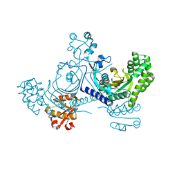
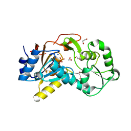
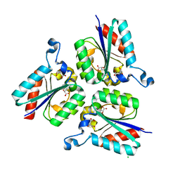
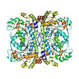
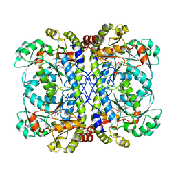
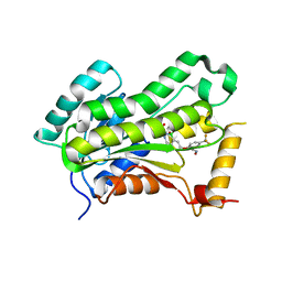
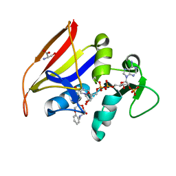
 | | Crystal structure of 2,3,4,5-tetrahydropyridine-2,6-dicarboxylate N-succinyltransferase from Legionella pneumophila Philadelphia 1 | | Descriptor: | 1,2-ETHANEDIOL, 2,3,4,5-tetrahydropyridine-2,6-dicarboxylate N-succinyltransferase | | Authors: | Seattle Structural Genomics Center for Infectious Disease (SSGCID) | | Deposit date: | 2018-02-28 | | Release date: | 2018-03-21 | | Last modified: | 2023-10-04 | | Method: | X-RAY DIFFRACTION (1.8 Å) | | Cite: | Crystal structure of 2,3,4,5-tetrahydropyridine-2,6-dicarboxylate N-succinyltransferase from Legionella pneumophila Philadelphia 1
to be published
|
|
6NMO
 | |
6NAB
 | |
6C0D
 | |
6BS7
 | |
6NQ4
 | |
6NUP
 | |
6CUM
 | |
3KM3
 | |
6CW5
 | |
6CV6
 | |
6CJA
 | |
6CJB
 | |
6CUQ
 | |
4EU1
 | |
5W07
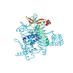
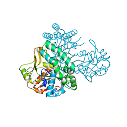
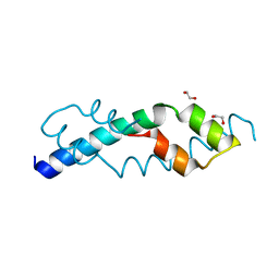
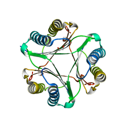
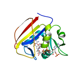
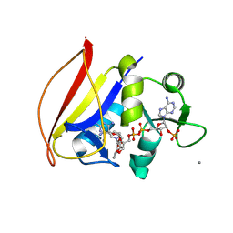
 | | CRYSTAL STRUCTURE OF THE INHA FROM MYCOBACTERIUM TUBERCULOSIS IN COMPLEX WITH AN12855, EBSI 4333. | | Descriptor: | (~{N}~{E})-~{N}-[[2-[[2-ethylsulfonyl-1,1-bis(oxidanyl)-3,4-dihydro-2,3,1$l^{4}-benzodiazaborinin-7-yl]oxy]-5-(trifluoromethyl)phenyl]methylidene]hydroxylamine, Enoyl-[acyl-carrier-protein] reductase [NADH] | | Authors: | Abendroth, J, Edwards, T.E, Lorimer, D. | | Deposit date: | 2017-05-30 | | Release date: | 2018-06-13 | | Last modified: | 2024-03-13 | | Method: | X-RAY DIFFRACTION (2.65 Å) | | Cite: | Discovery of a cofactor-independent inhibitor of Mycobacterium tuberculosis InhA
To Be Published
|
|
4HVJ
 | |
7K62
 | |
7K6A
 | |
7K69
 | |
7K68
 | |
7K6C
 | |
7JFN
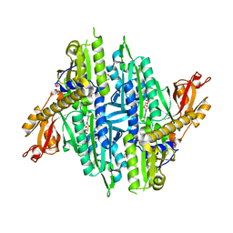
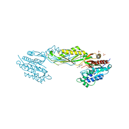
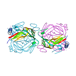
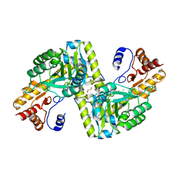
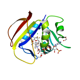
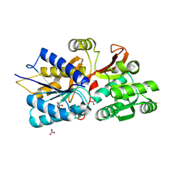 | | Crystal Structure of Leucine-, isoleucine-, valine-, threonine-, and alanine-binding protein from Brucella ovis, closed conformation | | Descriptor: | 1,2-ETHANEDIOL, NITRATE ION, POTASSIUM ION, ... | | Authors: | Seattle Structural Genomics Center for Infectious Disease (SSGCID) | | Deposit date: | 2020-07-17 | | Release date: | 2020-07-29 | | Last modified: | 2023-10-18 | | Method: | X-RAY DIFFRACTION (1.7 Å) | | Cite: | Crystal Structure of Leucine-, isoleucine-, valine-, threonine-, and alanine-binding protein from Brucella ovis, closed conformation
to be published
|
|
7K74
 | |


