Loading
PDBj
MenuPDBj@FacebookPDBj@TwitterPDBj@YouTubewwPDB FoundationwwPDB
RCSB PDBPDBeBMRBAdv. SearchSearch help
Search by PDB author
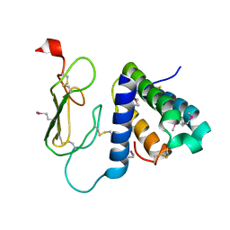
3AQF
DownloadVisualize
BU of 3aqf by Molmil
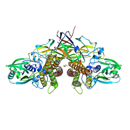
Crystal structure of the human CRLR/RAMP2 extracellular complex
Descriptor: Calcitonin gene-related peptide type 1 receptor, Receptor activity-modifying protein 2
Authors:Kusano, S, Kukimono-Niino, M, Shirouzu, M, Shindo, T, Yokoyama, S.
Deposit date:2010-10-29
Release date:2011-11-02
Last modified:2012-07-25
Method:X-RAY DIFFRACTION (2.6 Å)
Cite:Structural basis for extracellular interactions between calcitonin receptor-like receptor and receptor activity-modifying protein 2 for adrenomedullin-specific binding
Protein Sci., 21, 2012
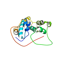
2E5S
DownloadVisualize
BU of 2e5s by Molmil
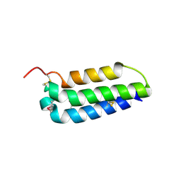
Solution structure of the zf-CCCHx2 domain of muscleblind-like 2, isoform 1 [Homo sapiens]
Descriptor: OTTHUMP00000018578, ZINC ION
Authors:Dang, W, Muto, Y, Inoue, M, Kigawa, T, Shirouzu, M, Terada, T, Yokoyama, S, RIKEN Structural Genomics/Proteomics Initiative (RSGI)
Deposit date:2006-12-22
Release date:2007-06-26
Last modified:2024-05-29
Method:SOLUTION NMR
Cite:Solution structure of the RNA binding domain in the human muscleblind-like protein 2
Protein Sci., 18, 2009
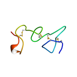
2DK4
DownloadVisualize
BU of 2dk4 by Molmil
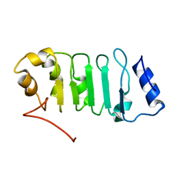
Solution structure of Splicing Factor Motif in Pre-mRNA splicing factor 18 (hPRP18)
Descriptor: Pre-mRNA-splicing factor 18
Authors:He, F, Muto, Y, Inoue, M, Kigawa, T, Shirouzu, M, Terada, T, Yokoyama, S, RIKEN Structural Genomics/Proteomics Initiative (RSGI)
Deposit date:2006-04-06
Release date:2006-10-06
Last modified:2024-05-01
Method:SOLUTION NMR
Cite:Solution structure of the splicing factor motif of the human Prp18 protein.
Proteins, 80, 2012
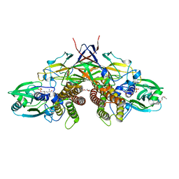
7WUX
DownloadVisualize
BU of 7wux by Molmil
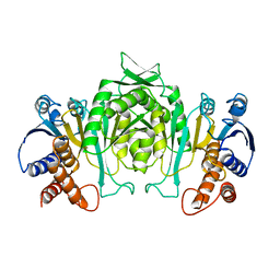
Crystal structure of AziU3/U2 complexed with (5S,6S)-O7-sulfo DADH from Streptomyces sahachiroi
Descriptor: (2S,5S,6S)-2,6-bis(azanyl)-5-oxidanyl-7-sulfooxy-heptanoic acid, 1-ETHOXY-2-(2-ETHOXYETHOXY)ETHANE, AziU2, ...
Authors:Kurosawa, S, Yoshida, A, Tomita, T, Nishiyama, M.
Deposit date:2022-02-09
Release date:2022-09-07
Last modified:2024-05-29
Method:X-RAY DIFFRACTION (1.8 Å)
Cite:Molecular Basis for Enzymatic Aziridine Formation via Sulfate Elimination.
J.Am.Chem.Soc., 144, 2022
7WUW
DownloadVisualize
BU of 7wuw by Molmil
Crystal structure of AziU3/U2 from Streptomyces sahachiroi
Descriptor: AziU2, AziU3, MAGNESIUM ION, ...
Authors:Kurosawa, S, Yoshida, A, Tomita, T, Nishiyama, M.
Deposit date:2022-02-09
Release date:2022-09-07
Last modified:2024-05-29
Method:X-RAY DIFFRACTION (1.75 Å)
Cite:Molecular Basis for Enzymatic Aziridine Formation via Sulfate Elimination.
J.Am.Chem.Soc., 144, 2022
2LWI
DownloadVisualize
BU of 2lwi by Molmil
Solution structure of H-RasT35S mutant protein in complex with Kobe2601
Descriptor: 2-(2,4-dinitrophenyl)-N-(4-fluorophenyl)hydrazinecarbothioamide, GTPase HRas, MAGNESIUM ION, ...
Authors:Araki, M, Tamura, A, Shima, F, Kataoka, T.
Deposit date:2012-08-01
Release date:2013-05-22
Last modified:2024-05-15
Method:SOLUTION NMR
Cite:In silico discovery of small-molecule Ras inhibitors that display antitumor activity by blocking the Ras-effector interaction.
Proc.Natl.Acad.Sci.USA, 110, 2013
1TLE
DownloadVisualize
BU of 1tle by Molmil
LE (LAMININ-TYPE EGF-LIKE) MODULE GIII4 IN SOLUTION AT PH 3.5 AND 290 K, NMR, 14 STRUCTURES
Descriptor: LAMININ
Authors:Baumgartner, R, Czisch, M, Mayer, U, Schl, E.P, Huber, R, Timpl, R, Holak, T.A.
Deposit date:1996-01-26
Release date:1997-02-12
Last modified:2022-03-02
Method:SOLUTION NMR
Cite:Structure of the nidogen binding LE module of the laminin gamma1 chain in solution.
J.Mol.Biol., 257, 1996
7D2O
DownloadVisualize
BU of 7d2o by Molmil
Solution structure of Gaussia Luciferase by NMR
Descriptor: Luciferase
Authors:Kobayashi, N, Wu, N, Kuroda, Y, Yamazaki, T.
Deposit date:2020-09-17
Release date:2020-12-02
Last modified:2023-06-14
Method:SOLUTION NMR
Cite:Solution structure of Gaussia Luciferase with five disulfide bonds and identification of a putative coelenterazine binding cavity by heteronuclear NMR.
Sci Rep, 10, 2020
2AYQ
DownloadVisualize
BU of 2ayq by Molmil
3-ISOPROPYLMALATE DEHYDROGENASE FROM THE MODERATE FACULTATIVE THERMOPHILE, BACILLUS COAGULANS
Descriptor: 3-ISOPROPYLMALATE DEHYDROGENASE
Authors:Tsuchiya, D, Takenaka, A.
Deposit date:1998-02-20
Release date:1998-05-27
Last modified:2024-05-22
Method:X-RAY DIFFRACTION (3 Å)
Cite:Crystal structure of 3-isopropylmalate dehydrogenase from the moderate facultative thermophile, Bacillus coagulans: two strategies for thermostabilization of protein structures.
J.Biochem.(Tokyo), 122, 1997
1JJ3
DownloadVisualize
BU of 1jj3 by Molmil
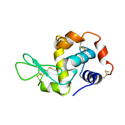
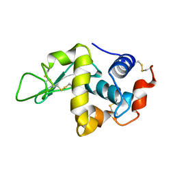
CRYSTAL STRUCTURE OF MONOCLINIC LYSOZYME GROWN AT PH 4.6
Descriptor: LYSOZYME
Authors:Datta, S, Biswal, B.K, Vijayan, M.
Deposit date:2001-07-03
Release date:2001-11-02
Last modified:2023-08-16
Method:X-RAY DIFFRACTION (1.9 Å)
Cite:The effect of stabilizing additives on the structure and hydration of proteins: a study involving tetragonal lysozyme.
Acta Crystallogr.,Sect.D, 57, 2001
1JJ1
DownloadVisualize
BU of 1jj1 by Molmil
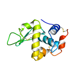
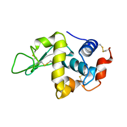
CRYSTAL STRUCTURE OF ORTHORHOMBIC LYSOZYME GROWN AT PH 4.6 IN PRESENCE OF 5% SORBITOL
Descriptor: LYSOZYME
Authors:Datta, S, Biswal, B.K, Vijayan, M.
Deposit date:2001-07-03
Release date:2001-11-02
Last modified:2023-08-16
Method:X-RAY DIFFRACTION (1.9 Å)
Cite:The effect of stabilizing additives on the structure and hydration of proteins: a study involving tetragonal lysozyme.
Acta Crystallogr.,Sect.D, 57, 2001
1JIS
DownloadVisualize
BU of 1jis by Molmil
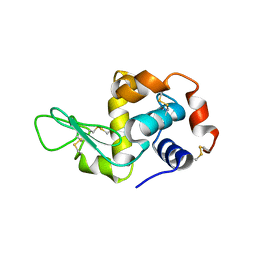
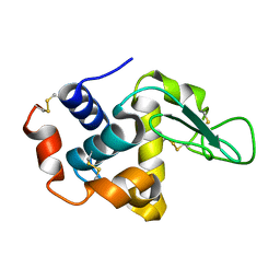
CRYSTAL STRUCTURE OF TETRAGONAL LYSOZYME GROWN AT PH 4.6
Descriptor: LYSOZYME
Authors:Datta, S, Biswal, B.K, Vijayan, M.
Deposit date:2001-07-03
Release date:2001-11-02
Last modified:2023-08-16
Method:X-RAY DIFFRACTION (1.9 Å)
Cite:The effect of stabilizing additives on the structure and hydration of proteins: a study involving tetragonal lysozyme.
Acta Crystallogr.,Sect.D, 57, 2001
1JIY
DownloadVisualize
BU of 1jiy by Molmil
CRYSTAL STRUCTURE OF TETRAGONAL LYSOZYME GROWN IN PRESENCE 20% SORBITOL
Descriptor: LYSOZYME
Authors:Datta, S, Biswal, B.K, Vijayan, M.
Deposit date:2001-07-03
Release date:2001-11-02
Last modified:2023-08-16
Method:X-RAY DIFFRACTION (1.9 Å)
Cite:The effect of stabilizing additives on the structure and hydration of proteins: a study involving tetragonal lysozyme.
Acta Crystallogr.,Sect.D, 57, 2001
1JJ0
DownloadVisualize
BU of 1jj0 by Molmil
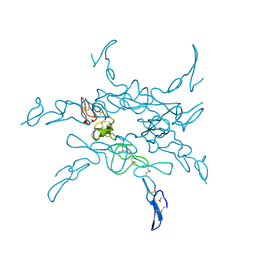
CRYSTAL STRUCTURE OF TETRAGONAL LYSOZYME GROWN IN PRESENCE of 30% SUCROSE
Descriptor: LYSOZYME, beta-D-fructofuranose-(2-1)-alpha-D-glucopyranose
Authors:Datta, S, Biswal, B.K, Vijayan, M.
Deposit date:2001-07-03
Release date:2001-11-02
Last modified:2023-08-16
Method:X-RAY DIFFRACTION (1.9 Å)
Cite:The effect of stabilizing additives on the structure and hydration of proteins: a study involving tetragonal lysozyme.
Acta Crystallogr.,Sect.D, 57, 2001
2ELL
DownloadVisualize
BU of 2ell by Molmil
Solution structure of the Leucine Rich Repeat of human Acidic leucine-rich nuclear phosphoprotein 32 family member B
Descriptor: Acidic leucine-rich nuclear phosphoprotein 32 family member B
Authors:Tochio, N, Koshiba, S, Watanabe, S, Harada, T, Umehara, T, Tanaka, A, Kigawa, T, Yokoyama, S, RIKEN Structural Genomics/Proteomics Initiative (RSGI)
Deposit date:2007-03-27
Release date:2008-04-01
Last modified:2024-05-01
Method:SOLUTION NMR
Cite:Solution structure of histone chaperone ANP32B: interaction with core histones H3-H4 through its acidic concave domain.
J.Mol.Biol., 401, 2010
2ZJ9
DownloadVisualize
BU of 2zj9 by Molmil
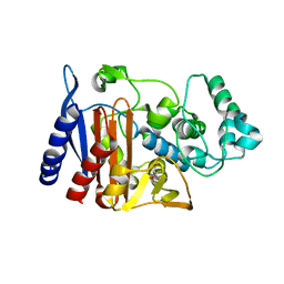
X-ray crystal structure of AmpC beta-Lactamase (AmpC(D)) from an Escherichia coli with a Tripeptide Deletion (Gly286 Ser287 Asp288) on the H10 Helix
Descriptor: AmpC, ISOPROPYL ALCOHOL, SODIUM ION
Authors:Yamaguchi, Y, Sato, G, Yamagata, Y, Wachino, J, Arakawa, Y, Kurosaki, H.
Deposit date:2008-02-29
Release date:2009-03-10
Last modified:2023-11-01
Method:X-RAY DIFFRACTION (1.7 Å)
Cite:Structure of AmpC beta-lactamase (AmpCD) from an Escherichia coli clinical isolate with a tripeptide deletion (Gly286-Ser287-Asp288) in the H10 helix
Acta Crystallogr.,Sect.F, 65, 2009
2C32
DownloadVisualize
BU of 2c32 by Molmil
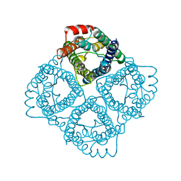
Co-axial association of recombinant eye lens aquaporin-0 observed in loosely packed 3D-crystals
Descriptor: LENS FIBER MAJOR INTRINSIC PROTEIN
Authors:Palanivelu, D.V, Schirmer, T.
Deposit date:2005-10-03
Release date:2005-12-07
Last modified:2023-12-13
Method:X-RAY DIFFRACTION (7.01 Å)
Cite:Co-Axial Association of Recombinant Eye Lens Aquaporin-0 Observed in Loosely Packed 3D-Crystals
J.Mol.Biol., 355, 2006

221371

건을2024-06-19부터공개중

PDB statisticsPDBj update infoContact PDBjnumon