5A2P
 
 | |
4K8U
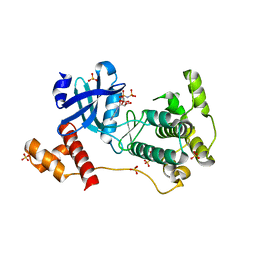
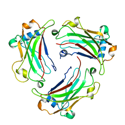 | | Crystal structure of TRAF4 TRAF domain | | 分子名称: | TNF receptor-associated factor 4 | | 著者 | Park, H.H, Yoon, J.H. | | 登録日 | 2013-04-18 | | 公開日 | 2014-01-15 | | 最終更新日 | 2024-03-20 | | 実験手法 | X-RAY DIFFRACTION (2.302 Å) | | 主引用文献 | Structure of the TRAF4 TRAF domain with a coiled-coil domain and its implications for the TRAF4 signalling pathway.
Acta Crystallogr.,Sect.D, 70, 2014
|
|
5EZ5
 | |
4UV7
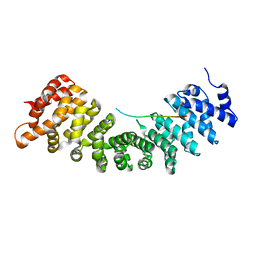
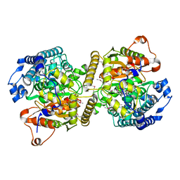
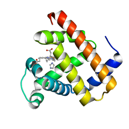
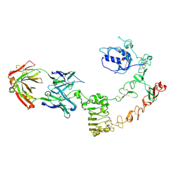 | | The complex structure of extracellular domain of EGFR and GC1118A | | 分子名称: | 2-acetamido-2-deoxy-beta-D-glucopyranose, 2-acetamido-2-deoxy-beta-D-glucopyranose-(1-4)-2-acetamido-2-deoxy-beta-D-glucopyranose, EPIDERMAL GROWTH FACTOR RECEPTOR, ... | | 著者 | Yoo, J.H, Cho, H.S. | | 登録日 | 2014-08-05 | | 公開日 | 2015-10-14 | | 最終更新日 | 2024-01-10 | | 実験手法 | X-RAY DIFFRACTION (2.1 Å) | | 主引用文献 | Gc1118, an Anti-Egfr Antibody with a Distinct Binding Epitope and Superior Inhibitory Activity Against High-Affinity Egfr Ligands.
Mol.Cancer Ther., 15, 2016
|
|
2XHS
 | |
4BFM
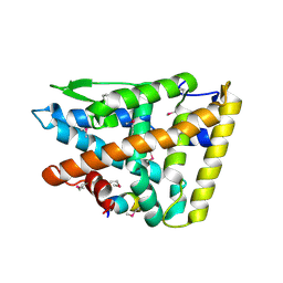
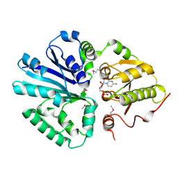
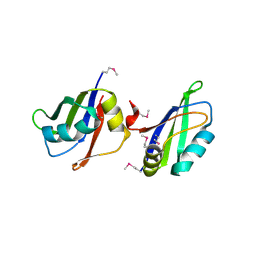
 | | The crystal structure of mouse PK38 | | 分子名称: | MATERNAL EMBRYONIC LEUCINE ZIPPER KINASE, PHOSPHOAMINOPHOSPHONIC ACID-ADENYLATE ESTER, SULFATE ION | | 著者 | Yoo, J.H, Cho, Y.S, Park, S.M, Cho, H.S. | | 登録日 | 2013-03-21 | | 公開日 | 2014-02-12 | | 最終更新日 | 2023-12-20 | | 実験手法 | X-RAY DIFFRACTION (2.35 Å) | | 主引用文献 | The Structures of the Kinase Domain and Uba Domain of Mpk38 Suggest the Activation Mechanism for Kinase Activity.
Acta Crystallogr.,Sect.D, 70, 2014
|
|
2C83
 | |
2C84
 | |
4B18
 | |
8D6Y
 | |
8D6W
 | |
8D6V
 | |
8D6X
 | |
2IY8
 | |
2IY7
 | |
6AIX
 | |
6AIY
 | |
6KVR
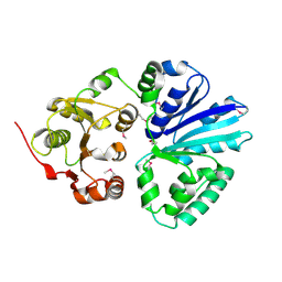
 | | Fatty acid amide hydrolase | | 分子名称: | Fatty acid amide hydrolase | | 著者 | Min, C.A, Yun, J.S, Chang, J.H. | | 登録日 | 2019-09-05 | | 公開日 | 2021-09-15 | | 実験手法 | X-RAY DIFFRACTION (2.2 Å) | | 主引用文献 | Comparison of Candida Albicans Fatty Acid Amide Hydrolase Structure with Homologous Amidase Signature Family Enzymes
Crystals, 9, 2019
|
|
5IM0
 | |
7VUS
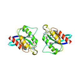
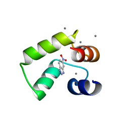
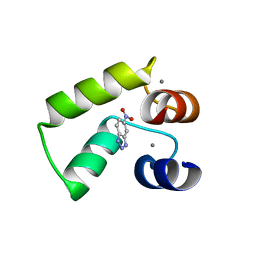 | | Crystal structure of AlleyCat9 with 5-nitro-benzotriazole | | 分子名称: | (4S)-2-METHYL-2,4-PENTANEDIOL, 5-nitro-1H-benzotriazole, AlleyCat, ... | | 著者 | Tame, J.R.H, Korendovych, I.V, Margheritis, E, Takahashi, K. | | 登録日 | 2021-11-04 | | 公開日 | 2022-07-27 | | 最終更新日 | 2024-05-29 | | 実験手法 | X-RAY DIFFRACTION (1.7 Å) | | 主引用文献 | NMR-guided directed evolution.
Nature, 610, 2022
|
|
7VUR
 | | Crystal structure of AlleyCat9 with calcium but no inhibitor | | 分子名称: | (4S)-2-METHYL-2,4-PENTANEDIOL, AlleyCat, CALCIUM ION | | 著者 | Margheritis, E, Takahashi, K, Korendovych, I.V, Tame, J.R.H. | | 登録日 | 2021-11-04 | | 公開日 | 2022-07-27 | | 最終更新日 | 2023-11-29 | | 実験手法 | X-RAY DIFFRACTION (1.7 Å) | | 主引用文献 | NMR-guided directed evolution.
Nature, 610, 2022
|
|
7VUC
 | |
7VUU
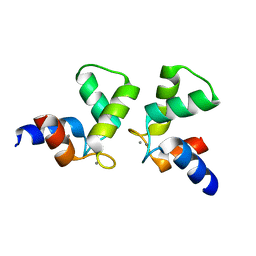
 | | Crystal structure of AlleyCat10 with inhibitor | | 分子名称: | 5-nitro-1H-benzotriazole, AlleyCat, CALCIUM ION | | 著者 | Tame, J.R.H, Korendovych, I.V, Margheritis, E, Takahashi, K. | | 登録日 | 2021-11-04 | | 公開日 | 2022-07-27 | | 最終更新日 | 2024-05-29 | | 実験手法 | X-RAY DIFFRACTION (1.95 Å) | | 主引用文献 | NMR-guided directed evolution.
Nature, 610, 2022
|
|
7VUT
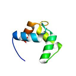
 | | Crystal structure of AlleyCat10 | | 分子名称: | AlleyCat10, CALCIUM ION | | 著者 | Tame, J.R.H, Korendovych, I.V, Margheritis, E, Takahashi, K. | | 登録日 | 2021-11-04 | | 公開日 | 2022-07-27 | | 最終更新日 | 2024-05-29 | | 実験手法 | X-RAY DIFFRACTION (1.7 Å) | | 主引用文献 | NMR-guided directed evolution.
Nature, 610, 2022
|
|
4BYA
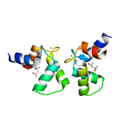
 | | Calmodulin, C-terminal domain, M144H mutant | | 分子名称: | CALCIUM ION, CALMODULIN, C-TERMINAL DOMAIN, ... | | 著者 | Moroz, Y.S, Wu, Y, van Nuland, N.A.J, Korendovych, I.V. | | 登録日 | 2013-07-18 | | 公開日 | 2014-06-18 | | 最終更新日 | 2024-06-19 | | 実験手法 | SOLUTION NMR | | 主引用文献 | New Tricks for Old Proteins: Single Mutations in a Nonenzymatic Protein Give Rise to Various Enzymatic Activities.
J.Am.Chem.Soc., 137, 2015
|
|


