1F4J
 
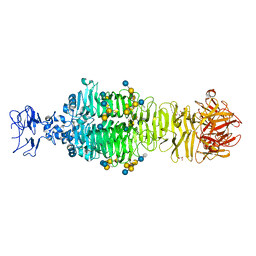
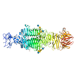
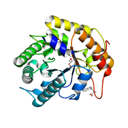
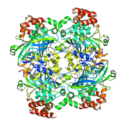 | | STRUCTURE OF TETRAGONAL CRYSTALS OF HUMAN ERYTHROCYTE CATALASE | | 分子名称: | CATALASE, PROTOPORPHYRIN IX CONTAINING FE | | 著者 | Safo, M.K, Musayev, F.N, Wu, S.H, Abraham, D.J, Ko, T.P. | | 登録日 | 2000-06-07 | | 公開日 | 2000-06-21 | | 最終更新日 | 2024-02-07 | | 実験手法 | X-RAY DIFFRACTION (2.4 Å) | | 主引用文献 | Structure of tetragonal crystals of human erythrocyte catalase.
Acta Crystallogr.,Sect.D, 57, 2001
|
|
1XHH
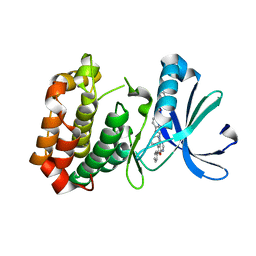
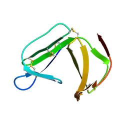 | | Solution Structure of porcine beta-microseminoprotein | | 分子名称: | beta-microseminoprotein | | 著者 | Wang, I, Lou, Y.C, Wu, K.P, Wu, S.H, Chang, W.C, Chen, C. | | 登録日 | 2004-09-20 | | 公開日 | 2005-03-20 | | 最終更新日 | 2022-03-02 | | 実験手法 | SOLUTION NMR | | 主引用文献 | Novel solution structure of porcine beta-microseminoprotein
J.Mol.Biol., 346, 2005
|
|
1QQW
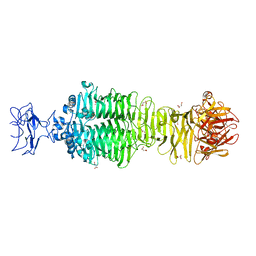
 | | CRYSTAL STRUCTURE OF HUMAN ERYTHROCYTE CATALASE | | 分子名称: | CATALASE, PROTOPORPHYRIN IX CONTAINING FE | | 著者 | Ko, T.P, Safo, M.K, Musayev, F.N, Wang, C, Wu, S.H, Abraham, D.J. | | 登録日 | 1999-06-09 | | 公開日 | 1999-06-14 | | 最終更新日 | 2024-02-14 | | 実験手法 | X-RAY DIFFRACTION (2.75 Å) | | 主引用文献 | Structure of human erythrocyte catalase.
Acta Crystallogr.,Sect.D, 56, 2000
|
|
1X37
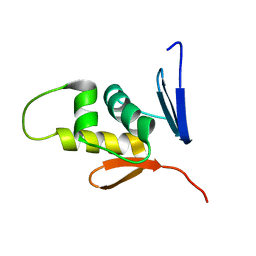
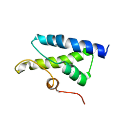 | | Structure of Bacillus subtilis Lon protease SSD domain | | 分子名称: | ATP-dependent protease La 1 | | 著者 | Wang, I, Lou, Y.C, Lo, S.C, Lee, Y.L, Wu, S.H, Chen, C. | | 登録日 | 2005-04-30 | | 公開日 | 2005-10-30 | | 最終更新日 | 2024-05-29 | | 実験手法 | SOLUTION NMR | | 主引用文献 | Structural basis and DNA binding property of SSD domain of Bacillus subtilis Lon protease
to be published
|
|
1JC6
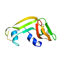
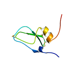 | | SOLUTION STRUCTURE OF BUNGARUS FACIATUS IX, A KUNITZ-TYPE CHYMOTRYPSIN INHIBITOR | | 分子名称: | VENOM BASIC PROTEASE INHIBITORS IX AND VIIIB | | 著者 | Chen, C, Hsu, C.H, Su, N.Y, Chiou, S.H, Wu, S.H. | | 登録日 | 2001-06-08 | | 公開日 | 2003-06-17 | | 最終更新日 | 2022-02-23 | | 実験手法 | SOLUTION NMR | | 主引用文献 | Solution structure of a Kunitz-type chymotrypsin inhibitor isolated from the elapid snake Bungarus fasciatus
J.BIOL.CHEM., 276, 2001
|
|
5JSD
 | | Crystal structure of phiAB6 tailspike in complex with five-repeated oligosaccharides of Acinetobacter baumannii surface polysaccharide | | 分子名称: | ACETIC ACID, MALONIC ACID, beta-D-galactopyranose-(1-3)-2-amino-2-deoxy-beta-D-galactopyranose-(1-3)-[5,7-bisacetamido-3,5,7,9-tetradeoxy-L-glycero-alpha-L-manno-non-2-ulopyranosonic acid-(2-6)-beta-D-glucopyranose-(1-6)]beta-D-galactopyranose-(1-3)-2-amino-2-deoxy-beta-D-galactopyranose-(1-3)-[beta-D-glucopyranose-(1-6)]beta-D-galactopyranose, ... | | 著者 | Lee, I.M, Tu, I.F, Huang, K.F, Wu, S.H. | | 登録日 | 2016-05-08 | | 公開日 | 2017-03-08 | | 最終更新日 | 2024-03-20 | | 実験手法 | X-RAY DIFFRACTION (1.48 Å) | | 主引用文献 | Structural basis for fragmenting the exopolysaccharide of Acinetobacter baumannii by bacteriophage Phi AB6 tailspike protein
Sci Rep, 7, 2017
|
|
5JS4
 | | Crystal structure of phiAB6 tailspike | | 分子名称: | MALONIC ACID, phiAB6 tailspike | | 著者 | Lee, I.M, Tu, I.F, Huang, K.F, Wu, S.H. | | 登録日 | 2016-05-07 | | 公開日 | 2017-03-08 | | 最終更新日 | 2024-03-20 | | 実験手法 | X-RAY DIFFRACTION (1.48 Å) | | 主引用文献 | Structural basis for fragmenting the exopolysaccharide of Acinetobacter baumannii by bacteriophage Phi AB6 tailspike protein
Sci Rep, 7, 2017
|
|
5JSE
 | | Crystal structure of phiAB6 tailspike in complex with three-repeated oligosaccharides of Acinetobacter baumannii surface polysaccharide | | 分子名称: | ACETIC ACID, MALONIC ACID, beta-D-galactopyranose-(1-3)-2-amino-2-deoxy-beta-D-galactopyranose-(1-3)-[5,7-bisacetamido-3,5,7,9-tetradeoxy-L-glycero-alpha-L-manno-non-2-ulopyranosonic acid-(2-6)-beta-D-glucopyranose-(1-6)]beta-D-galactopyranose-(1-3)-2-amino-2-deoxy-beta-D-galactopyranose-(1-3)-[beta-D-glucopyranose-(1-6)]beta-D-galactopyranose, ... | | 著者 | Lee, I.M, Tu, I.F, Huang, K.F, Wu, S.H. | | 登録日 | 2016-05-08 | | 公開日 | 2017-03-08 | | 最終更新日 | 2024-03-20 | | 実験手法 | X-RAY DIFFRACTION (1.89 Å) | | 主引用文献 | Structural basis for fragmenting the exopolysaccharide of Acinetobacter baumannii by bacteriophage Phi AB6 tailspike protein
Sci Rep, 7, 2017
|
|
8IQ9
 | | Crystal structure of trimeric K2-2 TSP in complex with tetrasaccharide and octasaccharide | | 分子名称: | 1,2-ETHANEDIOL, ACETYL GROUP, K2-2 TSP, ... | | 著者 | Ye, T.J, Ko, T.P, Huang, K.F, Wu, S.H. | | 登録日 | 2023-03-16 | | 公開日 | 2024-02-21 | | 最終更新日 | 2024-04-03 | | 実験手法 | X-RAY DIFFRACTION (1.58 Å) | | 主引用文献 | Klebsiella pneumoniae K2 capsular polysaccharide degradation by a bacteriophage depolymerase does not require trimer formation.
Mbio, 15, 2024
|
|
8IQE
 | | Crystal structure of tetrameric K2-2 TSP | | 分子名称: | GLYCEROL, K2-VCL6 TSP | | 著者 | Ye, T.J, Huang, K.F, Tu, I.F, Lee, I.M, Chang, Y.P, Wu, S.H. | | 登録日 | 2023-03-16 | | 公開日 | 2024-02-21 | | 最終更新日 | 2024-04-03 | | 実験手法 | X-RAY DIFFRACTION (2.17 Å) | | 主引用文献 | Klebsiella pneumoniae K2 capsular polysaccharide degradation by a bacteriophage depolymerase does not require trimer formation.
Mbio, 15, 2024
|
|
5WSL
 | | Structural studies of keratinase from Meiothermus taiwanensis WR-220 | | 分子名称: | CALCIUM ION, SULFATE ION, keratinase | | 著者 | Ho, M.C, Wu, S.H, Chen, M.Y, Tu, I.F. | | 登録日 | 2016-12-07 | | 公開日 | 2017-10-18 | | 最終更新日 | 2023-11-08 | | 実験手法 | X-RAY DIFFRACTION (1.5 Å) | | 主引用文献 | The discovery of novel heat-stable keratinases from Meiothermus taiwanensis WR-220 and other extremophiles
Sci Rep, 7, 2017
|
|
7W1D
 | | Crystal structure of Klebsiella pneumoniae K1 capsule-specific polysaccharide lyase in a C2 crystal form | | 分子名称: | CARBONATE ION, CITRIC ACID, K1 LYASE | | 著者 | Tu, I.F, Ko, T.P, Huang, K.F, Wu, S.H. | | 登録日 | 2021-11-19 | | 公開日 | 2022-05-18 | | 最終更新日 | 2024-05-29 | | 実験手法 | X-RAY DIFFRACTION (2.78 Å) | | 主引用文献 | Structural and biological insights into Klebsiella pneumoniae surface polysaccharide degradation by a bacteriophage K1 lyase: implications for clinical use.
J.Biomed.Sci., 29, 2022
|
|
7W1E
 | | Crystal structure of Klebsiella pneumoniae K1 capsule-specific polysaccharide lyase in complex with products | | 分子名称: | 2,6-anhydro-4,5-O-[(1R)-1-carboxyethylidene]-3-deoxy-L-threo-hex-2-enonic acid, 3-O-acetyl-6-deoxy-alpha-L-galactopyranose-(1-3)-beta-D-glucopyranose, GLYCEROL, ... | | 著者 | Tu, I.F, Huang, K.F, Wu, S.H. | | 登録日 | 2021-11-19 | | 公開日 | 2022-05-18 | | 最終更新日 | 2024-05-29 | | 実験手法 | X-RAY DIFFRACTION (1.46 Å) | | 主引用文献 | Structural and biological insights into Klebsiella pneumoniae surface polysaccharide degradation by a bacteriophage K1 lyase: implications for clinical use.
J.Biomed.Sci., 29, 2022
|
|
7W1C
 | | Crystal structure of Klebsiella pneumoniae K1 capsule-specific polysaccharide lyase in a P1 crystal form | | 分子名称: | (2S)-2-hydroxybutanedioic acid, GLYCEROL, IMIDAZOLE, ... | | 著者 | Tu, I.F, Huang, K.F, Wu, S.H. | | 登録日 | 2021-11-19 | | 公開日 | 2022-05-18 | | 最終更新日 | 2024-05-29 | | 実験手法 | X-RAY DIFFRACTION (1.48 Å) | | 主引用文献 | Structural and biological insights into Klebsiella pneumoniae surface polysaccharide degradation by a bacteriophage K1 lyase: implications for clinical use.
J.Biomed.Sci., 29, 2022
|
|
7VT7
 | | Crystal structure of CBM deleted MtGlu5 in complex with CBI | | 分子名称: | Endoglucanase H, GLYCEROL, TRYPTOPHAN, ... | | 著者 | Ye, T.J, Ko, P.T, Huang, K.F, Wu, S.H. | | 登録日 | 2021-10-28 | | 公開日 | 2022-09-07 | | 最終更新日 | 2023-11-29 | | 実験手法 | X-RAY DIFFRACTION (1.53 Å) | | 主引用文献 | Synergic action of an inserted carbohydrate-binding module in a glycoside hydrolase family 5 endoglucanase.
Acta Crystallogr D Struct Biol, 78, 2022
|
|
7VT8
 | | Crystal structure of MtGlu5 from Meiothermus taiwanensis WR-220 | | 分子名称: | Endoglucanase H, SULFATE ION, beta-D-glucopyranose | | 著者 | Ye, T.J, Ko, P.T, Huang, K.F, Wu, S.H. | | 登録日 | 2021-10-28 | | 公開日 | 2022-09-07 | | 最終更新日 | 2023-11-29 | | 実験手法 | X-RAY DIFFRACTION (2.99 Å) | | 主引用文献 | Synergic action of an inserted carbohydrate-binding module in a glycoside hydrolase family 5 endoglucanase.
Acta Crystallogr D Struct Biol, 78, 2022
|
|
7VT5
 | | Crystal structure of CBM deleted MtGlu5 from Meiothermus taiwanensis WR-220 | | 分子名称: | Endoglucanase H, METHIONINE, TRYPTOPHAN | | 著者 | Ye, T.J, Ko, P.T, Huang, K.F, Wu, S.H. | | 登録日 | 2021-10-28 | | 公開日 | 2022-09-07 | | 最終更新日 | 2023-11-29 | | 実験手法 | X-RAY DIFFRACTION (1.46 Å) | | 主引用文献 | Synergic action of an inserted carbohydrate-binding module in a glycoside hydrolase family 5 endoglucanase.
Acta Crystallogr D Struct Biol, 78, 2022
|
|
7VT4
 | | Crystal structure of mutant E393Q of MtGlu5 | | 分子名称: | Endoglucanase H, GLYCEROL, SULFATE ION | | 著者 | Ye, T.J, Ko, P.T, Huang, K.F, Wu, S.H. | | 登録日 | 2021-10-28 | | 公開日 | 2022-09-07 | | 最終更新日 | 2023-11-29 | | 実験手法 | X-RAY DIFFRACTION (1.93 Å) | | 主引用文献 | Synergic action of an inserted carbohydrate-binding module in a glycoside hydrolase family 5 endoglucanase.
Acta Crystallogr D Struct Biol, 78, 2022
|
|
7VT6
 | | Crystal structure of CBM deleted MtGlu5 in complex with BGC. | | 分子名称: | Endoglucanase H, GLYCEROL, TRYPTOPHAN, ... | | 著者 | Ye, T.J, Ko, P.T, Huang, K.F, Wu, S.H. | | 登録日 | 2021-10-28 | | 公開日 | 2022-09-07 | | 最終更新日 | 2023-11-29 | | 実験手法 | X-RAY DIFFRACTION (1.53 Å) | | 主引用文献 | Synergic action of an inserted carbohydrate-binding module in a glycoside hydrolase family 5 endoglucanase.
Acta Crystallogr D Struct Biol, 78, 2022
|
|
3M11
 | | Crystal Structure of Aurora A Kinase complexed with inhibitor | | 分子名称: | 1-(4-{2-[(5,6-diphenylfuro[2,3-d]pyrimidin-4-yl)amino]ethyl}phenyl)-3-phenylurea, Serine/threonine-protein kinase 6 | | 著者 | Wu, J.S, Leou, J.S, Coumar, M.S, Hsieh, H.P, Wu, S.Y. | | 登録日 | 2010-03-03 | | 公開日 | 2011-03-09 | | 最終更新日 | 2023-11-01 | | 実験手法 | X-RAY DIFFRACTION (2.75 Å) | | 主引用文献 | Fast-forwarding hit to lead: aurora and epidermal growth factor receptor kinase inhibitor lead identification
J.Med.Chem., 53, 2010
|
|
2M87
 | |
2HKY
 | |
4X09
 | | Structure of human RNase 6 in complex with sulphate anions | | 分子名称: | GLYCEROL, Ribonuclease K6, SULFATE ION | | 著者 | Prats-Ejarque, G, Arranz-Trullen, J, Blanco, J.A, Pulido, D, Moussaoui, M, Boix, E. | | 登録日 | 2014-11-21 | | 公開日 | 2016-04-06 | | 最終更新日 | 2024-01-10 | | 実験手法 | X-RAY DIFFRACTION (1.722 Å) | | 主引用文献 | The first crystal structure of human RNase 6 reveals a novel substrate-binding and cleavage site arrangement.
Biochem.J., 473, 2016
|
|
1JLZ
 | | Solution Structure of a K+-Channel Blocker from the Scorpion Toxin of Tityus cambridgei | | 分子名称: | Tityustoxin alpha-KTx | | 著者 | Wang, I, Wu, S.-H, Chang, H.-K, Shieh, R.-C, Yu, H.-M, Chen, C. | | 登録日 | 2001-07-17 | | 公開日 | 2002-02-06 | | 最終更新日 | 2022-02-23 | | 実験手法 | SOLUTION NMR | | 主引用文献 | Solution structure of a K(+)-channel blocker from the scorpion Tityus cambridgei.
Protein Sci., 11, 2002
|
|
1M58
 | | Solution Structure of Cytotoxic RC-RNase2 | | 分子名称: | RC-RNase2 ribonuclease | | 著者 | Hsu, C.-H, Liao, Y.-D, Wu, S.-H, Chen, C. | | 登録日 | 2002-07-09 | | 公開日 | 2003-01-09 | | 最終更新日 | 2022-12-21 | | 実験手法 | SOLUTION NMR | | 主引用文献 | 1H, 15N and 13C resonance assignments and secondary structure determination of the RC-RNase 2 from oocytes of bullfrog Rana catesbeiana.
J.Biomol.Nmr, 19, 2001
|
|


