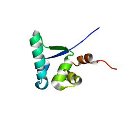
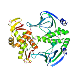
1XJH
 
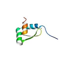 | | NMR structure of the redox switch domain of the E. coli Hsp33 | | 分子名称: | 33 kDa chaperonin, ZINC ION | | 著者 | Won, H.S, Low, L.Y, De Guzman, R.N, Martinez-Yamout, M.A, Jakob, U, Dyson, H.J. | | 登録日 | 2004-09-23 | | 公開日 | 2004-10-05 | | 最終更新日 | 2024-05-22 | | 実験手法 | SOLUTION NMR | | 主引用文献 | The Zinc-dependent Redox Switch Domain of the Chaperone Hsp33 has a Novel Fold
J.Mol.Biol., 341, 2004
|
|
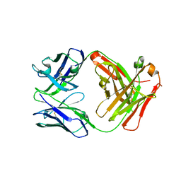
8QMO
 | | Cryo-EM structure of the benzo[a]pyrene-bound Hsp90-XAP2-AHR complex | | 分子名称: | ADENOSINE-5'-DIPHOSPHATE, AH receptor-interacting protein, Aryl hydrocarbon receptor, ... | | 著者 | Kwong, H.S, Grandvuillemin, L, Sirounian, S, Ancelin, A, Lai-Kee-Him, J, Carivenc, C, Lancey, C, Ragan, T.J, Hesketh, E.L, Bourguet, W, Gruszczyk, J. | | 登録日 | 2023-09-24 | | 公開日 | 2024-01-10 | | 最終更新日 | 2024-02-28 | | 実験手法 | ELECTRON MICROSCOPY (2.76 Å) | | 主引用文献 | Structural Insights into the Activation of Human Aryl Hydrocarbon Receptor by the Environmental Contaminant Benzo[a]pyrene and Structurally Related Compounds.
J.Mol.Biol., 436, 2024
|
|
7X89
 | | Tid1 | | 分子名称: | DnaJ homolog subfamily A member 3, mitochondrial | | 著者 | Jang, J, Lee, S.H, Kang, D.H, Sim, D.W, Jo, K.S, Ryu, H, Kim, E.H, Ryu, K.S, Lee, J.H, Kim, J.H, Won, H.S. | | 登録日 | 2022-03-11 | | 公開日 | 2023-03-22 | | 最終更新日 | 2024-05-15 | | 実験手法 | SOLUTION NMR | | 主引用文献 | Structural studies on the J-domain and GF-motif of the mitochondrial Hsp40, Tid1
To Be Published
|
|
5J4G
 | | Crystal structure of the C-terminally His6-tagged HP0902, an uncharacterized protein from Helicobacter pylori 26695 | | 分子名称: | Uncharacterized protein | | 著者 | Sim, D.W, Lee, W.C, Kim, H.Y, Kim, J.H, Won, H.S. | | 登録日 | 2016-04-01 | | 公開日 | 2017-02-08 | | 最終更新日 | 2023-11-08 | | 実験手法 | X-RAY DIFFRACTION (2.6 Å) | | 主引用文献 | Structural identification of the lipopolysaccharide-binding capability of a cupin-family protein from Helicobacter pylori
FEBS Lett., 590, 2016
|
|
6IWS
 | | Solution structure of the J-domain of Tid1, a Mitochondrial Hsp40/DnaJ Protein | | 分子名称: | DnaJ homolog subfamily A member 3, mitochondrial | | 著者 | Sim, D.W, Jo, K.S, Won, H.S, Kim, J.H. | | 登録日 | 2018-12-06 | | 公開日 | 2019-12-11 | | 最終更新日 | 2024-05-01 | | 実験手法 | SOLUTION NMR | | 主引用文献 | Solution structure of the J-domain of Tid1, a Mitochondrial Hsp40/DnaJ Protein
To Be Published
|
|
2MC4
 | |
5GZ0
 | | Crystal structure of FM329, a recombinant Fab adopted from cetuximab | | 分子名称: | FM329 heavy chain, FM329 light chain | | 著者 | Sim, D.W, Kim, J.H, Kim, Y.P, Won, H.S. | | 登録日 | 2016-09-26 | | 公開日 | 2017-10-04 | | 最終更新日 | 2023-11-08 | | 実験手法 | X-RAY DIFFRACTION (1.7 Å) | | 主引用文献 | Crystal structure of FM329, a recombinant Fab adopted from cetuximab
To Be Published
|
|
5I76
 | | Crystal structure of FM318, a recombinant Fab adopted from cetuximab | | 分子名称: | FM318_heavy_cahin, FM318_light_chain | | 著者 | Sim, D.W, Kim, J.H, Seok, S.H, Seo, M.D, Kim, Y.P, Won, H.S. | | 登録日 | 2016-02-16 | | 公開日 | 2016-12-07 | | 最終更新日 | 2023-11-08 | | 実験手法 | X-RAY DIFFRACTION (1.922 Å) | | 主引用文献 | Bacterial production and structure-functional validation of a recombinant antigen-binding fragment (Fab) of an anti-cancer therapeutic antibody targeting epidermal growth factor receptor.
Appl.Microbiol.Biotechnol., 100, 2016
|
|
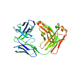
7ZUB
 | | Cryo-EM structure of the indirubin-bound Hsp90-XAP2-AHR complex | | 分子名称: | (3~{Z})-3-(3-oxidanylidene-1~{H}-indol-2-ylidene)-1~{H}-indol-2-one, ADENOSINE-5'-DIPHOSPHATE, AH receptor-interacting protein, ... | | 著者 | Gruszczyk, J, Savva, C.G, Lai-Kee-Him, J, Bous, J, Ancelin, A, Kwong, H.S, Grandvuillemin, L, Bourguet, W. | | 登録日 | 2022-05-12 | | 公開日 | 2022-11-23 | | 最終更新日 | 2024-07-24 | | 実験手法 | ELECTRON MICROSCOPY (2.85 Å) | | 主引用文献 | Cryo-EM structure of the agonist-bound Hsp90-XAP2-AHR cytosolic complex.
Nat Commun, 13, 2022
|
|
5J4F
 | | Crystal structure of the N-terminally His6-tagged HP0902, an uncharacterized protein from Helicobacter pylori 26695 | | 分子名称: | Uncharacterized protein | | 著者 | Sim, D.-W, Lee, W.-C, Kim, H.Y, Kim, J.-H, Won, H.-S. | | 登録日 | 2016-04-01 | | 公開日 | 2017-02-08 | | 最終更新日 | 2023-11-08 | | 実験手法 | X-RAY DIFFRACTION (1.4 Å) | | 主引用文献 | Structural identification of the lipopolysaccharide-binding capability of a cupin-family protein from Helicobacter pylori
FEBS Lett., 590, 2016
|
|
6IRP
 | |
2MX0
 | | Solution structure of HP0268 from Helicobacter pylori | | 分子名称: | Uncharacterized protein HP_0268 | | 著者 | Lee, K.Y. | | 登録日 | 2014-12-05 | | 公開日 | 2015-12-09 | | 最終更新日 | 2024-05-15 | | 実験手法 | SOLUTION NMR | | 主引用文献 | Structure-based functional identification of Helicobacter pylori HP0268 as a nuclease with both DNA nicking and RNase activities
Nucleic Acids Res., 43, 2015
|
|
5YCL
 | |
4N9I
 | | Crystal Structure of Transcription regulation protein CRP complexed with cGMP | | 分子名称: | CYCLIC GUANOSINE MONOPHOSPHATE, Catabolite gene activator | | 著者 | Lee, B.-J, Seok, S.-H, Im, H, Yoon, H.-J. | | 登録日 | 2013-10-21 | | 公開日 | 2014-07-09 | | 最終更新日 | 2023-11-08 | | 実験手法 | X-RAY DIFFRACTION (2.19 Å) | | 主引用文献 | Structures of inactive CRP species reveal the atomic details of the allosteric transition that discriminates cyclic nucleotide second messengers.
Acta Crystallogr.,Sect.D, 70, 2014
|
|
4N9H
 | | Crystal structure of Transcription regulation Protein CRP | | 分子名称: | Catabolite gene activator | | 著者 | Lee, B.J, Seok, S.H, Im, H, Yoon, H.J. | | 登録日 | 2013-10-21 | | 公開日 | 2014-07-09 | | 実験手法 | X-RAY DIFFRACTION (2.2 Å) | | 主引用文献 | Structures of inactive CRP species reveal the atomic details of the allosteric transition that discriminates cyclic nucleotide second messengers.
Acta Crystallogr.,Sect.D, 70, 2014
|
|
7PZF
 | |
7Q3T
 | |
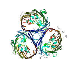
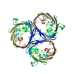
6RCK
 | | Crystal structure of the OmpK36 GD insertion chimera from Klebsiella pneumonia | | 分子名称: | (2S)-3-{[{[(2S)-2,3-DIHYDROXYPROPYL]OXY}(HYDROXY)PHOSPHORYL]OXY}-2-[(6E)-HEXADEC-6-ENOYLOXY]PROPYL (8E)-OCTADEC-8-ENOATE, (HYDROXYETHYLOXY)TRI(ETHYLOXY)OCTANE, LAURYL DIMETHYLAMINE-N-OXIDE, ... | | 著者 | Beis, K, Romano, M, Kwong, J. | | 登録日 | 2019-04-11 | | 公開日 | 2019-09-11 | | 最終更新日 | 2024-01-24 | | 実験手法 | X-RAY DIFFRACTION (2.029 Å) | | 主引用文献 | OmpK36-mediated Carbapenem resistance attenuates ST258 Klebsiella pneumoniae in vivo.
Nat Commun, 10, 2019
|
|
6RCP
 | |
6RD3
 | |
7SZI
 | | Cryo-EM structure of OmpK36-TraN mating pair stabilization proteins from carbapenem-resistant Klebsiella pneumoniae | | 分子名称: | OmpK36, TraN | | 著者 | Beltran, L.C, Seddon, C, Beis, K, Frankel, G, Egelman, E.H. | | 登録日 | 2021-11-27 | | 公開日 | 2022-06-08 | | 最終更新日 | 2022-07-13 | | 実験手法 | ELECTRON MICROSCOPY (2.7 Å) | | 主引用文献 | Mating pair stabilization mediates bacterial conjugation species specificity.
Nat Microbiol, 7, 2022
|
|
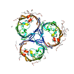
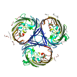
8QUQ
 | | Crystal structure of Ompk36 GD at 3500 eV based on spherical harmonics absorption corrections | | 分子名称: | OmpK36, SULFATE ION | | 著者 | Duman, R, Wagner, A, Beis, K, Wong, J. | | 登録日 | 2023-10-16 | | 公開日 | 2024-06-19 | | 実験手法 | X-RAY DIFFRACTION (2.34 Å) | | 主引用文献 | Ray-tracing analytical absorption correction for X-ray crystallography based on tomographic reconstructions.
J.Appl.Crystallogr., 57, 2024
|
|
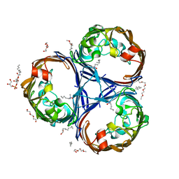
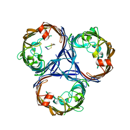
8QUR
 | | Crystal structure of Ompk36 GD at 3500 eV with no absorption corrections | | 分子名称: | OmpK36, SULFATE ION | | 著者 | Duman, R, Wagner, A, Beis, K, Wong, J. | | 登録日 | 2023-10-16 | | 公開日 | 2024-06-19 | | 実験手法 | X-RAY DIFFRACTION (2.34 Å) | | 主引用文献 | Ray-tracing analytical absorption correction for X-ray crystallography based on tomographic reconstructions.
J.Appl.Crystallogr., 57, 2024
|
|
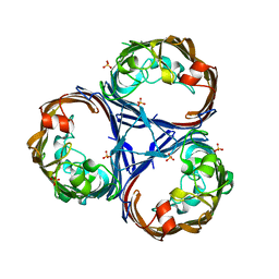
8QVS
 | | Crystal structure of Ompk36 GD at 3500 eV based on a combination of spherical harmonics and analytical absorption corrections | | 分子名称: | OmpK36, SULFATE ION | | 著者 | Duman, R, Wagner, A, Beis, K, Wong, J. | | 登録日 | 2023-10-18 | | 公開日 | 2024-06-19 | | 実験手法 | X-RAY DIFFRACTION (2.34 Å) | | 主引用文献 | Ray-tracing analytical absorption correction for X-ray crystallography based on tomographic reconstructions.
J.Appl.Crystallogr., 57, 2024
|
|
6YO1
 | | Crystal structure of ribonuclease A solved by vanadium SAD phasing | | 分子名称: | Ribonuclease pancreatic, URIDINE-2',3'-VANADATE | | 著者 | El Omari, K, Mohamad, N, Bountra, K, Duman, R, Romano, M, Schlegel, K, Kwong, H, Mykhaylyk, V, Olesen, C.E, Moller, J.V, Bublitz, M, Beis, K, Wagner, A. | | 登録日 | 2020-04-14 | | 公開日 | 2020-11-04 | | 最終更新日 | 2020-12-02 | | 実験手法 | X-RAY DIFFRACTION (1.9 Å) | | 主引用文献 | Experimental phasing with vanadium and application to nucleotide-binding membrane proteins.
Iucrj, 7, 2020
|
|


