3SS2
 
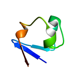
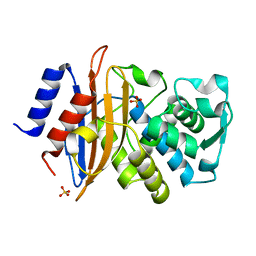
 | | Neutron structure of perdeuterated rubredoxin using 48 hours 3rd pass data | | 分子名称: | FE (III) ION, Rubredoxin | | 著者 | Munshi, P, Chung, C.-L, Blakeley, M.P, Weiss, K.L, Myles, D.A.A, Meilleur, F. | | 登録日 | 2011-07-07 | | 公開日 | 2011-12-28 | | 最終更新日 | 2023-09-13 | | 実験手法 | NEUTRON DIFFRACTION (1.75 Å) | | 主引用文献 | Rapid visualization of hydrogen positions in protein neutron crystallographic structures.
Acta Crystallogr.,Sect.D, 68, 2012
|
|
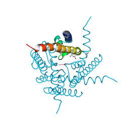
5G18
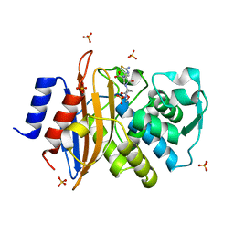 | | Direct Observation of Active-site Protonation States in a Class A beta lactamase with a monobactam substrate | | 分子名称: | 2-({[(1Z)-1-(2-amino-1,3-thiazol-4-yl)-2-oxo-2-{[(2S,3S)-1-oxo-3-(sulfoamino)butan-2-yl]amino}ethylidene]amino}oxy)-2-methylpropanoic acid, BETA-LACTAMASE CTX-M-97, SULFATE ION | | 著者 | Vandavasi, V.G, Weiss, K.L, Parks, J.M, Cooper, J.B, Ginell, S.L, Coates, L. | | 登録日 | 2016-03-23 | | 公開日 | 2016-11-09 | | 最終更新日 | 2019-10-23 | | 実験手法 | X-RAY DIFFRACTION (1.1 Å) | | 主引用文献 | Active-Site Protonation States in an Acyl-Enzyme Intermediate of a Class A beta-Lactamase with a Monobactam Substrate.
Antimicrob. Agents Chemother., 61, 2017
|
|
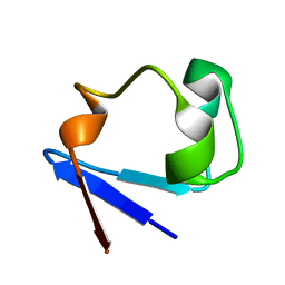
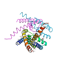
3RYG
 | | 128 hours neutron structure of perdeuterated rubredoxin | | 分子名称: | FE (III) ION, Rubredoxin | | 著者 | Munshi, P, Chung, C.-L, Weiss, K.L, Blakeley, M.P, Myles, D.A.A, Meilleur, F. | | 登録日 | 2011-05-11 | | 公開日 | 2011-12-28 | | 最終更新日 | 2023-09-13 | | 実験手法 | NEUTRON DIFFRACTION (1.75 Å) | | 主引用文献 | Rapid visualization of hydrogen positions in protein neutron crystallographic structures.
Acta Crystallogr.,Sect.D, 68, 2012
|
|
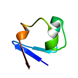
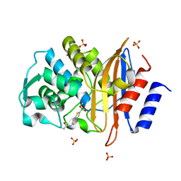
3RZ6
 | | Neutron structure of perdeuterated rubredoxin using 40 hours 1st pass data | | 分子名称: | FE (III) ION, Rubredoxin | | 著者 | Munshi, P, Chung, C.-L, Weiss, K.L, Blakeley, M.P, Myles, D.A.A, Meilleur, F. | | 登録日 | 2011-05-11 | | 公開日 | 2011-12-28 | | 最終更新日 | 2023-09-13 | | 実験手法 | NEUTRON DIFFRACTION (1.75 Å) | | 主引用文献 | Rapid visualization of hydrogen positions in protein neutron crystallographic structures.
Acta Crystallogr.,Sect.D, 68, 2012
|
|
3RZT
 | | Neutron structure of perdeuterated rubredoxin using rapid (14 hours) data | | 分子名称: | FE (III) ION, Rubredoxin | | 著者 | Munshi, P, Chung, C.-L, Weiss, K.L, Blakeley, M.P, Myles, D.A.A, Meilleur, F. | | 登録日 | 2011-05-12 | | 公開日 | 2011-12-28 | | 最終更新日 | 2023-09-13 | | 実験手法 | NEUTRON DIFFRACTION (1.7504 Å) | | 主引用文献 | Rapid visualization of hydrogen positions in protein neutron crystallographic structures.
Acta Crystallogr.,Sect.D, 68, 2012
|
|
5A93
 | | 293K Joint X-ray Neutron with Cefotaxime: EXPLORING THE MECHANISM OF BETA-LACTAM RING PROTONATION IN THE CLASS A BETA-LACTAMASE ACYLATION MECHANISM USING NEUTRON AND X-RAY CRYSTALLOGRAPHY | | 分子名称: | BETA-LACTAMASE CTX-M-97, CEFOTAXIME, C3' cleaved, ... | | 著者 | Vandavasi, V.G, Weiss, K.L, Cooper, J.B, Erskine, P.T, Tomanicek, S.J, Ostermann, A, Schrader, T.E, Ginell, S.L, Coates, L. | | 登録日 | 2015-07-17 | | 公開日 | 2015-12-16 | | 最終更新日 | 2024-01-10 | | 実験手法 | NEUTRON DIFFRACTION (1.598 Å), X-RAY DIFFRACTION | | 主引用文献 | Exploring the Mechanism of Beta-Lactam Ring Protonation in the Class a Beta-Lactamase Acylation Mechanism Using Neutron and X-Ray Crystallography.
J.Med.Chem., 59, 2016
|
|
5A91
 | | 15K X-ray ligand free: Exploring the Mechanism of beta-Lactam Ring Protonation in the Class A beta-lactamase Acylation Mechanism Using Neutron and X-ray Crystallography | | 分子名称: | Beta-lactamase Toho-1, SULFATE ION | | 著者 | Vandavasi, V.G, Weiss, K.L, Cooper, J.B, Erskine, P.T, Tomanicek, S.J, Ostermann, A, Schrader, T.E, Ginell, S.L, Coates, L. | | 登録日 | 2015-07-17 | | 公開日 | 2015-12-16 | | 最終更新日 | 2024-05-08 | | 実験手法 | X-RAY DIFFRACTION (1.2 Å) | | 主引用文献 | Exploring the Mechanism of Beta-Lactam Ring Protonation in the Class a Beta-Lactamase Acylation Mechanism Using Neutron and X-Ray Crystallography.
J.Med.Chem., 59, 2016
|
|
5A92
 | | 15K X-ray structure with Cefotaxime: Exploring the Mechanism of beta- Lactam Ring Protonation in the Class A beta-lactamase Acylation Mechanism Using Neutron and X-ray Crystallography | | 分子名称: | BETA-LACTAMASE CTX-M-97, CEFOTAXIME, C3' cleaved, ... | | 著者 | Vandavasi, V.G, Weiss, K.L, Cooper, J.B, Erskine, P.T, Tomanicek, S.J, Ostermann, A, Schrader, T.E, Ginell, S.L, Coates, L. | | 登録日 | 2015-07-17 | | 公開日 | 2015-12-16 | | 最終更新日 | 2018-10-03 | | 実験手法 | X-RAY DIFFRACTION (1.05 Å) | | 主引用文献 | Exploring the Mechanism of Beta-Lactam Ring Protonation in the Class a Beta-Lactamase Acylation Mechanism Using Neutron and X-Ray Crystallography.
J.Med.Chem., 59, 2016
|
|
5A90
 | | 100K Neutron Ligand Free: Exploring the Mechanism of beta-Lactam Ring Protonation in the Class A beta-lactamase Acylation Mechanism Using Neutron and X-ray Crystallography | | 分子名称: | BETA-LACTAMASE CTX-M-97 | | 著者 | Vandavasi, V.G, Weiss, K.L, Cooper, J.B, Erskine, P.T, Tomanicek, S.J, Ostermann, A, Schrader, T.E, Ginell, S.L, Coates, L. | | 登録日 | 2015-07-17 | | 公開日 | 2015-12-16 | | 最終更新日 | 2024-05-08 | | 実験手法 | NEUTRON DIFFRACTION (1.7 Å) | | 主引用文献 | Exploring the Mechanism of Beta-Lactam Ring Protonation in the Class a Beta-Lactamase Acylation Mechanism Using Neutron and X-Ray Crystallography.
J.Med.Chem., 59, 2016
|
|
2XQZ
 | | Neutron structure of the perdeuterated Toho-1 R274N R276N double mutant beta-lactamase | | 分子名称: | BETA-LACTAMSE TOHO-1 | | 著者 | Tomanicek, S.J, Wang, K.K, Weiss, K.L, Blakeley, M.P, Cooper, J, Chen, Y, Coates, L. | | 登録日 | 2010-09-08 | | 公開日 | 2010-12-22 | | 最終更新日 | 2024-05-08 | | 実験手法 | NEUTRON DIFFRACTION (2.1 Å) | | 主引用文献 | The Active Site Protonation States of Perdeuterated Toho-1 Beta-Lactamase Determined by Neutron Diffraction Support a Role for Glu166 as the General Base in Acylation.
FEBS Lett., 585, 2011
|
|
2XR0
 | | Room temperature X-ray structure of the perdeuterated Toho-1 R274N R276N double mutant beta-lactamase | | 分子名称: | SULFATE ION, TOHO-1 BETA-LACTAMASE | | 著者 | Tomanicek, S.J, Wang, K.K, Weiss, K.L, Blakeley, M.P, Cooper, J, Chen, Y, Coates, L. | | 登録日 | 2010-09-08 | | 公開日 | 2010-12-22 | | 最終更新日 | 2023-12-20 | | 実験手法 | X-RAY DIFFRACTION (2.2 Å) | | 主引用文献 | The Active Site Protonation States of Perdeuterated Toho-1 Beta-Lactamase Determined by Neutron Diffraction Support a Role for Glu166 as the General Base in Acylation.
FEBS Lett., 585, 2011
|
|
5KMW
 | | TOHO1 Beta lactamase mutant E166A/R274N/R276N -benzyl penicillin complex | | 分子名称: | Beta-lactamase Toho-1, OPEN FORM - PENICILLIN G, PENICILLIN G, ... | | 著者 | Coates, L, Langan, P.S, Vandavasi, V.G, Weiss, K.L, Cooper, J.B, Ginell, S.L. | | 登録日 | 2016-06-27 | | 公開日 | 2017-03-01 | | 最終更新日 | 2019-12-04 | | 実験手法 | X-RAY DIFFRACTION (1.1 Å) | | 主引用文献 | TOHO1 Beta lactamase mutant E166A/R274N/R276N -benzyl penicillin complex
to be published
|
|
6C7A
 | | Conformational Changes in a Class A Beta lactamase that Prime it for Catalysis | | 分子名称: | Beta-lactamase Toho-1, SULFATE ION | | 著者 | Coates, L, Langan, P.S, Vandavasi, V.G, Cooper, S.J, Weiss, K.L, Ginell, S.L, Parks, J.M. | | 登録日 | 2018-01-22 | | 公開日 | 2018-03-21 | | 最終更新日 | 2024-03-13 | | 実験手法 | X-RAY DIFFRACTION (1.05 Å) | | 主引用文献 | Substrate Binding Induces Conformational Changes in a Class A Beta-lactamase That Prime It for Catalysis
Acs Catalysis, 8, 2018
|
|
6C78
 | | Substrate Binding Induces Conformational Changes In A Class A Beta Lactamase That Primes It For Catalysis | | 分子名称: | Beta-lactamase Toho-1 | | 著者 | Langan, P.S, Vandavasi, V.G, Cooper, S.J, Weiss, K.L, Ginell, S.L, Parks, J.M, Coates, L. | | 登録日 | 2018-01-22 | | 公開日 | 2018-03-21 | | 最終更新日 | 2024-03-13 | | 実験手法 | NEUTRON DIFFRACTION (1.75 Å) | | 主引用文献 | Substrate Binding Induces Conformational Changes in a Class A Beta-lactamase That Prime It for Catalysis
Acs Catalysis, 8, 2018
|
|
6C79
 | | Conformational Changes in a Class A Beta lactamase that Prime it for Catalysis | | 分子名称: | (6R,7R)-3-(acetyloxymethyl)-7-[[(2Z)-2-(2-amino-1,3-thiazol-4-yl)-2-methoxyimino-ethanoyl]amino]-8-oxo-5-thia-1-azabicy clo[4.2.0]oct-2-ene-2-carboxylic acid, Beta-lactamase Toho-1, SULFATE ION | | 著者 | Coates, L, Langan, P.S, Vandavasi, V.G, Cooper, S.J, Weiss, K.L, Ginell, S.L, Parks, J.M. | | 登録日 | 2018-01-22 | | 公開日 | 2018-03-21 | | 最終更新日 | 2024-03-13 | | 実験手法 | X-RAY DIFFRACTION (1.1 Å) | | 主引用文献 | Substrate Binding Induces Conformational Changes in a Class A Beta-lactamase That Prime It for Catalysis
Acs Catalysis, 8, 2018
|
|
6UFE
 | | The structure of a potassium selective ion channel at atomic resolution | | 分子名称: | (4S)-2-METHYL-2,4-PENTANEDIOL, POTASSIUM ION, Transporter | | 著者 | Langan, P.S, Vandavasi, V.G, Sullivan, B, Afonine, P.V, Weiss, K.L. | | 登録日 | 2019-09-24 | | 公開日 | 2020-08-05 | | 最終更新日 | 2023-10-11 | | 実験手法 | X-RAY DIFFRACTION (1.2 Å) | | 主引用文献 | The structure of a potassium-selective ion channel reveals a hydrophobic gate regulating ion permeation.
Iucrj, 7, 2020
|
|
6U58
 | | Toho1 Beta Lactamase Glu166Gln Mutant | | 分子名称: | Beta-lactamase, SULFATE ION | | 著者 | Langan, P.S, Sullivan, B, Weiss, K.L. | | 登録日 | 2019-08-27 | | 公開日 | 2020-02-19 | | 最終更新日 | 2024-04-03 | | 実験手法 | NEUTRON DIFFRACTION (1.9 Å), X-RAY DIFFRACTION | | 主引用文献 | Probing the Role of the Conserved Residue Glu166 in a Class A Beta-Lactamase Using Neutron and X-ray Protein Crystallography
Acta Crystallogr.,Sect.D, 76, 2020
|
|
6DZ1
 | | Studies of Ion Transport in K+ Channels | | 分子名称: | (4S)-2-METHYL-2,4-PENTANEDIOL, POTASSIUM ION, Potassium channel protein | | 著者 | Langan, P.S, Vandavasi, V.G, Weiss, K.L, Wagner, A, Duman, R, El Omari, K, Afonine, P.V, Coates, L. | | 登録日 | 2018-07-02 | | 公開日 | 2018-11-14 | | 最終更新日 | 2024-03-13 | | 実験手法 | X-RAY DIFFRACTION (2.26 Å) | | 主引用文献 | Anomalous X-ray diffraction studies of ion transport in K+channels.
Nat Commun, 9, 2018
|
|
6V8Y
 | | Structure of a Sodium Potassium ion Channel | | 分子名称: | (4S)-2-METHYL-2,4-PENTANEDIOL, POTASSIUM ION, Potassium channel protein, ... | | 著者 | Roy, R, Weiss, K.L, Coates, L. | | 登録日 | 2019-12-12 | | 公開日 | 2021-04-14 | | 最終更新日 | 2023-10-11 | | 実験手法 | X-RAY DIFFRACTION (1.53 Å) | | 主引用文献 | Structural plasticity of the selectivity filter in a nonselective ion channel.
Iucrj, 8, 2021
|
|
4BD0
 | | X-ray structure of a perdeuterated Toho-1 R274N R276N double mutant Beta-lactamase in complex with a fully deuterated boronic acid (BZB) | | 分子名称: | BENZO[B]THIOPHENE-2-BORONIC ACID, BETA-LACTAMASE TOHO-1, SULFATE ION | | 著者 | Tomanicek, S.J, Weiss, K.L, Standaert, R.F, Ostermann, A, Schrader, T.E, Ng, J.D, Coates, L. | | 登録日 | 2012-10-04 | | 公開日 | 2013-01-09 | | 最終更新日 | 2023-12-20 | | 実験手法 | X-RAY DIFFRACTION (1.207 Å) | | 主引用文献 | Neutron and X-Ray Crystal Structures of a Perdeuterated Enzyme Inhibitor Complex Reveal the Catalytic Proton Network of the Toho-1 Beta-Lactamase for the Acylation Reaction.
J.Biol.Chem., 288, 2013
|
|
4BD1
 | | Neutron structure of a perdeuterated Toho-1 R274N R276N double mutant Beta-lactamase in complex with a fully deuterated boronic acid (BZB) | | 分子名称: | BENZO[B]THIOPHENE-2-BORONIC ACID, TOHO-1 BETA-LACTAMASE | | 著者 | Tomanicek, S.J, Weiss, K.L, Standaert, R.F, Ostermann, A, Schrader, T.E, Ng, J.D, Coates, L. | | 登録日 | 2012-10-04 | | 公開日 | 2013-01-16 | | 最終更新日 | 2017-03-22 | | 実験手法 | NEUTRON DIFFRACTION (2.002 Å) | | 主引用文献 | Neutron and X-Ray Crystal Structures of a Perdeuterated Enzyme Inhibitor Complex Reveal the Catalytic Proton Network of the Toho-1 Beta-Lactamase for the Acylation Reaction.
J.Biol.Chem., 288, 2013
|
|
4C3Q
 | | Neutron structure of a perdeuterated Toho-1 R274N R276N double mutant Beta-lactamase in complex with a fully deuterated boronic acid (BZB) at 100K | | 分子名称: | BENZO[B]THIOPHENE-2-BORONIC ACID, BETA-LACTAMASE TOHO-1 | | 著者 | Coates, L, Tomanicek, S.J, Schrader, T, Weiss, K.L, Ng, J.D, Ostermann, A. | | 登録日 | 2013-08-26 | | 公開日 | 2014-07-09 | | 最終更新日 | 2017-03-22 | | 実験手法 | NEUTRON DIFFRACTION (2.2 Å) | | 主引用文献 | Cryogenic Neutron Protein Crystallography: Routine Methods and Potential Benefits
J.Appl.Crystallogr., 47, 2014
|
|
7KKU
 | | X-ray Counterpart to Neutron Structure of Oxidized Human MnSOD | | 分子名称: | MANGANESE (III) ION, PHOSPHATE ION, Superoxide dismutase [Mn], ... | | 著者 | Azadmanesh, J, Lutz, W.E, Coates, L, Weiss, K.L, Borgstahl, G.E.O. | | 登録日 | 2020-10-28 | | 公開日 | 2021-04-21 | | 最終更新日 | 2023-10-18 | | 実験手法 | X-RAY DIFFRACTION (2.02 Å) | | 主引用文献 | Direct detection of coupled proton and electron transfers in human manganese superoxide dismutase.
Nat Commun, 12, 2021
|
|
7KLB
 | | X-ray Counterpart to Neutron Structure of Reduced Human MnSOD | | 分子名称: | MANGANESE (II) ION, PHOSPHATE ION, POTASSIUM ION, ... | | 著者 | Azadmanesh, J, Lutz, W.E, Coates, L, Weiss, K.L, Borgstahl, G.E.O. | | 登録日 | 2020-10-29 | | 公開日 | 2021-04-21 | | 最終更新日 | 2023-11-15 | | 実験手法 | X-RAY DIFFRACTION (2.16 Å) | | 主引用文献 | Direct detection of coupled proton and electron transfers in human manganese superoxide dismutase.
Nat Commun, 12, 2021
|
|
7KKS
 | | Neutron structure of Oxidized Human MnSOD | | 分子名称: | MANGANESE (III) ION, Superoxide dismutase [Mn], mitochondrial | | 著者 | Azadmanesh, J, Lutz, W.E, Coates, L, Weiss, K.L, Borgstahl, G.E.O. | | 登録日 | 2020-10-28 | | 公開日 | 2021-04-21 | | 最終更新日 | 2023-10-18 | | 実験手法 | NEUTRON DIFFRACTION (2.2 Å) | | 主引用文献 | Direct detection of coupled proton and electron transfers in human manganese superoxide dismutase.
Nat Commun, 12, 2021
|
|


