1RBF
 
 | |
1RBI
 | |
1RBC
 | |
1RBG
 | |
1RBE
 | |
1RBH
 | |
1RBD
 | |
1D5D
 | |
2RLN
 | |
1D5H
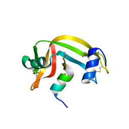
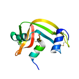
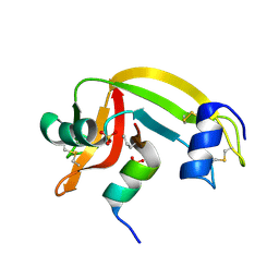
 | | Rnase s(f8a). mutant ribonucleasE S. | | 分子名称: | RNASE S, S PEPTIDE, SULFATE ION | | 著者 | Ratnaparkhi, G.S, Varadarajan, R. | | 登録日 | 1999-10-07 | | 公開日 | 1999-10-20 | | 最終更新日 | 2018-03-14 | | 実験手法 | X-RAY DIFFRACTION (2.25 Å) | | 主引用文献 | Thermodynamic and structural studies of cavity formation in proteins suggest that loss of packing interactions rather than the hydrophobic effect dominates the observed energetics.
Biochemistry, 39, 2000
|
|
1D5E
 | |
1FEV
 | |
7X7N
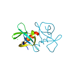
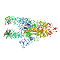 | | 3D model of the 3-RBD up single trimeric spike protein of SARS-CoV2 in the presence of synthetic peptide SIH-5. | | 分子名称: | 2-acetamido-2-deoxy-beta-D-glucopyranose, Spike glycoprotein, Synthetic peptide SIH-5 | | 著者 | Khatri, B, Pramanick, I, Malladi, S.K, Rajmani, R.S, Kumar, S, Ghosh, P, Sengupta, N, Rahisuddin, R, Kumaran, S, Ringe, R.P, Varadarajan, R, Dutta, S, Chatterjee, J. | | 登録日 | 2022-03-10 | | 公開日 | 2022-04-27 | | 最終更新日 | 2022-11-16 | | 実験手法 | ELECTRON MICROSCOPY (4.47 Å) | | 主引用文献 | A dimeric proteomimetic prevents SARS-CoV-2 infection by dimerizing the spike protein.
Nat.Chem.Biol., 18, 2022
|
|
1A19
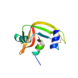
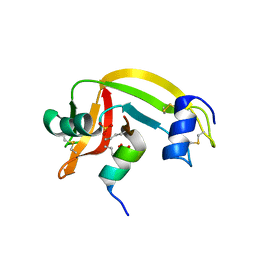
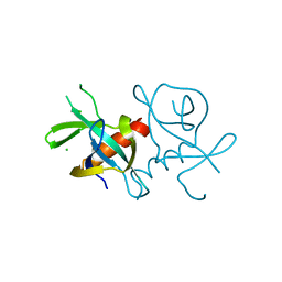
 | | BARSTAR (FREE), C82A MUTANT | | 分子名称: | BARSTAR | | 著者 | Ratnaparkhi, G.S, Varadarajan, R. | | 登録日 | 1997-12-25 | | 公開日 | 1998-04-08 | | 最終更新日 | 2024-05-22 | | 実験手法 | X-RAY DIFFRACTION (2.76 Å) | | 主引用文献 | Discrepancies between the NMR and X-ray structures of uncomplexed barstar: analysis suggests that packing densities of protein structures determined by NMR are unreliable.
Biochemistry, 37, 1998
|
|
2RNS
 | | REFINEMENT OF THE CRYSTAL STRUCTURE OF RIBONUCLEASE S. COMPARISON WITH AND BETWEEN THE VARIOUS RIBONUCLEASE A STRUCTURES | | 分子名称: | RIBONUCLEASE S, SULFATE ION | | 著者 | Kim, E.E, Varadarajan, R, Wyckoff, H.W, Richards, F.M. | | 登録日 | 1992-02-19 | | 公開日 | 1994-01-31 | | 最終更新日 | 2024-10-30 | | 実験手法 | X-RAY DIFFRACTION (1.6 Å) | | 主引用文献 | Refinement of the crystal structure of ribonuclease S. Comparison with and between the various ribonuclease A structures.
Biochemistry, 31, 1992
|
|
1KEB
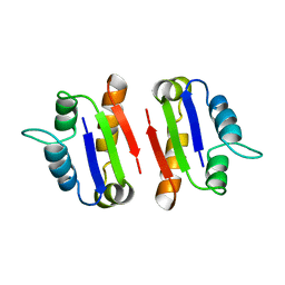
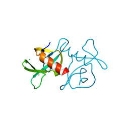
 | | Crystal Structure of Double Mutant M37L,P40S E.coli Thioredoxin | | 分子名称: | COPPER (II) ION, Thioredoxin 1 | | 著者 | Rudresh, Jain, R, Dani, V, Mitra, A, Srivastava, S, Sarma, S.P, Varadarajan, R, Ramakumar, S. | | 登録日 | 2001-11-15 | | 公開日 | 2002-11-13 | | 最終更新日 | 2023-08-16 | | 実験手法 | X-RAY DIFFRACTION (1.8 Å) | | 主引用文献 | Structural Consequences of Replacement of an alpha-helical Pro Residue in E.coli Thioredoxin
PROTEIN ENG., 15, 2002
|
|
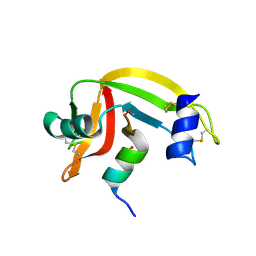
1Z3L
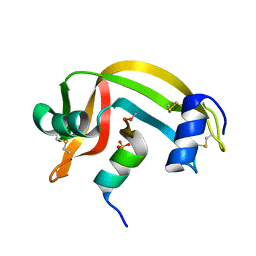
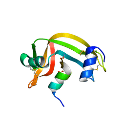
 | | X-Ray Crystal Structure of a Mutant Ribonuclease S (F8Anb) | | 分子名称: | Ribonuclease pancreatic, S-Peptide, S-Protein, ... | | 著者 | Das, M, Vasudeva Rao, B, Ghosh, S, Varadarajan, R. | | 登録日 | 2005-03-14 | | 公開日 | 2005-03-29 | | 最終更新日 | 2023-11-15 | | 実験手法 | X-RAY DIFFRACTION (1.8 Å) | | 主引用文献 | Attempts to delineate the relative contributions of changes in hydrophobicity and packing to changes in stability of ribonuclease S mutants.
Biochemistry, 44, 2005
|
|
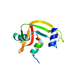
1Z3P
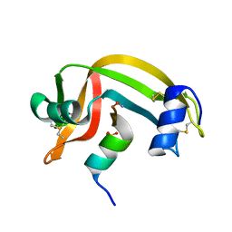
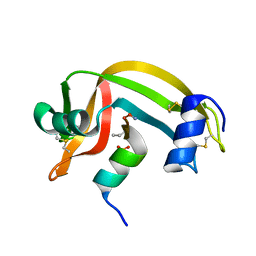
 | | X-Ray crystal structure of a mutant Ribonuclease S (M13Nva) | | 分子名称: | Ribonuclease pancreatic, S-Peptide, S-Protein, ... | | 著者 | Das, M, Rao, B.V, Ghosh, S, Varadarajan, R. | | 登録日 | 2005-03-14 | | 公開日 | 2005-03-29 | | 最終更新日 | 2023-10-25 | | 実験手法 | X-RAY DIFFRACTION (2 Å) | | 主引用文献 | Attempts to delineate the relative contributions of changes in hydrophobicity and packing to changes in stability of ribonuclease S mutants.
Biochemistry, 44, 2005
|
|
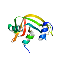
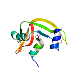
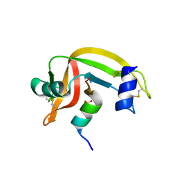
1Z3M
 | | Crystal structure of mutant Ribonuclease S (F8Nva) | | 分子名称: | Ribonuclease pancreatic, S-Peptide, S-protein, ... | | 著者 | Das, M, Vasudeva Rao, B, Ghosh, S, Varadarajan, R. | | 登録日 | 2005-03-14 | | 公開日 | 2005-03-29 | | 最終更新日 | 2023-11-15 | | 実験手法 | X-RAY DIFFRACTION (2 Å) | | 主引用文献 | Attempts to delineate the relative contributions of changes in hydrophobicity and packing to changes in stability of ribonuclease S mutants.
Biochemistry, 44, 2005
|
|
7EPG
 | |
7EPI
 | |
7EPJ
 | |
1J7Z
 | |
1J81
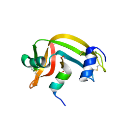
 | | Osmolyte Stabilization of RNase | | 分子名称: | RIBONUCLEASE PANCREATIC, SULFATE ION | | 著者 | Ratnaparkhi, G.S, Varadarajan, R. | | 登録日 | 2001-05-19 | | 公開日 | 2001-06-06 | | 最終更新日 | 2017-11-29 | | 実験手法 | X-RAY DIFFRACTION (2.2 Å) | | 主引用文献 | Osmolytes stabilize ribonuclease S by stabilizing its fragments S protein and S peptide to compact folding-competent states.
J.Biol.Chem., 276, 2001
|
|
1J80
 | | Osmolyte Stabilization of RNase | | 分子名称: | RIBONUCLEASE PANCREATIC, SULFATE ION | | 著者 | Ratnaparkhi, G.S, Varadarajan, R. | | 登録日 | 2001-05-19 | | 公開日 | 2001-06-06 | | 最終更新日 | 2024-10-30 | | 実験手法 | X-RAY DIFFRACTION (2.1 Å) | | 主引用文献 | Osmolytes stabilize ribonuclease S by stabilizing its fragments S protein and S peptide to compact folding-competent states.
J.Biol.Chem., 276, 2001
|
|


