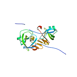
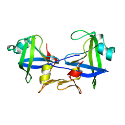
1Z96
 
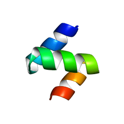 | | Crystal structure of the Mud1 UBA domain | | 分子名称: | UBA-domain protein mud1 | | 著者 | Trempe, J.-F, Brown, N.R, Lowe, E.D, Noble, M.E.M, Gordon, C, Campbell, I.D, Johnson, L.N, Endicott, J.A. | | 登録日 | 2005-03-31 | | 公開日 | 2005-10-04 | | 最終更新日 | 2024-02-14 | | 実験手法 | X-RAY DIFFRACTION (1.8 Å) | | 主引用文献 | Mechanism of Lys48-linked polyubiquitin chain recognition by the Mud1 UBA domain
Embo J., 24, 2005
|
|
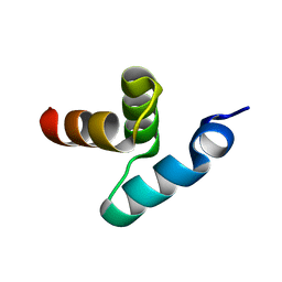
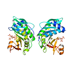
2BWE
 | | The crystal structure of the complex between the UBA and UBL domains of Dsk2 | | 分子名称: | DSK2 | | 著者 | Lowe, E.D, Hasan, N, Trempe, J.-F, Fonso, L, Noble, M.E.M, Endicott, J.A, Johnson, L.N, Brown, N.R. | | 登録日 | 2005-07-13 | | 公開日 | 2006-01-25 | | 最終更新日 | 2023-12-13 | | 実験手法 | X-RAY DIFFRACTION (3.1 Å) | | 主引用文献 | Structures of the Dsk2 Ubl and Uba Domains and Their Complex.
Acta Crystallogr.,Sect.D, 62, 2006
|
|
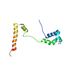
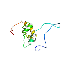
2BWB
 | | Crystal structure of the UBA domain of Dsk2 from S. cerevisiae | | 分子名称: | UBIQUITIN-LIKE PROTEIN DSK2 | | 著者 | Lowe, E.D, Hasan, N, Trempe, J.-F, Fonso, L, Noble, M.E.M, Endicott, J.A, Johnson, L.N, Brown, N.R. | | 登録日 | 2005-07-13 | | 公開日 | 2006-01-25 | | 最終更新日 | 2024-05-08 | | 実験手法 | X-RAY DIFFRACTION (2.3 Å) | | 主引用文献 | Structures of the Dsk2 Ubl and Uba Domains and Their Complex.
Acta Crystallogr.,Sect.D, 62, 2006
|
|
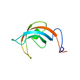
2BWF
 | | Crystal structure of the UBL domain of Dsk2 from S. cerevisiae | | 分子名称: | FORMIC ACID, UBIQUITIN-LIKE PROTEIN DSK2 | | 著者 | Lowe, E.D, Hasan, N, Trempe, J.-F, Fonso, L, Noble, M.E.M, Endicott, J.A, Johnson, L.N, Brown, N.R. | | 登録日 | 2005-07-13 | | 公開日 | 2006-01-25 | | 最終更新日 | 2023-12-13 | | 実験手法 | X-RAY DIFFRACTION (1.15 Å) | | 主引用文献 | Structures of the Dsk2 Ubl and Uba Domains and Their Complex.
Acta Crystallogr.,Sect.D, 62, 2006
|
|
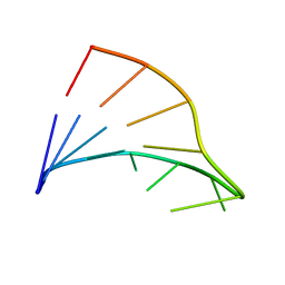
1L1P
 | | Solution Structure of the PPIase Domain from E. coli Trigger Factor | | 分子名称: | trigger factor | | 著者 | Kozlov, G, Trempe, J.-F, Perreault, A, Wong, M, Denisov, A, Ghandi, S, Gehring, K, Ekiel, I, Montreal-Kingston Bacterial Structural Genomics Initiative (BSGI) | | 登録日 | 2002-02-19 | | 公開日 | 2003-06-24 | | 最終更新日 | 2024-05-22 | | 実験手法 | SOLUTION NMR | | 主引用文献 | Solution Structure of the Closed Form of a Peptidyl-Prolyl Isomerase Reveals the Mechanism of Protein Folding
To be Published
|
|
5KES
 | | Solution structure of the yeast Ddi1 HDD domain | | 分子名称: | DNA damage-inducible protein 1 | | 著者 | Trempe, J.-F, Ratcliffe, C, Veverka, V, Saskova, K, Gehring, K. | | 登録日 | 2016-06-10 | | 公開日 | 2016-10-05 | | 最終更新日 | 2024-05-15 | | 実験手法 | SOLUTION NMR | | 主引用文献 | Structural studies of the yeast DNA damage-inducible protein Ddi1 reveal domain architecture of this eukaryotic protein family.
Sci Rep, 6, 2016
|
|
1FC8
 | |
4Z2Z
 | |
5VSX
 | |
5VSZ
 | |
2X5N
 | | Crystal Structure of the SpRpn10 VWA domain | | 分子名称: | 26S PROTEASOME REGULATORY SUBUNIT RPN10, SULFATE ION | | 著者 | Riedinger, C, Boehringer, J, Trempe, J.-F, Lowe, E.D, Brown, N.R, Gehring, K, Noble, M.E.M, Gordon, C, Endicott, J.A. | | 登録日 | 2010-02-10 | | 公開日 | 2010-08-25 | | 最終更新日 | 2024-05-08 | | 実験手法 | X-RAY DIFFRACTION (1.3 Å) | | 主引用文献 | The Structure of Rpn10 and its Interactions with Polyubiquitin Chains and the Proteasome Subunit Rpn12.
J.Biol.Chem., 285, 2010
|
|
9BJA
 | |
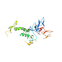
1G9L
 | | SOLUTION STRUCTURE OF THE PABC DOMAIN OF HUMAN POLY(A) BINDING PROTEIN | | 分子名称: | POLYADENYLATE-BINDING PROTEIN 1 | | 著者 | Kozlov, G, Trempe, J.-F, Khaleghpour, K, Kahvejian, A, Ekiel, I, Gehring, K. | | 登録日 | 2000-11-24 | | 公開日 | 2001-03-14 | | 最終更新日 | 2024-05-22 | | 実験手法 | SOLUTION NMR | | 主引用文献 | Structure and function of the C-terminal PABC domain of human poly(A)-binding protein.
Proc.Natl.Acad.Sci.USA, 98, 2001
|
|
6NBX
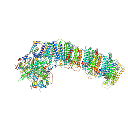 | | T.elongatus NDH (data-set 2) | | 分子名称: | IRON/SULFUR CLUSTER, NAD(P)H-quinone oxidoreductase chain 4 1, NAD(P)H-quinone oxidoreductase subunit 1, ... | | 著者 | Laughlin, T.G, Bayne, A, Trempe, J.-F, Savage, D.F, Davies, K.M. | | 登録日 | 2018-12-10 | | 公開日 | 2019-02-27 | | 最終更新日 | 2019-12-18 | | 実験手法 | ELECTRON MICROSCOPY (3.5 Å) | | 主引用文献 | Structure of the complex I-like molecule NDH of oxygenic photosynthesis.
Nature, 566, 2019
|
|
6NBQ
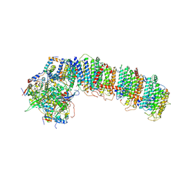 | | T.elongatus NDH (data-set 1) | | 分子名称: | IRON/SULFUR CLUSTER, NAD(P)H-quinone oxidoreductase chain 4 1, NAD(P)H-quinone oxidoreductase subunit 2, ... | | 著者 | Laughlin, T.G, Bayne, A, Trempe, J.-F, Savage, D.F, Davies, K.M. | | 登録日 | 2018-12-09 | | 公開日 | 2019-02-27 | | 最終更新日 | 2019-12-18 | | 実験手法 | ELECTRON MICROSCOPY (3.1 Å) | | 主引用文献 | Structure of the complex I-like molecule NDH of oxygenic photosynthesis.
Nature, 566, 2019
|
|
6NBY
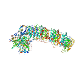 | | T.elongatus NDH (composite model) | | 分子名称: | IRON/SULFUR CLUSTER, NAD(P)H-quinone oxidoreductase chain 4 1, NAD(P)H-quinone oxidoreductase subunit 1, ... | | 著者 | Laughlin, T.G, Bayne, A, Trempe, J.-F, Savage, D.F, Davies, K.M. | | 登録日 | 2018-12-10 | | 公開日 | 2019-02-27 | | 最終更新日 | 2020-04-15 | | 実験手法 | ELECTRON MICROSCOPY (3.1 Å) | | 主引用文献 | Structure of the complex I-like molecule NDH of oxygenic photosynthesis.
Nature, 566, 2019
|
|
4K95
 | | Crystal Structure of Parkin | | 分子名称: | E3 ubiquitin-protein ligase parkin, ZINC ION | | 著者 | Seirafi, M, Menade, M, Sauve, V, Kozlov, G, Trempe, J.-F, Nagar, B, Gehring, K. | | 登録日 | 2013-04-19 | | 公開日 | 2013-05-15 | | 最終更新日 | 2023-09-20 | | 実験手法 | X-RAY DIFFRACTION (6.499 Å) | | 主引用文献 | Structure of parkin reveals mechanisms for ubiquitin ligase activation.
Science, 340, 2013
|
|
4K7D
 | | Crystal Structure of Parkin C-terminal RING domains | | 分子名称: | CHLORIDE ION, E3 ubiquitin-protein ligase parkin, MALONATE ION, ... | | 著者 | Sauve, V, Trempe, J.-F, Menade, M, Gehring, K. | | 登録日 | 2013-04-17 | | 公開日 | 2013-05-15 | | 最終更新日 | 2024-02-28 | | 実験手法 | X-RAY DIFFRACTION (2.8 Å) | | 主引用文献 | Structure of parkin reveals mechanisms for ubiquitin ligase activation.
Science, 340, 2013
|
|
4RGH
 | |


