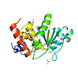
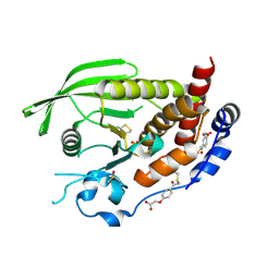
9DJ9
 
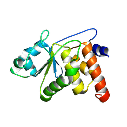 | | HUMAN VH1-RELATED DUAL-SPECIFICITY PHOSPHATASE (VHR) in distinct apo form | | 分子名称: | Dual specificity protein phosphatase 3 | | 著者 | Keedy, D.A, Lemberikman, A.M, Isiorho, E.A, Aleshin, A.E, Wu, J, Lambert, L.J, Cosford, N.D.P, Tautz, L. | | 登録日 | 2024-09-06 | | 公開日 | 2024-09-25 | | 最終更新日 | 2025-04-23 | | 実験手法 | X-RAY DIFFRACTION (1.924 Å) | | 主引用文献 | Fragment Screening Identifies Novel Allosteric Binders and Binding Sites in the VHR ( DUSP3 ) Phosphatase.
Acs Omega, 10, 2025
|
|
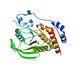
3F81
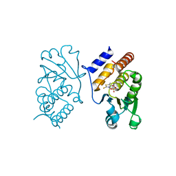 | | Interaction of VHR with SA3 | | 分子名称: | 2-[(5~{E})-5-[[3-[4-(2-fluoranylphenoxy)phenyl]-1-phenyl-pyrazol-4-yl]methylidene]-4-oxidanylidene-2-sulfanylidene-1,3-thiazolidin-3-yl]ethanesulfonic acid, Dual specificity protein phosphatase 3 | | 著者 | Wu, S, Mutelin, T, Tautz, L. | | 登録日 | 2008-11-11 | | 公開日 | 2009-11-10 | | 最終更新日 | 2023-09-06 | | 実験手法 | X-RAY DIFFRACTION (1.9 Å) | | 主引用文献 | Multidentate small-molecule inhibitors of vaccinia H1-related (VHR) phosphatase decrease proliferation of cervix cancer cells.
J.Med.Chem., 52, 2009
|
|
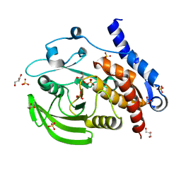
8TK2
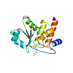 | | HUMAN VH1-RELATED DUAL-SPECIFICITY PHOSPHATASE (VHR) complexed with 2-((2,4-difluorobenzyl)amino)-2-oxoacetic acid | | 分子名称: | DI(HYDROXYETHYL)ETHER, DIMETHYL SULFOXIDE, Dual specificity protein phosphatase 3, ... | | 著者 | Aleshin, A.E, Wu, J, Lambert, L.J, Cosford, N.D.P, Tautz, L. | | 登録日 | 2023-07-25 | | 公開日 | 2024-08-14 | | 最終更新日 | 2025-02-26 | | 実験手法 | X-RAY DIFFRACTION (1.7 Å) | | 主引用文献 | Fragment Screening Identifies Novel Allosteric Binders and Binding Sites in the VHR ( DUSP3 ) Phosphatase.
Acs Omega, 10, 2025
|
|
8TK5
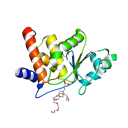 | | HUMAN VH1-RELATED DUAL-SPECIFICITY PHOSPHATASE (VHR) complexed with HEPES | | 分子名称: | 3,6,9,12,15,18,21-HEPTAOXATRICOSANE-1,23-DIOL, 4-(2-HYDROXYETHYL)-1-PIPERAZINE ETHANESULFONIC ACID, DI(HYDROXYETHYL)ETHER, ... | | 著者 | Aleshin, A.E, Wu, J, Lambert, L.J, Cosford, N.D.P, Tautz, L. | | 登録日 | 2023-07-25 | | 公開日 | 2024-08-14 | | 最終更新日 | 2025-02-26 | | 実験手法 | X-RAY DIFFRACTION (1.9 Å) | | 主引用文献 | Fragment Screening Identifies Novel Allosteric Binders and Binding Sites in the VHR ( DUSP3 ) Phosphatase.
Acs Omega, 10, 2025
|
|
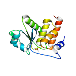
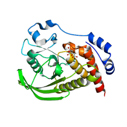
8TK3
 | | HUMAN VH1-RELATED DUAL-SPECIFICITY PHOSPHATASE (VHR) having oxidized catalytic cysteine and complexed with 6-(difluoromethyl)pyrimidin-4-ol at two allosteric sites | | 分子名称: | 6-(difluoromethyl)pyrimidin-4-ol, DIMETHYL SULFOXIDE, Dual specificity protein phosphatase 3 | | 著者 | Aleshin, A.E, Wu, J, Lambert, L.J, Cosford, N.D.P, Tautz, L. | | 登録日 | 2023-07-25 | | 公開日 | 2024-08-14 | | 最終更新日 | 2025-02-26 | | 実験手法 | X-RAY DIFFRACTION (2 Å) | | 主引用文献 | Fragment Screening Identifies Novel Allosteric Binders and Binding Sites in the VHR ( DUSP3 ) Phosphatase.
Acs Omega, 10, 2025
|
|
8TK6
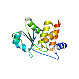 | | HUMAN VH1-RELATED DUAL-SPECIFICITY PHOSPHATASE (VHR) in apo form | | 分子名称: | Dual specificity protein phosphatase 3 | | 著者 | Aleshin, A.E, Wu, J, Lambert, L.J, Cosford, N.D.P, Tautz, L. | | 登録日 | 2023-07-25 | | 公開日 | 2024-08-14 | | 最終更新日 | 2025-02-26 | | 実験手法 | X-RAY DIFFRACTION (1.65 Å) | | 主引用文献 | Fragment Screening Identifies Novel Allosteric Binders and Binding Sites in the VHR ( DUSP3 ) Phosphatase.
Acs Omega, 10, 2025
|
|
8TK4
 | | HUMAN VH1-RELATED DUAL-SPECIFICITY PHOSPHATASE (VHR) complexed with phosphate | | 分子名称: | Dual specificity protein phosphatase 3, PHOSPHATE ION | | 著者 | Aleshin, A.E, Wu, J, Lambert, L.J, Cosford, N.D.P, Tautz, L. | | 登録日 | 2023-07-25 | | 公開日 | 2024-08-14 | | 最終更新日 | 2025-02-26 | | 実験手法 | X-RAY DIFFRACTION (1.8 Å) | | 主引用文献 | Fragment Screening Identifies Novel Allosteric Binders and Binding Sites in the VHR ( DUSP3 ) Phosphatase.
Acs Omega, 10, 2025
|
|
2F46
 | |
6YID
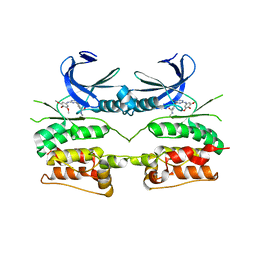 | | Crystal structure of ULK2 in complex with SBI-0206965 | | 分子名称: | 2-({5-bromo-2-[(3,4,5-trimethoxyphenyl)amino]pyrimidin-4-yl}oxy)-N-methylbenzene-1-carboximidic acid, Serine/threonine-protein kinase ULK2 | | 著者 | Chaikuad, A, Ren, H, Bakas, N.A, Lambert, L.J, Cosford, N.D.P, Knapp, S, Structural Genomics Consortium (SGC) | | 登録日 | 2020-04-01 | | 公開日 | 2020-06-17 | | 最終更新日 | 2024-01-24 | | 実験手法 | X-RAY DIFFRACTION (2.7 Å) | | 主引用文献 | Design, Synthesis, and Characterization of an Orally Active Dual-Specific ULK1/2 Autophagy Inhibitor that Synergizes with the PARP Inhibitor Olaparib for the Treatment of Triple-Negative Breast Cancer.
J.Med.Chem., 63, 2020
|
|
1ZC0
 | |
3O4U
 | |
9EEZ
 | | STEP (PTPN5) with active-site disulfide bond and covalent ligand bound to distal C505 and C518 | | 分子名称: | DIMETHYL SULFOXIDE, SULFATE ION, Tyrosine-protein phosphatase non-receptor type 5, ... | | 著者 | Guerrero, L, Ebrahim, A, Riley, B.T, Keedy, D.A. | | 登録日 | 2024-11-19 | | 公開日 | 2024-12-11 | | 最終更新日 | 2025-05-07 | | 実験手法 | X-RAY DIFFRACTION (1.6 Å) | | 主引用文献 | Three STEPs forward: A trio of unexpected structures of PTPN5.
Biorxiv, 2025
|
|
9EEY
 | |
9EEX
 | |
3O4S
 | |
3O4T
 | |


