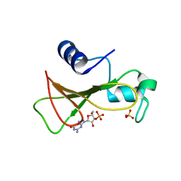
4HAA
 
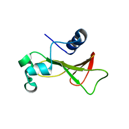 | | Structure of Ribonuclease Binase Glu43Ala/Phe81Ala Mutant | | 分子名称: | Ribonuclease | | 著者 | Polyakov, K.M, Trofimov, A.A, Mitchevich, V.A, Dorovatovskii, P.V, Schulga, A.A, Makarov, A.A, Tkach, E.N, Goncharuk, D.A. | | 登録日 | 2012-09-26 | | 公開日 | 2012-10-17 | | 最終更新日 | 2023-09-20 | | 実験手法 | X-RAY DIFFRACTION (1.9 Å) | | 主引用文献 | Structure and functional studies of the ribonuclease binase Glu43Ala/Phe81Ala mutant.
Acta Crystallogr.,Sect.D, 69, 2013
|
|
1FW7
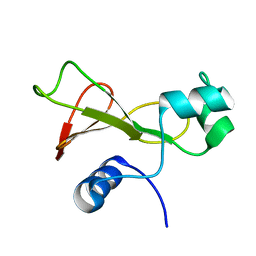 | |
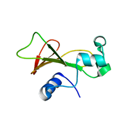
1GOV
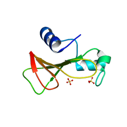 | | RIBONUCLEASE BI(G SPECIFIC ENDONUCLEASE) COMPLEXED WITH SULFATE IONS | | 分子名称: | RIBONUCLEASE, SULFATE ION | | 著者 | Polyakov, K.M, Lebedev, A.A, Pavlovsky, A.G, Sanishvili, R.G, Dodson, G.G. | | 登録日 | 2001-10-26 | | 公開日 | 2001-11-29 | | 最終更新日 | 2024-05-08 | | 実験手法 | X-RAY DIFFRACTION (2 Å) | | 主引用文献 | The Structure of Substrate-Free Microbial Ribonuclease Binase and of its Complexes with 3'Gmp and Sulfate Ions
Acta Crystallogr.,Sect.D, 58, 2002
|
|
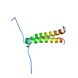
2KPF
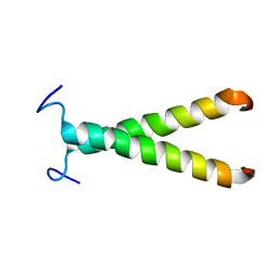
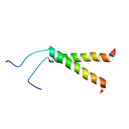 | | Spatial structure of the dimeric transmembrane domain of glycophorin A in bicelles soluton | | 分子名称: | Glycophorin-A | | 著者 | Mineev, K.S, Bocharov, E.V, Goncharuk, M.V, Arseniev, A.S, Volynsky, P.E, Efremov, R.G. | | 登録日 | 2009-10-13 | | 公開日 | 2010-09-22 | | 最終更新日 | 2024-05-01 | | 実験手法 | SOLUTION NMR | | 主引用文献 | Dimeric structure of the transmembrane domain of glycophorin a in lipidic and detergent environments.
Acta Naturae, 3, 2011
|
|
1GOY
 | | HYDROLASE(ENDORIBONUCLEASE)RIBONUCLEASE BI(G SPECIFIC ENDONUCLEASE) (E.C.3.1.27.-) COMPLEXED WITH GUANOSINE-3'-PHOSPHATE (3'-GMP) | | 分子名称: | GUANOSINE-3'-MONOPHOSPHATE, RIBONUCLEASE, SULFATE ION | | 著者 | Polyakov, K.M, Lebedev, A.A, Pavlovsky, A.G, Sanishvili, R.G, Dodson, G.G. | | 登録日 | 2001-10-26 | | 公開日 | 2001-11-29 | | 最終更新日 | 2024-05-08 | | 実験手法 | X-RAY DIFFRACTION (2 Å) | | 主引用文献 | The Structure of Substrate-Free Microbial Ribonuclease Binase and of its Complexes with 3'Gmp and Sulfate Ions
Acta Crystallogr.,Sect.D, 58, 2002
|
|
1GOU
 | |
2J5D
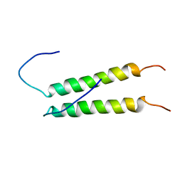
 | | NMR structure of BNIP3 transmembrane domain in lipid bicelles | | 分子名称: | BCL2/ADENOVIRUS E1B 19 KDA PROTEIN-INTERACTING PROTEIN 3 | | 著者 | Bocharov, E.V, Pustovalova, Y.E, Volynsky, P.E, Maslennikov, I.V, Goncharuk, M.V, Ermolyuk, Y.S, Arseniev, A.S. | | 登録日 | 2006-09-14 | | 公開日 | 2007-04-17 | | 最終更新日 | 2024-05-15 | | 実験手法 | SOLUTION NMR | | 主引用文献 | Unique dimeric structure of BNip3 transmembrane domain suggests membrane permeabilization as a cell death trigger.
J. Biol. Chem., 282, 2007
|
|
2K1L
 | |
2K1K
 | |
2K9Y
 | |
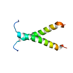
2KPE
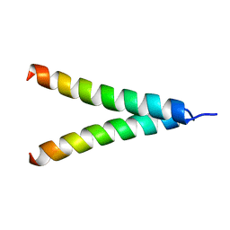 | | Refined structure of Glycophorin A transmembrane segment dimer in DPC micelles | | 分子名称: | Glycophorin-A | | 著者 | Mineev, K.S, Bocharov, E.V, Goncharuk, M.V, Arseniev, A.S, Volynsky, P.E, Efremov, R.G. | | 登録日 | 2009-10-13 | | 公開日 | 2010-09-22 | | 最終更新日 | 2024-05-01 | | 実験手法 | SOLUTION NMR | | 主引用文献 | Dimeric structure of the transmembrane domain of glycophorin a in lipidic and detergent environments.
Acta Naturae, 3, 2011
|
|


