Loading
PDBj
メニューPDBj@FacebookPDBj@TwitterPDBj@YouTubewwPDB FoundationwwPDB
RCSB PDBPDBeBMRBAdv. SearchSearch help
Search by PDB author
6GUA
DownloadVisualize
BU of 6gua by Molmil
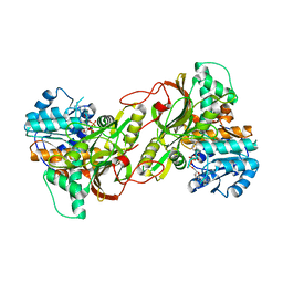
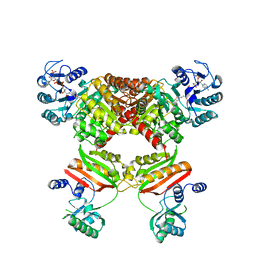
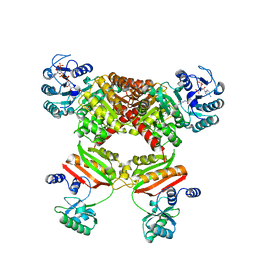
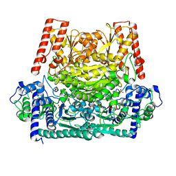
Xylulose 5-phosphate phosphoketolase from Lactococcus lactis
分子名称: MAGNESIUM ION, PHOSPHATE ION, Probable phosphoketolase, ...
著者Scheidig, A.J, Szedlacsek, S.E.
登録日2018-06-19
公開日2019-05-22
最終更新日2024-01-17
実験手法SOLUTION SCATTERING (1.95 Å), X-RAY DIFFRACTION
主引用文献Crystal structure of a xylulose 5-phosphate phosphoketolase. Insights into the substrate specificity for xylulose 5-phosphate.
J.Struct.Biol., 207, 2019
1CA0
DownloadVisualize
BU of 1ca0 by Molmil
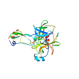
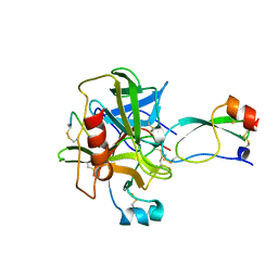
BOVINE CHYMOTRYPSIN COMPLEXED TO APPI
分子名称: BOVINE CHYMOTRYPSIN, PROTEASE INHIBITOR DOMAIN OF ALZHEIMER'S AMYLOID BETA-PROTEIN PRECURSOR
著者Scheidig, A.J, Kossiakoff, A.A.
登録日1997-01-23
公開日1997-07-23
最終更新日2024-10-30
実験手法X-RAY DIFFRACTION (2.1 Å)
主引用文献Crystal structures of bovine chymotrypsin and trypsin complexed to the inhibitor domain of Alzheimer's amyloid beta-protein precursor (APPI) and basic pancreatic trypsin inhibitor (BPTI): engineering of inhibitors with altered specificities.
Protein Sci., 6, 1997
4W8G
DownloadVisualize
BU of 4w8g by Molmil
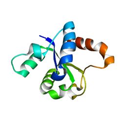
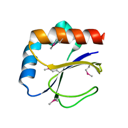
Crystal structure of the TIR domain of the Toll-related Receptor TRR-2 from the lower metazoan Hydra magnipapillata (crystal form I)
分子名称: Toll-receptor-related 2
著者Weisse, R.H.-J, Scheidig, A.J.
登録日2014-08-24
公開日2015-09-09
最終更新日2024-01-10
実験手法X-RAY DIFFRACTION (1.95 Å)
主引用文献Crystal structure of the TIR domain of the Toll-related Receptor TRR-2 from the lower metazoan Hydra magnipapillata (crystal form I)
To Be Published
821P
DownloadVisualize
BU of 821p by Molmil
THREE-DIMENSIONAL STRUCTURES AND PROPERTIES OF A TRANSFORMING AND A NONTRANSFORMING GLYCINE-12 MUTANT OF P21H-RAS
分子名称: C-H-RAS P21 PROTEIN, MAGNESIUM ION, PHOSPHOAMINOPHOSPHONIC ACID-GUANYLATE ESTER
著者Scheidig, A.J, Krengel, U, Pai, E.F, Kabsch, W, Wittinghofer, A, Goody, R.S.
登録日1993-03-29
公開日1994-01-31
最終更新日2024-02-14
実験手法X-RAY DIFFRACTION (1.5 Å)
主引用文献Three-dimensional structures and properties of a transforming and a nontransforming glycine-12 mutant of p21H-ras.
Biochemistry, 32, 1993
4PNC
DownloadVisualize
BU of 4pnc by Molmil
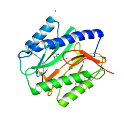
E. COLI METHIONINE AMINOPEPTIDASE IN COMPLEX WITH INHIBITOR 7-METHOXY-2-METHYLEN-3,4-DIHYDRONAPHTHALEN-1(2H)-ONE
分子名称: (2S)-7-methoxy-2-methyl-3,4-dihydronaphthalen-1(2H)-one, COBALT (II) ION, Methionine aminopeptidase, ...
著者Scheidig, A.J, Altmeyer, M, Klein, C.D.
登録日2014-05-23
公開日2014-07-23
最終更新日2023-12-27
実験手法X-RAY DIFFRACTION (1.54 Å)
主引用文献Beta-aminoketones as prodrugs for selective irreversible inhibitors of type-1 methionine aminopeptidases.
Bioorg.Med.Chem.Lett., 24, 2014
5OJG
DownloadVisualize
BU of 5ojg by Molmil
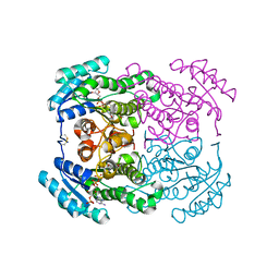
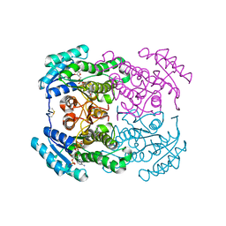
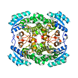
Crystal structure of the dehydrogenase/reductase SDR family member 4 (DHRS4) from Caenorhabditis elegans
分子名称: Dehydrogenase/reductase SDR family member 4, NADP NICOTINAMIDE-ADENINE-DINUCLEOTIDE PHOSPHATE, butane-2,3-dione
著者Scheidig, A.J, Faust, A, Ebert, B, Maser, E, Kisiela, M.
登録日2017-07-21
公開日2017-11-22
最終更新日2024-01-17
実験手法X-RAY DIFFRACTION (1.9 Å)
主引用文献Crystal structure and catalytic characterization of the dehydrogenase/reductase SDR family member 4 (DHRS4) from Caenorhabditis elegans.
FEBS J., 285, 2018
5OJI
DownloadVisualize
BU of 5oji by Molmil
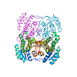
Crystal structure of the dehydrogenase/reductase SDR family member 4 (DHRS4) from Caenorhabditis elegans
分子名称: Dehydrogenase/reductase SDR family member 4, ISATIN, NADP NICOTINAMIDE-ADENINE-DINUCLEOTIDE PHOSPHATE
著者Scheidig, A.J, Faust, A, Ebert, B, Maser, E, Kisiela, M.
登録日2017-07-21
公開日2017-11-22
最終更新日2024-01-17
実験手法X-RAY DIFFRACTION (1.6 Å)
主引用文献Crystal structure and catalytic characterization of the dehydrogenase/reductase SDR family member 4 (DHRS4) from Caenorhabditis elegans.
FEBS J., 285, 2018
1CBW
DownloadVisualize
BU of 1cbw by Molmil
BOVINE CHYMOTRYPSIN COMPLEXED TO BPTI
分子名称: BOVINE CHYMOTRYPSIN, BPTI, SULFATE ION
著者Hynes, T.R, Scheidig, A.J, Kossiakoff, A.A.
登録日1996-12-22
公開日1997-07-23
最終更新日2023-08-09
実験手法X-RAY DIFFRACTION (2.6 Å)
主引用文献Crystal structures of bovine chymotrypsin and trypsin complexed to the inhibitor domain of Alzheimer's amyloid beta-protein precursor (APPI) and basic pancreatic trypsin inhibitor (BPTI): engineering of inhibitors with altered specificities.
Protein Sci., 6, 1997
6S65
DownloadVisualize
BU of 6s65 by Molmil
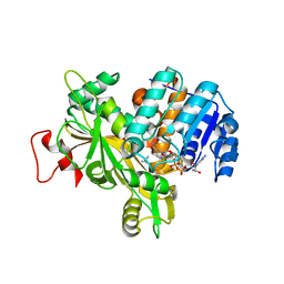
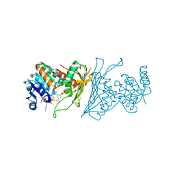
Crystal Structure of the Homospermidine Synthase (HSS) variant N135F from Blastochloris viridis in Complex with NAD
分子名称: Homospermidine synthase, NICOTINAMIDE-ADENINE-DINUCLEOTIDE
著者Helfrich, F, Scheidig, A.J.
登録日2019-07-02
公開日2020-07-15
最終更新日2024-01-24
実験手法X-RAY DIFFRACTION (1.75 Å)
主引用文献Crystal Structure of the Homospermidine Synthase (HSS) variant N135F from Blastochloris viridis in Complex with NAD
To Be Published
2BME
DownloadVisualize
BU of 2bme by Molmil
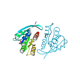
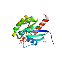
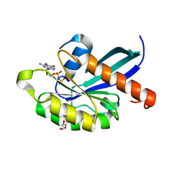
high resolution structure of GppNHp-bound human Rab4a
分子名称: 2-AMINO-2-HYDROXYMETHYL-PROPANE-1,3-DIOL, BETA-MERCAPTOETHANOL, MAGNESIUM ION, ...
著者Scheidig, A.J, Huber, S.K.
登録日2005-03-13
公開日2005-04-25
最終更新日2023-12-13
実験手法X-RAY DIFFRACTION (1.57 Å)
主引用文献High Resolution Crystal Structures of Human Rab4A in its Active and Inactive Conformations.
FEBS Lett., 579, 2005
2BMD
DownloadVisualize
BU of 2bmd by Molmil
high resolution structure of GDP-bound human Rab4a
分子名称: GLYCEROL, GUANOSINE-5'-DIPHOSPHATE, RAS-RELATED PROTEIN RAB4A
著者Scheidig, A.J, Huber, S.K.
登録日2005-03-13
公開日2005-04-25
最終更新日2024-05-08
実験手法X-RAY DIFFRACTION (1.8 Å)
主引用文献High Resolution Crystal Structures of Human Rab4A in its Active and Inactive Conformations.
FEBS Lett., 579, 2005
4W8H
DownloadVisualize
BU of 4w8h by Molmil
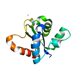
Crystal structure of the TIR domain of the Toll-related Receptor TRR-2 from the lower metazoan Hydra magnipapillata (crystal form II)
分子名称: CHLORIDE ION, Toll-receptor-related 2, hexa-His tag
著者Weisse, R.H.-J, Scheidig, A.J.
登録日2014-08-24
公開日2015-09-09
最終更新日2024-01-10
実験手法X-RAY DIFFRACTION (1.14 Å)
主引用文献Crystal structure of the TIR domain of the Toll-related Receptor TRR-2 from the lower metazoan Hydra magnipapillata (crystal form II)
To Be Published
4XYM
DownloadVisualize
BU of 4xym by Molmil
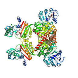
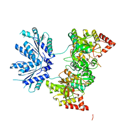
Ca. Korarchaeum cryptofilum dinucleotide forming Acetyl-coenzyme A synthetase 1 in complex with coenzyme A, Ca-AMPCP and HgCl+
分子名称: CALCIUM ION, CHLORIDE ION, COENZYME A, ...
著者Weisse, R.H.-J, Scheidig, A.J.
登録日2015-02-02
公開日2016-01-27
最終更新日2024-05-08
実験手法X-RAY DIFFRACTION (1.9 Å)
主引用文献Structure of NDP-forming Acetyl-CoA synthetase ACD1 reveals a large rearrangement for phosphoryl transfer.
Proc.Natl.Acad.Sci.USA, 113, 2016
4XZ3
DownloadVisualize
BU of 4xz3 by Molmil
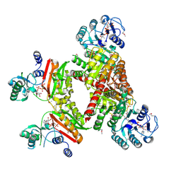
Ca. Korarchaeum cryptofilum dinucleotide forming Acetyl-coenzyme A synthetase 1 (Se-Met derivative) in complex with coenzyme A and Mg-AMPPCP, phosphohistidine segment pointing towards nucleotide binding site
分子名称: Acyl-CoA synthetase (NDP forming), COENZYME A, MAGNESIUM ION, ...
著者Weisse, R.H.-J, Scheidig, A.J.
登録日2015-02-03
公開日2016-01-27
最終更新日2016-02-10
実験手法X-RAY DIFFRACTION (2.398 Å)
主引用文献Structure of NDP-forming Acetyl-CoA synthetase ACD1 reveals a large rearrangement for phosphoryl transfer.
Proc.Natl.Acad.Sci.USA, 113, 2016
4XYL
DownloadVisualize
BU of 4xyl by Molmil
Ca. Korarchaeum cryptofilum ACD1 in complex with coenzyme A
分子名称: CHLORIDE ION, COENZYME A, alpha subunit of Acyl-CoA synthetase (NDP forming), ...
著者Weisse, R.H.-J, Scheidig, A.J.
登録日2015-02-02
公開日2016-01-27
最終更新日2024-01-10
実験手法X-RAY DIFFRACTION (1.95 Å)
主引用文献Structure of NDP-forming Acetyl-CoA synthetase ACD1 reveals a large rearrangement for phosphoryl transfer.
Proc.Natl.Acad.Sci.USA, 113, 2016
2NP7
DownloadVisualize
BU of 2np7 by Molmil
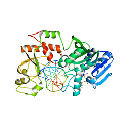
Crystal structure of the adenine-specific DNA methyltransferase M.TaqI complexed with the cofactor analog AETA and a 10 bp DNA containing an abasic site analog at the target position and pyrrolo-dC at the target base partner position
分子名称: 5'-D(*GP*AP*CP*AP*(4PC)P*CP*GP*(6MA)P*AP*C)-3', 5'-D(*GP*TP*TP*CP*GP*(3DR)P*TP*GP*TP*C)-3', 5'-DEOXY-5'-[2-(AMINO)ETHYLTHIO]ADENOSINE, ...
著者Lenz, T, Scheidig, A.J, Weinhold, E.
登録日2006-10-26
公開日2007-11-13
最終更新日2023-08-30
実験手法X-RAY DIFFRACTION (1.9 Å)
主引用文献Caught in the act II: Visualization of an intermediate in the DNA base-flipping pathway induced by the adenine-specific DNA methyltransferase M.TaqI
To be Published
2NP6
DownloadVisualize
BU of 2np6 by Molmil
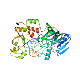
Crystal structure of the adenine-specific DNA methyltransferase M.TaqI complexed with the cofactor analog AETA and a 10 bp DNA containing an abasic site analog at the target position
分子名称: 5'-D(*GP*AP*CP*AP*TP*CP*GP*(6MA)P*AP*C)-3', 5'-D(*GP*TP*TP*CP*GP*(3DR)P*TP*GP*TP*C)-3', 5'-DEOXY-5'-[2-(AMINO)ETHYLTHIO]ADENOSINE, ...
著者Lenz, T, Scheidig, A.J, Weinhold, E.
登録日2006-10-26
公開日2007-11-13
最終更新日2023-08-30
実験手法X-RAY DIFFRACTION (2.1 Å)
主引用文献Caught in the act II: Visualization of an intermediate in the DNA base-flipping pathway induced by the adenine-specific DNA methyltransferase M.TaqI
To be Published
6Y87
DownloadVisualize
BU of 6y87 by Molmil
Crystal Structure of the Homospermidine Synthase (HSS) from Pseudomonas aeruginosa in Complex with NAD and PUT
分子名称: 1,4-DIAMINOBUTANE, Homospermidine synthase, NICOTINAMIDE-ADENINE-DINUCLEOTIDE
著者Helfrich, F, Scheidig, A.J.
登録日2020-03-03
公開日2021-03-31
最終更新日2024-01-24
実験手法X-RAY DIFFRACTION (2.15 Å)
主引用文献Structural and catalytic characterization of Blastochloris viridis and Pseudomonas aeruginosa homospermidine synthases supports the essential role of cation-pi interaction.
Acta Crystallogr D Struct Biol, 77, 2021
5HBR
DownloadVisualize
BU of 5hbr by Molmil
Ca. Korarchaeum cryptofilum dinucleotide forming Acetyl-coenzyme A synthetase 1 in complex with phosphate and coenzyme A
分子名称: COENZYME A, MAGNESIUM ION, PHOSPHATE ION, ...
著者Weisse, R.H.-J, Scheidig, A.J.
登録日2016-01-02
公開日2016-01-27
最終更新日2024-05-08
実験手法X-RAY DIFFRACTION (1.995 Å)
主引用文献Structure of NDP-forming Acetyl-CoA synthetase ACD1 reveals a large rearrangement for phosphoryl transfer.
Proc.Natl.Acad.Sci.USA, 113, 2016
3LQF
DownloadVisualize
BU of 3lqf by Molmil
Crystal structure of the short-chain dehydrogenase Galactitol-Dehydrogenase (GatDH) of Rhodobacter sphaeroides in complex with NAD and erythritol
分子名称: Galactitol dehydrogenase, MAGNESIUM ION, MESO-ERYTHRITOL, ...
著者Carius, Y, Christian, H, Faust, A, Kornberger, P, Zander, U, Klink, B.U, Kohring, G.W, Giffhorn, F, Scheidig, A.J.
登録日2010-02-09
公開日2010-04-21
最終更新日2024-04-03
実験手法X-RAY DIFFRACTION (1.8 Å)
主引用文献Structural insight into substrate differentiation of the sugar-metabolizing enzyme galactitol dehydrogenase from Rhodobacter sphaeroides D.
J.Biol.Chem., 285, 2010
2WDZ
DownloadVisualize
BU of 2wdz by Molmil
Crystal structure of the short chain dehydrogenase Galactitol- Dehydrogenase (GatDH) of Rhodobacter sphaeroides in complex with NAD+ and 1,2-Pentandiol
分子名称: (2S)-pentane-1,2-diol, MAGNESIUM ION, NICOTINAMIDE-ADENINE-DINUCLEOTIDE, ...
著者Carius, Y, Christian, H, Faust, A, Kornberger, P, Kohring, G.W, Giffhorn, F, Scheidig, A.J.
登録日2009-03-27
公開日2010-03-31
最終更新日2024-05-01
実験手法X-RAY DIFFRACTION (1.95 Å)
主引用文献Structural Insight Into Substrate Differentiation of the Sugar-Metabolizing Enzyme Galactitol Dehydrogenase from Rhodobacter Sphaeroides D.
J.Biol.Chem., 285, 2010
4KOA
DownloadVisualize
BU of 4koa by Molmil
Crystal Structure Analysis of 1,5-anhydro-D-fructose reductase from Sinorhizobium meliloti
分子名称: 1,5-anhydro-D-fructose reductase, NADPH DIHYDRO-NICOTINAMIDE-ADENINE-DINUCLEOTIDE PHOSPHATE
著者Schu, M, Faust, A, Stosik, B, Kohring, G.-W, Giffhorn, F, Scheidig, A.J.
登録日2013-05-11
公開日2013-08-07
最終更新日2023-09-20
実験手法X-RAY DIFFRACTION (1.93 Å)
主引用文献The structure of substrate-free 1,5-anhydro-D-fructose reductase from Sinorhizobium meliloti 1021 reveals an open enzyme conformation.
Acta Crystallogr.,Sect.F, 69, 2013
3DML
DownloadVisualize
BU of 3dml by Molmil
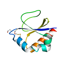
Crystal structure of the periplasmic thioredoxin SoxS from Paracoccus pantotrophus (reduced form)
分子名称: Putative uncharacterized protein
著者Carius, Y, Friedrich, C.G, Scheidig, A.J.
登録日2008-07-01
公開日2008-08-26
最終更新日2024-10-16
実験手法X-RAY DIFFRACTION (1.9 Å)
主引用文献Crystal structure of the periplasmic thioredoxin SoxS from Paracoccus pantotrophus indicates a dual Trx/Grx functionality for activation of chemotrophic sulfur oxidation in vivo
To be Published
3D4T
DownloadVisualize
BU of 3d4t by Molmil
Crystal structure of the periplasmic thioredoxin SoxS from Paracoccus pantotrophus (oxidized form)
分子名称: BETA-MERCAPTOETHANOL, Putative uncharacterized protein
著者Carius, Y, Friedrich, C.G, Scheidig, A.J.
登録日2008-05-15
公開日2008-07-15
最終更新日2024-04-03
実験手法X-RAY DIFFRACTION (2.05 Å)
主引用文献Crystal structure of the periplasmic thioredoxin SoxS from Paracoccus pantotrophus indicates a dual Trx/Grx functionality for activation of chemotrophic sulfur oxidation in vivo
To be Published
1X6V
DownloadVisualize
BU of 1x6v by Molmil
The crystal structure of human 3'-phosphoadenosine-5'-phosphosulfate synthetase 1
分子名称: ADENOSINE-5'-DIPHOSPHATE, Bifunctional 3'-phosphoadenosine 5'-phosphosulfate synthetase 1, CHLORIDE ION
著者Harjes, S, Bayer, P, Scheidig, A.J.
登録日2004-08-12
公開日2005-03-22
最終更新日2023-08-23
実験手法X-RAY DIFFRACTION (1.75 Å)
主引用文献The Crystal Structure of Human PAPS Synthetase 1 Reveals Asymmetry in Substrate Binding
J.Mol.Biol., 347, 2005


1234>

226707

件を2024-10-30に公開中

PDB statisticsPDBj update infoContact PDBjnumon