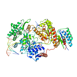
8P1P
 
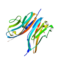
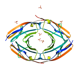
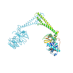 | | USP28 in complex with AZ1 | | 分子名称: | 2-[[5-bromanyl-2-[[4-fluoranyl-3-(trifluoromethyl)phenyl]methoxy]phenyl]methylamino]ethanol, CHLORIDE ION, DIMETHYL SULFOXIDE, ... | | 著者 | Sauer, F, Karal-Nair, R, Kisker, C. | | 登録日 | 2023-05-12 | | 公開日 | 2024-05-22 | | 実験手法 | X-RAY DIFFRACTION (2.76 Å) | | 主引用文献 | USP28 in complex with AZ1
To Be Published
|
|
6ZVA
 | |
8P14
 | |
8P1Q
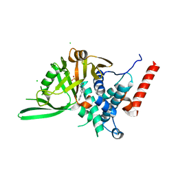
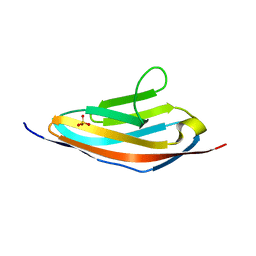
 | | USP28 in complex with FT206 | | 分子名称: | 3-azanyl-N-[(2S)-6-[(1S,5R)-3,8-diazabicyclo[3.2.1]octan-3-yl]-1,2,3,4-tetrahydronaphthalen-2-yl]-6-methyl-thieno[2,3-b]pyridine-2-carboxamide, DIMETHYL SULFOXIDE, Ubiquitin carboxyl-terminal hydrolase 28 | | 著者 | Sauer, F, Karal Nair, R, Kisker, C. | | 登録日 | 2023-05-12 | | 公開日 | 2024-05-22 | | 実験手法 | X-RAY DIFFRACTION (2.79 Å) | | 主引用文献 | USP28 in complex with FT206
To Be Published
|
|
8P19
 | |
3KNB
 | |
3Q5O
 | |
3QP3
 | |
3PUC
 | |
6TUN
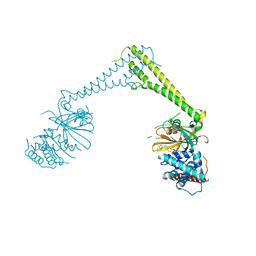
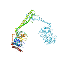
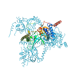
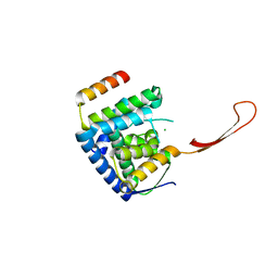 | | Helicase domain complex | | 分子名称: | CDK-activating kinase assembly factor MAT1, CHLORIDE ION, General transcription and DNA repair factor IIH helicase subunit XPD | | 著者 | Sauer, F, Kisker, C. | | 登録日 | 2020-01-07 | | 公開日 | 2020-11-11 | | 最終更新日 | 2024-05-15 | | 実験手法 | X-RAY DIFFRACTION (2.07 Å) | | 主引用文献 | In TFIIH the Arch domain of XPD is mechanistically essential for transcription and DNA repair.
Nat Commun, 11, 2020
|
|
4F14
 | |
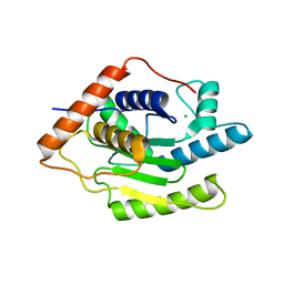
6H4I
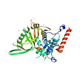
 | | Usp28 catalytic domain apo | | 分子名称: | SULFATE ION, Ubiquitin carboxyl-terminal hydrolase 28 | | 著者 | Klemm, T.A, Sauer, F, Kisker, C. | | 登録日 | 2018-07-21 | | 公開日 | 2019-03-27 | | 最終更新日 | 2019-05-15 | | 実験手法 | X-RAY DIFFRACTION (3.22 Å) | | 主引用文献 | Differential Oligomerization of the Deubiquitinases USP25 and USP28 Regulates Their Activities.
Mol.Cell, 74, 2019
|
|
6H4H
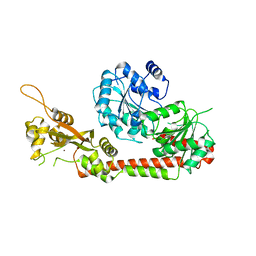
 | | Usp28 catalytic domain variant E593D in complex with UbPA | | 分子名称: | Polyubiquitin-B, SULFATE ION, Ubiquitin carboxyl-terminal hydrolase 28, ... | | 著者 | Klemm, T.A, Sauer, F, Kisker, C. | | 登録日 | 2018-07-21 | | 公開日 | 2019-03-27 | | 最終更新日 | 2019-05-15 | | 実験手法 | X-RAY DIFFRACTION (3.5 Å) | | 主引用文献 | Differential Oligomerization of the Deubiquitinases USP25 and USP28 Regulates Their Activities.
Mol.Cell, 74, 2019
|
|
6H4K
 | |
6H4L
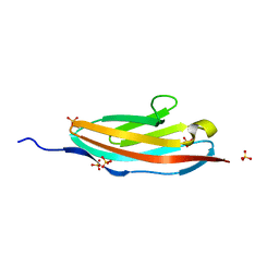
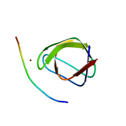
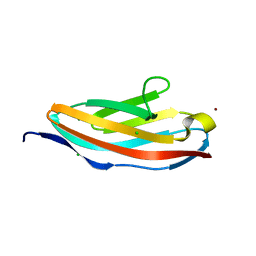 | | Structure of Titin M4 trigonal form | | 分子名称: | CHLORIDE ION, Titin, ZINC ION | | 著者 | Sauer, F, Wilmanns, M. | | 登録日 | 2018-07-21 | | 公開日 | 2019-08-07 | | 最終更新日 | 2020-02-19 | | 実験手法 | X-RAY DIFFRACTION (1.6 Å) | | 主引用文献 | Structural diversity in the atomic resolution 3D fingerprint of the titin M-band segment.
Plos One, 14, 2019
|
|
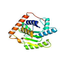
6H4J
 | | Usp25 catalytic domain | | 分子名称: | CHLORIDE ION, Ubiquitin carboxyl-terminal hydrolase 25 | | 著者 | Klemm, T.A, Sauer, F, Kisker, C. | | 登録日 | 2018-07-21 | | 公開日 | 2019-03-27 | | 最終更新日 | 2024-01-17 | | 実験手法 | X-RAY DIFFRACTION (3.07 Å) | | 主引用文献 | Differential Oligomerization of the Deubiquitinases USP25 and USP28 Regulates Their Activities.
Mol.Cell, 74, 2019
|
|
5B5Q
 | |
5LST
 | |
8REV
 | |
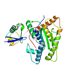
6FDU
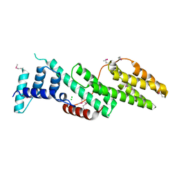
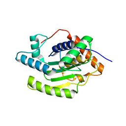
 | | Structure of Chlamydia trachomatis effector protein Cdu1 bound to Compound 3 | | 分子名称: | (2~{S},3~{S})-2-[[(2~{S})-2-[3,5-bis(chloranyl)phenyl]-2-(dimethylamino)ethanoyl]amino]-~{N}-[[2-(iminomethyl)pyrimidin-4-yl]methyl]-3-methyl-pentanamide, CHLORIDE ION, Deubiquitinase and deneddylase Dub1 | | 著者 | Ramirez, Y, Kisker, C, Altmann, E. | | 登録日 | 2017-12-26 | | 公開日 | 2018-08-15 | | 最終更新日 | 2024-01-17 | | 実験手法 | X-RAY DIFFRACTION (2.3 Å) | | 主引用文献 | Structural Basis of Substrate Recognition and Covalent Inhibition of Cdu1 from Chlamydia trachomatis.
ChemMedChem, 13, 2018
|
|
6FDQ
 | |
6FDK
 | |
6HCI
 | |


