2G60
 
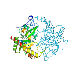
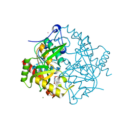
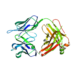 | | Structure of anti-FLAG M2 Fab domain | | 分子名称: | anti-FLAG M2 Fab heavy chain, anti-FLAG M2 Fab light chain | | 著者 | Roosild, T.P. | | 登録日 | 2006-02-23 | | 公開日 | 2006-09-12 | | 最終更新日 | 2017-10-18 | | 実験手法 | X-RAY DIFFRACTION (1.85 Å) | | 主引用文献 | Structure of anti-FLAG M2 Fab domain and its use in the stabilization of engineered membrane proteins.
Acta Crystallogr.,Sect.F, 62, 2006
|
|
3L9W
 | |
1LSS
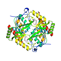
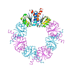
 | | KTN Mja218 CRYSTAL STRUCTURE IN COMPLEX WITH NAD+ | | 分子名称: | NICOTINAMIDE-ADENINE-DINUCLEOTIDE, Trk system potassium uptake protein trkA homolog | | 著者 | Roosild, T.P, Miller, S, Booth, I.R, Choe, S. | | 登録日 | 2002-05-18 | | 公開日 | 2002-07-03 | | 最終更新日 | 2024-02-14 | | 実験手法 | X-RAY DIFFRACTION (2.3 Å) | | 主引用文献 | A mechanism of regulating transmembrane potassium flux through a ligand-mediated conformational switch.
Cell(Cambridge,Mass.), 109, 2002
|
|
1LSU
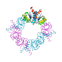
 | | KTN Bsu222 Crystal Structure in Complex with NADH | | 分子名称: | 1,4-DIHYDRONICOTINAMIDE ADENINE DINUCLEOTIDE, Conserved hypothetical protein yuaA | | 著者 | Roosild, T.P, Miller, S, Booth, I.R, Choe, S. | | 登録日 | 2002-05-18 | | 公開日 | 2002-07-03 | | 最終更新日 | 2024-02-14 | | 実験手法 | X-RAY DIFFRACTION (2.85 Å) | | 主引用文献 | A mechanism of regulating transmembrane potassium flux through a ligand-mediated conformational switch.
Cell(Cambridge,Mass.), 109, 2002
|
|
3EUE
 | |
3EUF
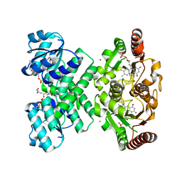
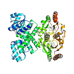
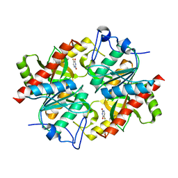
 | | Crystal structure of BAU-bound human uridine phosphorylase 1 | | 分子名称: | 1-((2-HYDROXYETHOXY)METHYL)-5-BENZYLPYRIMIDINE-2,4(1H,3H)-DIONE, PHOSPHATE ION, Uridine phosphorylase 1 | | 著者 | Roosild, T.P. | | 登録日 | 2008-10-10 | | 公開日 | 2009-03-31 | | 最終更新日 | 2023-09-06 | | 実験手法 | X-RAY DIFFRACTION (1.9 Å) | | 主引用文献 | Implications of the structure of human uridine phosphorylase 1 on the development of novel inhibitors for improving the therapeutic window of fluoropyrimidine chemotherapy.
Bmc Struct.Biol., 9, 2009
|
|
3EYW
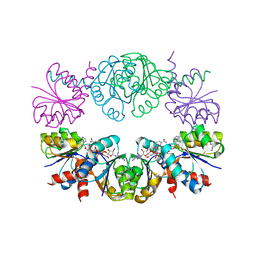
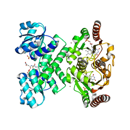
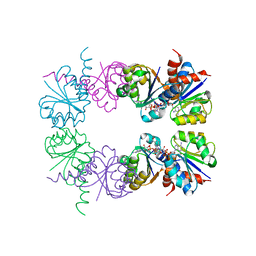
 | | Crystal structure of the C-terminal domain of E. coli KefC in complex with KefF | | 分子名称: | C-terminal domain of Glutathione-regulated potassium-efflux system protein kefC fused to full length Glutathione-regulated potassium-efflux system ancillary protein kefF, FLAVIN MONONUCLEOTIDE, MAGNESIUM ION, ... | | 著者 | Roosild, T.P. | | 登録日 | 2008-10-22 | | 公開日 | 2009-06-30 | | 最終更新日 | 2023-09-06 | | 実験手法 | X-RAY DIFFRACTION (2.4 Å) | | 主引用文献 | KTN (RCK) Domains Regulate K(+) Channels and Transporters by Controlling the Dimer-Hinge Conformation.
Structure, 17, 2009
|
|
1YGM
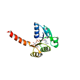
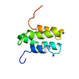 | | NMR structure of Mistic | | 分子名称: | hypothetical protein BSU31320 | | 著者 | Roosild, T.P, Greenwald, J, Vega, M, Castronovo, S, Riek, R, Choe, S. | | 登録日 | 2005-01-05 | | 公開日 | 2005-03-01 | | 最終更新日 | 2024-05-22 | | 実験手法 | SOLUTION NMR | | 主引用文献 | NMR structure of Mistic, a membrane-integrating protein for membrane protein expression.
Science, 307, 2005
|
|
3L9X
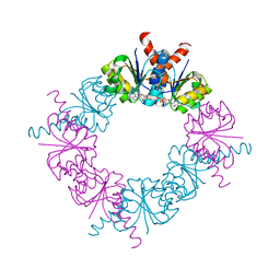
 | | KefC C-terminal domain in complex with KefF and ESG | | 分子名称: | FLAVIN MONONUCLEOTIDE, Glutathione-regulated potassium-efflux system protein kefC, linker, ... | | 著者 | Roosild, T.P. | | 登録日 | 2010-01-05 | | 公開日 | 2010-11-17 | | 最終更新日 | 2023-09-06 | | 実験手法 | X-RAY DIFFRACTION (2.1 Å) | | 主引用文献 | Mechanism of ligand-gated potassium efflux in bacterial pathogens.
Proc.Natl.Acad.Sci.USA, 107, 2010
|
|
3NBQ
 | |
3P0F
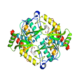
 | | Structure of hUPP2 in an inactive conformation with bound 5-benzylacyclouridine | | 分子名称: | 1-((2-HYDROXYETHOXY)METHYL)-5-BENZYLPYRIMIDINE-2,4(1H,3H)-DIONE, COBALT (II) ION, MAGNESIUM ION, ... | | 著者 | Roosild, T.P, Castronovo, S, Villoso, A. | | 登録日 | 2010-09-28 | | 公開日 | 2011-09-07 | | 最終更新日 | 2023-09-06 | | 実験手法 | X-RAY DIFFRACTION (1.54 Å) | | 主引用文献 | A novel structural mechanism for redox regulation of uridine phosphorylase 2 activity.
J.Struct.Biol., 176, 2011
|
|
3P0E
 | | Structure of hUPP2 in an active conformation with bound 5-benzylacyclouridine | | 分子名称: | 1-((2-HYDROXYETHOXY)METHYL)-5-BENZYLPYRIMIDINE-2,4(1H,3H)-DIONE, PHOSPHATE ION, Uridine phosphorylase 2 | | 著者 | Roosild, T.P, Castronovo, S, Villoso, A. | | 登録日 | 2010-09-28 | | 公開日 | 2011-09-07 | | 最終更新日 | 2024-02-21 | | 実験手法 | X-RAY DIFFRACTION (2 Å) | | 主引用文献 | A novel structural mechanism for redox regulation of uridine phosphorylase 2 activity.
J.Struct.Biol., 176, 2011
|
|
2HMW
 | |
2HMT
 | |
2HMS
 | |
2HMU
 | |
2HMV
 | |


