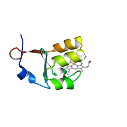
5MWJ
 
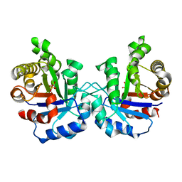
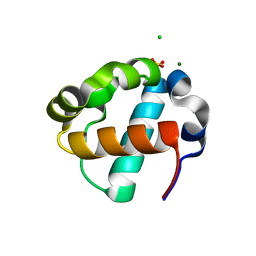
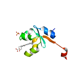
 | | Structure Enabled Discovery of a Stapled Peptide Inhibitor to Target the Oncogenic Transcriptional Repressor TLE1 | | 分子名称: | DIMETHYL SULFOXIDE, Transducin-like enhancer protein 1, pepide inhibtor | | 著者 | McGrath, S, Tortorici, M, Vidler, L, Drouin, L, Westwood, I, Gimeson, P, Van Montfort, R, Hoelder, S. | | 登録日 | 2017-01-18 | | 公開日 | 2017-04-05 | | 最終更新日 | 2024-01-17 | | 実験手法 | X-RAY DIFFRACTION (2.04 Å) | | 主引用文献 | Structure-Enabled Discovery of a Stapled Peptide Inhibitor to Target the Oncogenic Transcriptional Repressor TLE1.
Chemistry, 23, 2017
|
|
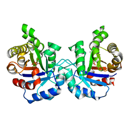
7TDY
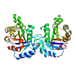
 | | The ubiquitin-associated domain of human thirty-eight negative kinase 1, flexibly fused to the 1TEL crystallization chaperone via a 2-glycine linker and crystallized at low protein concentration | | 分子名称: | FORMIC ACID, Transcription factor ETV6,Non-receptor tyrosine-protein kinase TNK1 | | 著者 | Nawarathnage, S, Bunn, D.R, Stewart, C, Doukev, T, Moody, J.D. | | 登録日 | 2022-01-03 | | 公開日 | 2023-01-11 | | 最終更新日 | 2024-04-03 | | 実験手法 | X-RAY DIFFRACTION (1.53 Å) | | 主引用文献 | Fusion crystallization reveals the behavior of both the 1TEL crystallization chaperone and the TNK1 UBA domain.
Structure, 31, 2023
|
|
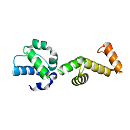
7TCY
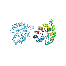
 | | The ubiquitin-associated domain of human thirty-eight negative kinase I | | 分子名称: | CHLORIDE ION, FORMIC ACID, MAGNESIUM ION, ... | | 著者 | Nawarathnage, S, Bunn, R.D, Stewart, C, Doukov, T, Moody, J.D. | | 登録日 | 2021-12-29 | | 公開日 | 2023-01-11 | | 最終更新日 | 2023-12-20 | | 実験手法 | X-RAY DIFFRACTION (1.54 Å) | | 主引用文献 | Fusion crystallization reveals the behavior of both the 1TEL crystallization chaperone and the TNK1 UBA domain.
Structure, 31, 2023
|
|
6GIQ
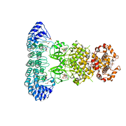
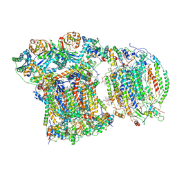
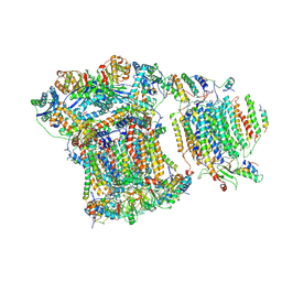 | | Saccharomyces cerevisiae respiratory supercomplex III2IV | | 分子名称: | (1R)-2-(dodecanoyloxy)-1-[(phosphonooxy)methyl]ethyl tetradecanoate, (1R)-2-(phosphonooxy)-1-[(tridecanoyloxy)methyl]ethyl pentadecanoate, (1R)-2-{[(S)-(2-aminoethoxy)(hydroxy)phosphoryl]oxy}-1-[(heptanoyloxy)methyl]ethyl octadecanoate, ... | | 著者 | Rathore, S, Berndtsson, J, Conrad, J, Ott, M. | | 登録日 | 2018-05-15 | | 公開日 | 2019-01-02 | | 最終更新日 | 2019-12-18 | | 実験手法 | ELECTRON MICROSCOPY (3.23 Å) | | 主引用文献 | Cryo-EM structure of the yeast respiratory supercomplex.
Nat. Struct. Mol. Biol., 26, 2019
|
|
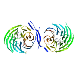
1O5X
 | | Plasmodium falciparum TIM complexed to 2-phosphoglycerate | | 分子名称: | 2-PHOSPHOGLYCERIC ACID, 3-HYDROXYPYRUVIC ACID, PHOSPHITE ION, ... | | 著者 | Parthasarathy, S, Eaazhisai, K, Balaram, H, Balaram, P, Murthy, M.R. | | 登録日 | 2003-10-06 | | 公開日 | 2004-01-13 | | 最終更新日 | 2023-10-25 | | 実験手法 | X-RAY DIFFRACTION (1.1 Å) | | 主引用文献 | Structure of Plasmodium falciparum Triose-phosphate Isomerase-2-Phosphoglycerate Complex at 1.1-A Resolution
J.Biol.Chem., 278, 2003
|
|
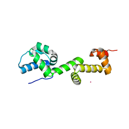
7U4W
 | | The ubiquitin-associated domain of human thirty-eight negative kinase-1 flexibly fused to the 1TEL crystallization chaperone via a 2-glycine linker and crystallized at traditional protein concentration | | 分子名称: | Transcription factor ETV6,Non-receptor tyrosine-protein kinase TNK1 | | 著者 | Nawarathnage, S, Pedroza Romo, M.J, Smith, T, Bunn, D, Stewart, C, Doukov, T, Moody, J.D. | | 登録日 | 2022-03-01 | | 公開日 | 2023-03-08 | | 最終更新日 | 2023-12-20 | | 実験手法 | X-RAY DIFFRACTION (2.1 Å) | | 主引用文献 | Fusion crystallization reveals the behavior of both the 1TEL crystallization chaperone and the TNK1 UBA domain.
Structure, 31, 2023
|
|
7U4Z
 | |
1LZO
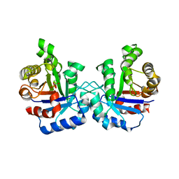 | | Plasmodium Falciparum Triosephosphate Isomerase-Phosphoglycolate Complex | | 分子名称: | 2-PHOSPHOGLYCOLIC ACID, Triosephosphate Isomerase | | 著者 | Parthasarathy, S, Balaram, H, Balaram, P, Murthy, M.R. | | 登録日 | 2002-06-11 | | 公開日 | 2003-01-28 | | 最終更新日 | 2024-02-14 | | 実験手法 | X-RAY DIFFRACTION (2.8 Å) | | 主引用文献 | Structure of the Plasmodium falciparum triosephosphate isomerase-phosphoglycolate complex in two crystal forms: characterization of catalytic loop open and closed conformations in the ligand-bound state
Biochemistry, 41, 2002
|
|
1M7P
 | | Plasmodium Falciparum Triosephosphate isomerase (PfTIM) compled to substrate analog glycerol-3-phosphate (G3P). | | 分子名称: | GLYCERALDEHYDE-3-PHOSPHATE, Triosephosphate Isomerase | | 著者 | Parthasarathy, S, Balaram, H, Balaram, P, Murthy, M.R.N. | | 登録日 | 2002-07-22 | | 公開日 | 2002-11-29 | | 最終更新日 | 2024-02-14 | | 実験手法 | X-RAY DIFFRACTION (2.4 Å) | | 主引用文献 | Structures of Plasmodium falciparum triosephosphate isomerase complexed to substrate analogues: observation of the catalytic loop in the open conformation in the ligand-bound state.
Acta Crystallogr.,Sect.D, 58, 2002
|
|
1M7O
 | | Plasmodium Falciparum Triosephosphate isomerase (PfTIM) compled to substrate analog 3-phosphoglycerate (3PG) | | 分子名称: | 3-PHOSPHOGLYCERIC ACID, Triosephosphate Isomerase | | 著者 | Parthasarathy, S, Balaram, H, Balaram, P, Murthy, M.R.N. | | 登録日 | 2002-07-22 | | 公開日 | 2002-11-29 | | 最終更新日 | 2024-02-14 | | 実験手法 | X-RAY DIFFRACTION (2.4 Å) | | 主引用文献 | Structures of Plasmodium falciparum triosephosphate isomerase complexed to substrate analogues: observation of the catalytic loop in the open conformation in the ligand-bound state.
Acta Crystallogr.,Sect.D, 58, 2002
|
|
1LYX
 | | Plasmodium Falciparum Triosephosphate Isomerase (PfTIM)-Phosphoglycolate complex | | 分子名称: | 2-PHOSPHOGLYCOLIC ACID, Triosephosphate Isomerase | | 著者 | Parthasarathy, S, Balaram, H, Balaram, P, Murthy, M.R. | | 登録日 | 2002-06-10 | | 公開日 | 2003-01-28 | | 最終更新日 | 2024-02-14 | | 実験手法 | X-RAY DIFFRACTION (1.9 Å) | | 主引用文献 | Structure of the Plasmodium falciparum triosephosphate isomerase-phosphoglycolate complex in two crystal forms: characterization of catalytic loop open and closed conformations in the ligand-bound state
Biochemistry, 41, 2002
|
|
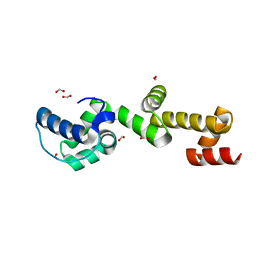
4HIL
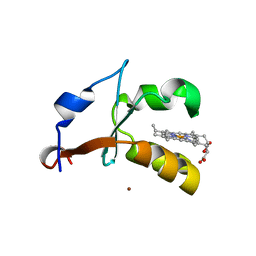
 | | 1.25A Resolution Structure of Rat Type B Cytochrome b5 | | 分子名称: | Cytochrome b5 type B, PROTOPORPHYRIN IX CONTAINING FE, SODIUM ION | | 著者 | Lovell, S, Battaile, K.P, Parthasarathy, S, Sun, N, Terzyan, S, Zhang, X, Rivera, M, Kuczera, K, Benson, D.R. | | 登録日 | 2012-10-11 | | 公開日 | 2013-10-16 | | 最終更新日 | 2023-09-20 | | 実験手法 | X-RAY DIFFRACTION (1.25 Å) | | 主引用文献 | 1.25A Resolution Structure of Rat Type B Cytochrome b5
To be Published
|
|
4HIN
 | | 2.4A Resolution Structure of Bovine Cytochrome b5 (S71L) | | 分子名称: | COPPER (II) ION, Cytochrome b5, PROTOPORPHYRIN IX CONTAINING FE | | 著者 | Lovell, S, Battaile, K.P, Parthasarathy, S, Sun, N, Terzyan, S, Zhang, X, Rivera, M, Kuczera, K, Benson, D.R. | | 登録日 | 2012-10-11 | | 公開日 | 2013-10-16 | | 最終更新日 | 2023-09-20 | | 実験手法 | X-RAY DIFFRACTION (2.4 Å) | | 主引用文献 | 2.4A Resolution Structure of Bovine Cytochrome b5 (S71L)
To be Published
|
|
3OZZ
 | |
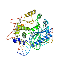
4OAV
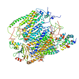
 | | Complete human RNase L in complex with 2-5A (5'-ppp heptamer), AMPPCP and RNA substrate. | | 分子名称: | (2R,3S,4R,5R)-5-(2,4-dioxo-3,4-dihydropyrimidin-1(2H)-yl)-4-hydroxy-2-({[(S)-hydroxy{[(2R,3S,4S)-4-hydroxy-2-(hydroxymethyl)tetrahydrofuran-3-yl]oxy}phosphoryl]oxy}methyl)tetrahydrofuran-3-yl dihydrogen phosphate, MAGNESIUM ION, PHOSPHOMETHYLPHOSPHONIC ACID ADENYLATE ESTER, ... | | 著者 | Han, Y, Donovan, J, Rath, S, Whitney, G, Chitrakar, A, Korennykh, A. | | 登録日 | 2014-01-06 | | 公開日 | 2014-03-12 | | 最終更新日 | 2023-09-20 | | 実験手法 | X-RAY DIFFRACTION (2.1 Å) | | 主引用文献 | Structure of human RNase L reveals the basis for regulated RNA decay in the IFN response.
Science, 343, 2014
|
|
4OAU
 | | Complete human RNase L in complex with biological activators. | | 分子名称: | 2-5A-dependent ribonuclease, ADENOSINE-5'-DIPHOSPHATE, MAGNESIUM ION, ... | | 著者 | Han, Y, Donovan, J, Rath, S, Whitney, G, Chitrakar, A, Korennykh, A. | | 登録日 | 2014-01-06 | | 公開日 | 2014-03-12 | | 最終更新日 | 2024-02-28 | | 実験手法 | X-RAY DIFFRACTION (2.6 Å) | | 主引用文献 | Structure of human RNase L reveals the basis for regulated RNA decay in the IFN response.
Science, 343, 2014
|
|
4S3N
 | | Crystal structure of human OAS3 domain I in complex with dsRNA | | 分子名称: | 2'-5'-oligoadenylate synthase 3, RNA (5'-R(*GP*GP*CP*UP*UP*UP*UP*GP*AP*CP*CP*UP*UP*UP*AP*UP*GP*AP*A)-3'), RNA (5'-R(*UP*UP*CP*AP*UP*AP*AP*AP*GP*GP*UP*CP*AP*AP*AP*AP*GP*CP*C)-3') | | 著者 | Donovan, J, Whitney, G, Rath, S, Korennykh, A. | | 登録日 | 2015-02-26 | | 公開日 | 2015-03-25 | | 最終更新日 | 2023-09-20 | | 実験手法 | X-RAY DIFFRACTION (2 Å) | | 主引用文献 | Structural mechanism of sensing long dsRNA via a noncatalytic domain in human oligoadenylate synthetase 3.
Proc.Natl.Acad.Sci.USA, 112, 2015
|
|
8E1F
 | | Sterile Alpha Motif Domain of Human Translocation ETS Leukemia, Non-Polymer Crystal Form | | 分子名称: | CHLORIDE ION, MAGNESIUM ION, SULFATE ION, ... | | 著者 | Wilson, E, Nawarathnage, S, Stewart, C, Brown, S, Bezzant, B, Bunn, D, Towne, N, Pedroza Romo, M.J, Moody, J.D. | | 登録日 | 2022-08-10 | | 公開日 | 2022-09-14 | | 最終更新日 | 2023-10-25 | | 実験手法 | X-RAY DIFFRACTION (2.16 Å) | | 主引用文献 | Sterile Alpha Motif Domain of Human Translocation ETS Leukemia, Non-Polymer Crystal Form
To Be Published
|
|
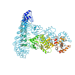
6YMX
 | | CIII2/CIV respiratory supercomplex from Saccharomyces cerevisiae | | 分子名称: | (1R)-2-(dodecanoyloxy)-1-[(phosphonooxy)methyl]ethyl tetradecanoate, (1R)-2-(phosphonooxy)-1-[(tridecanoyloxy)methyl]ethyl pentadecanoate, (1R)-2-{[(S)-(2-aminoethoxy)(hydroxy)phosphoryl]oxy}-1-[(heptanoyloxy)methyl]ethyl octadecanoate, ... | | 著者 | Berndtsson, J, Rathore, S, Ott, M. | | 登録日 | 2020-04-10 | | 公開日 | 2020-09-09 | | 最終更新日 | 2021-03-24 | | 実験手法 | ELECTRON MICROSCOPY (3.17 Å) | | 主引用文献 | Respiratory supercomplexes enhance electron transport by decreasing cytochrome c diffusion distance.
Embo Rep., 21, 2020
|
|
6YMY
 | | Cytochrome c oxidase from Saccharomyces cerevisiae | | 分子名称: | (2R,5S,11R,14R)-5,8,11-trihydroxy-2-(nonanoyloxy)-5,11-dioxido-16-oxo-14-[(propanoyloxy)methyl]-4,6,10,12,15-pentaoxa-5,11-diphosphanonadec-1-yl undecanoate, 1,2-DIACYL-SN-GLYCERO-3-PHOSHOCHOLINE, COPPER (II) ION, ... | | 著者 | Berndtsson, J, Rathore, S, Ott, M. | | 登録日 | 2020-04-10 | | 公開日 | 2020-09-09 | | 最終更新日 | 2021-03-24 | | 実験手法 | ELECTRON MICROSCOPY (3.41 Å) | | 主引用文献 | Respiratory supercomplexes enhance electron transport by decreasing cytochrome c diffusion distance.
Embo Rep., 21, 2020
|
|
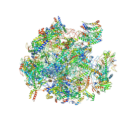
5OOL
 | | Structure of a native assembly intermediate of the human mitochondrial ribosome with unfolded interfacial rRNA | | 分子名称: | 16S ribosomal RNA, 39S ribosomal protein L10, mitochondrial, ... | | 著者 | Brown, A, Rathore, S, Kimanius, D, Aibara, S, Bai, X.C, Rorbach, J, Amunts, A, Ramakrishnan, V. | | 登録日 | 2017-08-08 | | 公開日 | 2017-09-13 | | 最終更新日 | 2023-03-15 | | 実験手法 | ELECTRON MICROSCOPY (3.06 Å) | | 主引用文献 | Structures of the human mitochondrial ribosome in native states of assembly.
Nat. Struct. Mol. Biol., 24, 2017
|
|
5OOM
 | | Structure of a native assembly intermediate of the human mitochondrial ribosome with unfolded interfacial rRNA | | 分子名称: | 16S ribosomal RNA, 39S ribosomal protein L10, mitochondrial, ... | | 著者 | Brown, A, Rathore, S, Kimanius, D, Aibara, S, Bai, X.C, Rorbach, J, Amunts, A, Ramakrishnan, V. | | 登録日 | 2017-08-08 | | 公開日 | 2017-09-13 | | 最終更新日 | 2023-03-15 | | 実験手法 | ELECTRON MICROSCOPY (3.03 Å) | | 主引用文献 | Structures of the human mitochondrial ribosome in native states of assembly.
Nat. Struct. Mol. Biol., 24, 2017
|
|
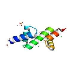
3LF5
 | | Structure of Human NADH cytochrome b5 oxidoreductase (Ncb5or) b5 Domain to 1.25A Resolution | | 分子名称: | Cytochrome b5 reductase 4, PROTOPORPHYRIN IX CONTAINING FE, SULFATE ION | | 著者 | Deng, B, Parthasarathy, S, Wang, W, Gibney, B.R, Battaile, K.P, Lovell, S, Benson, D.R, Zhu, H. | | 登録日 | 2010-01-15 | | 公開日 | 2010-07-14 | | 最終更新日 | 2023-09-06 | | 実験手法 | X-RAY DIFFRACTION (1.25 Å) | | 主引用文献 | Study of the individual cytochrome b5 and cytochrome b5 reductase domains of Ncb5or reveals a unique heme pocket and a possible role of the CS domain.
J.Biol.Chem., 285, 2010
|
|
2MXU
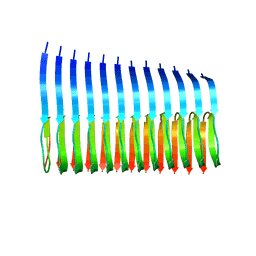 | | 42-Residue Beta Amyloid Fibril | | 分子名称: | Amyloid beta A4 protein | | 著者 | Xiao, Y, Ma, B, McElheny, D, Parthasarathy, S, Long, F, Hoshi, M, Nussinov, R, Ishii, Y. | | 登録日 | 2015-01-14 | | 公開日 | 2015-05-06 | | 最終更新日 | 2024-05-01 | | 実験手法 | SOLID-STATE NMR | | 主引用文献 | A beta (1-42) fibril structure illuminates self-recognition and replication of amyloid in Alzheimer's disease.
Nat.Struct.Mol.Biol., 22, 2015
|
|
7JG1
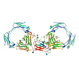 | | Dimeric Immunoglobin A (dIgA) | | 分子名称: | 2-acetamido-2-deoxy-beta-D-glucopyranose, 2-acetamido-2-deoxy-beta-D-glucopyranose-(1-4)-2-acetamido-2-deoxy-beta-D-glucopyranose, Igh protein, ... | | 著者 | Kumar Bharathkar, S, Parker, B.P, Malyutin, A.G, Stadtmueller, B.M. | | 登録日 | 2020-07-18 | | 公開日 | 2020-11-11 | | 実験手法 | ELECTRON MICROSCOPY (3.3 Å) | | 主引用文献 | The structures of Secretory and dimeric Immunoglobulin A.
Elife, 9, 2020
|
|


