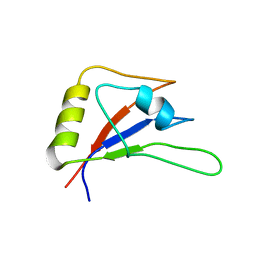
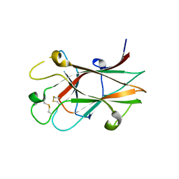
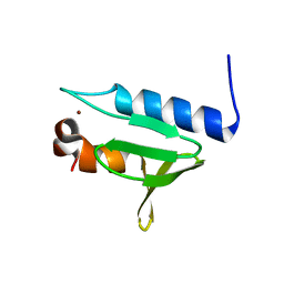
3YGS
 
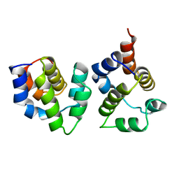 | | APAF-1 CARD IN COMPLEX WITH PRODOMAIN OF PROCASPASE-9 | | 分子名称: | APOPTOTIC PROTEASE ACTIVATING FACTOR 1, PROCASPASE 9 | | 著者 | Qin, H, Srinivasula, S, Wu, G, Fernandes-Alnemri, T, Alnemri, E, Shi, Y. | | 登録日 | 1999-05-08 | | 公開日 | 2000-04-19 | | 最終更新日 | 2023-12-27 | | 実験手法 | X-RAY DIFFRACTION (2.5 Å) | | 主引用文献 | Structural basis of procaspase-9 recruitment by the apoptotic protease-activating factor 1.
Nature, 399, 1999
|
|
2LW8
 | |
2MDK
 | |
2KOU
 | | DICER LIKE protein | | 分子名称: | Dicer-like protein 4 | | 著者 | Qin, H, Song, J, Yuan, Y.A. | | 登録日 | 2009-09-30 | | 公開日 | 2010-02-16 | | 最終更新日 | 2024-05-29 | | 実験手法 | SOLUTION NMR | | 主引用文献 | Structure of the Arabidopsis thaliana DCL4 DUF283 domain reveals a noncanonical double-stranded RNA-binding fold for protein-protein interaction
Rna, 2010
|
|
5ZSY
 | |
5ZSW
 | |
8U1T
 | | SARS-CoV-2 Envelope Protein Transmembrane Domain: Dimeric Structure Determined by Solid-State NMR | | 分子名称: | Envelope small membrane protein | | 著者 | Zhang, R, Qin, H, Prasad, R, Fu, R, Zhou, H.X, Cross, T. | | 登録日 | 2023-09-02 | | 公開日 | 2023-11-15 | | 最終更新日 | 2024-05-15 | | 実験手法 | SOLID-STATE NMR | | 主引用文献 | Dimeric Transmembrane Structure of the SARS-CoV-2 E Protein.
Commun Biol, 6, 2023
|
|
2L0J
 | | Solid State NMR structure of the M2 proton channel from Influenza A Virus in hydrated lipid bilayer | | 分子名称: | Matrix protein 2 | | 著者 | Sharma, M, Yi, M, Dong, H, Qin, H, Peterson, E, Busath, D.D, Zhou, H.X, Cross, T.A. | | 登録日 | 2010-07-08 | | 公開日 | 2010-11-03 | | 最終更新日 | 2024-05-01 | | 実験手法 | SOLID-STATE NMR | | 主引用文献 | Insight into the mechanism of the influenza a proton channel from a structure in a lipid bilayer.
Science, 330, 2010
|
|
2MBE
 | |
3GXU
 | |
3CKH
 | | Crystal structure of Eph A4 receptor | | 分子名称: | Ephrin type-A receptor 4 | | 著者 | Shi, J.H, Song, J.X. | | 登録日 | 2008-03-15 | | 公開日 | 2008-09-23 | | 最終更新日 | 2023-11-01 | | 実験手法 | X-RAY DIFFRACTION (2.8 Å) | | 主引用文献 | Crystal Structure and NMR Binding Reveal That Two Small Molecule Antagonists Target the High Affinity Ephrin-binding Channel of the EphA4 Receptor.
J.Biol.Chem., 283, 2008
|
|
2N6E
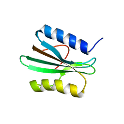 | |
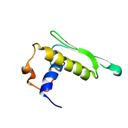
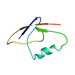
2YGS
 | | CARD DOMAIN FROM APAF-1 | | 分子名称: | APOPTOTIC PROTEASE ACTIVATING FACTOR 1 | | 著者 | Shi, Y. | | 登録日 | 1999-05-08 | | 公開日 | 2000-04-19 | | 最終更新日 | 2023-12-27 | | 実験手法 | X-RAY DIFFRACTION (1.6 Å) | | 主引用文献 | Structural basis of procaspase-9 recruitment by the apoptotic protease-activating factor 1.
Nature, 399, 1999
|
|
8DHT
 | | Crystal structure of a typeIII Rubisco | | 分子名称: | 3-PHOSPHOGLYCERIC ACID, ACETATE ION, GLYCEROL, ... | | 著者 | Qingqiu, H. | | 登録日 | 2022-06-28 | | 公開日 | 2022-11-30 | | 最終更新日 | 2023-11-15 | | 実験手法 | X-RAY DIFFRACTION (1.699 Å) | | 主引用文献 | Crystal structure of a type III Rubisco in complex with its product 3-phosphoglycerate.
Proteins, 91, 2023
|
|
5X57
 | | Structure of GAR domain of ACF7 | | 分子名称: | Microtubule-actin cross-linking factor 1, isoforms 1/2/3/5, NICKEL (II) ION | | 著者 | Yang, F, Wang, T, Zhang, Y, Wu, X.Y. | | 登録日 | 2017-02-15 | | 公開日 | 2017-07-05 | | 最終更新日 | 2024-03-27 | | 実験手法 | X-RAY DIFFRACTION (1.45 Å) | | 主引用文献 | ACF7 regulates inflammatory colitis and intestinal wound response by orchestrating tight junction dynamics.
Nat Commun, 8, 2017
|
|
8H41
 | | Crystal structure of a decarboxylase from Trichosporon moniliiforme in complex with o-nitrophenol | | 分子名称: | MAGNESIUM ION, O-NITROPHENOL, Salicylate decarboxylase | | 著者 | Gao, J, Zhao, Y.P, Li, Q, Liu, W.D, Sheng, X. | | 登録日 | 2022-10-09 | | 公開日 | 2023-08-16 | | 実験手法 | X-RAY DIFFRACTION (1.78 Å) | | 主引用文献 | A Combined Computational-Experimental Study on the Substrate Binding and Reaction Mechanism of Salicylic Acid Decarboxylase
Catalysts, 12, 2022
|
|
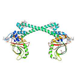
5XBW
 | | The structure of BrlR | | 分子名称: | Probable transcriptional regulator | | 著者 | Wang, F, Qing, H, Gu, L. | | 登録日 | 2017-03-21 | | 公開日 | 2018-05-02 | | 最終更新日 | 2023-11-22 | | 実験手法 | X-RAY DIFFRACTION (3.109 Å) | | 主引用文献 | BrlR from Pseudomonas aeruginosa is a receptor for both cyclic di-GMP and pyocyanin.
Nat Commun, 9, 2018
|
|
5XBI
 | |
5XBT
 | | The structure of BrlR bound to c-di-GMP | | 分子名称: | 9,9'-[(2R,3R,3aS,5S,7aR,9R,10R,10aS,12S,14aR)-3,5,10,12-tetrahydroxy-5,12-dioxidooctahydro-2H,7H-difuro[3,2-d:3',2'-j][1,3,7,9,2,8]tetraoxadiphosphacyclododecine-2,9-diyl]bis(2-amino-1,9-dihydro-6H-purin-6-one), DI(HYDROXYETHYL)ETHER, GLYCEROL, ... | | 著者 | Wang, F, Qing, H, Gu, L. | | 登録日 | 2017-03-21 | | 公開日 | 2018-05-02 | | 最終更新日 | 2024-03-27 | | 実験手法 | X-RAY DIFFRACTION (2.495 Å) | | 主引用文献 | BrlR from Pseudomonas aeruginosa is a receptor for both cyclic di-GMP and pyocyanin.
Nat Commun, 9, 2018
|
|


