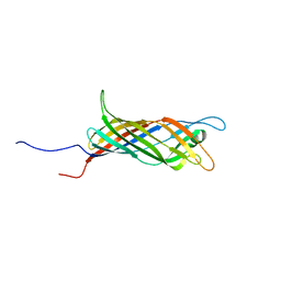
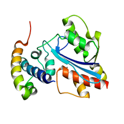
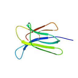
1UAP
 
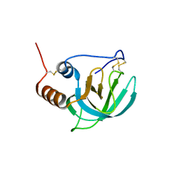 | | NMR structure of the NTR domain from human PCOLCE1 | | 分子名称: | Procollagen C-proteinase enhancer protein | | 著者 | Liepinsh, E, Banyai, L, Pintacuda, G, Trexler, M, Patthy, L, Otting, G. | | 登録日 | 2003-03-14 | | 公開日 | 2003-07-15 | | 最終更新日 | 2023-12-27 | | 実験手法 | SOLUTION NMR | | 主引用文献 | NMR Structure of the Netrin-like Domain (NTR) of Human Type I Procollagen C-Proteinase Enhancer Defines Structural Consensus of NTR Domains and Assesses Potential Proteinase Inhibitory Activity and Ligand Binding.
J.Biol.Chem., 278, 2003
|
|
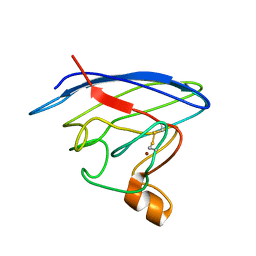
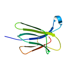
2AXD
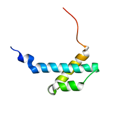 | | solution structure of the theta subunit of escherichia coli DNA polymerase III in complex with the epsilon subunit | | 分子名称: | DNA polymerase III, theta subunit | | 著者 | Keniry, M.A, Park, A.Y, Owen, E.A, Hamdan, S.M, Pintacuda, G, Otting, G, Dixon, N.E. | | 登録日 | 2005-09-05 | | 公開日 | 2006-07-04 | | 最終更新日 | 2024-05-29 | | 実験手法 | SOLUTION NMR | | 主引用文献 | Structure of the theta subunit of Escherichia coli DNA polymerase III in complex with the epsilon subunit
J.Bacteriol., 188, 2006
|
|
6EKA
 | | Solid-state MAS NMR structure of the HELLF prion amyloid fibrils | | 分子名称: | Podospora anserina S mat+ genomic DNA chromosome 3, supercontig 2 | | 著者 | Martinez, D, Daskalov, A, Andreas, L, Bardiaux, B, Coustou, V, Stanek, J, Berbon, M, Noubhani, M, Kauffmann, B, Wall, J.S, Pintacuda, G, Saupe, S.J, Habenstein, B, Loquet, A. | | 登録日 | 2017-09-25 | | 公開日 | 2018-10-10 | | 最終更新日 | 2024-06-19 | | 実験手法 | SOLID-STATE NMR | | 主引用文献 | Structural and molecular basis of cross-seeding barriers in amyloids
Proc.Natl.Acad.Sci.USA, 118, 2021
|
|
5MWV
 | | Solid-state NMR Structure of outer membrane protein G in lipid bilayers | | 分子名称: | Outer membrane protein G | | 著者 | Retel, J.S, Nieuwkoop, A.J, Hiller, M, Higman, V.A, Barbet-Massin, E, Stanek, J, Andreas, L.B, Franks, W.T, van Rossum, B.-J, Vinothkumar, K.R, Handel, L, de Palma, G.G, Bardiaux, B, Pintacuda, G, Emsley, L, Kuelbrandt, W, Oschkinat, H. | | 登録日 | 2017-01-20 | | 公開日 | 2017-12-27 | | 最終更新日 | 2024-05-15 | | 実験手法 | SOLID-STATE NMR | | 主引用文献 | Structure of outer membrane protein G in lipid bilayers.
Nat Commun, 8, 2017
|
|
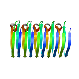
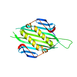
5JXV
 | | Solid-state MAS NMR structure of immunoglobulin beta 1 binding domain of protein G (GB1) | | 分子名称: | Immunoglobulin G-binding protein G | | 著者 | Andreas, L.B, Jaudzems, K, Stanek, J, Lalli, D, Bertarello, A, Le Marchand, T, Cala-De Paepe, D, Kotelovica, S, Akopjana, I, Knott, B, Wegner, S, Engelke, F, Lesage, A, Emsley, L, Tars, K, Herrmann, T, Pintacuda, G. | | 登録日 | 2016-05-13 | | 公開日 | 2016-08-10 | | 最終更新日 | 2024-06-19 | | 実験手法 | SOLID-STATE NMR | | 主引用文献 | Structure of fully protonated proteins by proton-detected magic-angle spinning NMR.
Proc.Natl.Acad.Sci.USA, 113, 2016
|
|
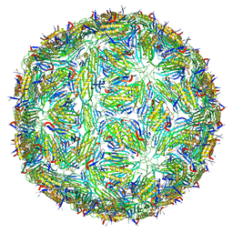
5JZR
 | | Solid-state MAS NMR structure of Acinetobacter phage 205 (AP205) coat protein in assembled capsid particles | | 分子名称: | Coat protein | | 著者 | Jaudzems, K, Andreas, L.B, Stanek, J, Lalli, D, Bertarello, A, Le Marchand, T, Cala-De Paepe, D, Kotelovica, S, Akopjana, I, Knott, B, Wegner, S, Engelke, F, Lesage, A, Emsley, L, Tars, K, Herrmann, T, Pintacuda, G. | | 登録日 | 2016-05-17 | | 公開日 | 2016-08-10 | | 最終更新日 | 2024-06-19 | | 実験手法 | SOLID-STATE NMR | | 主引用文献 | Structure of fully protonated proteins by proton-detected magic-angle spinning NMR.
Proc.Natl.Acad.Sci.USA, 113, 2016
|
|
6QAM
 | |
6QWR
 | |
2LU5
 | | Structure and chemical shifts of Cu(I),Zn(II) superoxide dismutase by solid-state NMR | | 分子名称: | COPPER (II) ION, Superoxide dismutase [Cu-Zn] | | 著者 | Knight, M.J, Pell, A.J, Bertini, I, Felli, I.C, Gonnelli, L, Pierattelli, R, Herrmann, T, Emsley, L, Pintacuda, G. | | 登録日 | 2012-06-08 | | 公開日 | 2012-06-27 | | 最終更新日 | 2023-06-14 | | 実験手法 | SOLID-STATE NMR | | 主引用文献 | Structure and backbone dynamics of a microcrystalline metalloprotein by solid-state NMR.
Proc.Natl.Acad.Sci.USA, 109, 2012
|
|
2N70
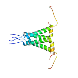 | | Two-fold symmetric structure of the 18-60 construct of S31N M2 from Influenza A in lipid bilayers | | 分子名称: | Matrix protein 2 | | 著者 | Andreas, L.B, Reese, M, Eddy, M.T, Gelev, V, Ni, Q, Miller, E.A, Emsley, L, Pintacuda, G, Chou, J.J, Griffin, R.G. | | 登録日 | 2015-09-01 | | 公開日 | 2015-09-23 | | 最終更新日 | 2024-05-15 | | 実験手法 | SOLID-STATE NMR | | 主引用文献 | Structure and Mechanism of the Influenza A M218-60 Dimer of Dimers.
J.Am.Chem.Soc., 137, 2015
|
|
1T3W
 | | Crystal Structure of the E.coli DnaG C-terminal domain (residues 434 to 581) | | 分子名称: | ACETIC ACID, DNA primase | | 著者 | Oakley, A.J, Loscha, K.V, Schaeffer, P.M, Liepinsh, E, Wilce, M.C.J, Otting, G, Dixon, N.E. | | 登録日 | 2004-04-28 | | 公開日 | 2004-11-02 | | 最終更新日 | 2016-09-28 | | 実験手法 | X-RAY DIFFRACTION (2.8 Å) | | 主引用文献 | Crystal and solution structures of the helicase-binding domain of Escherichia coli primase
J.Biol.Chem., 280, 2005
|
|
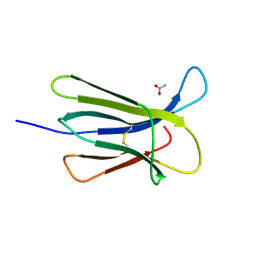
2XY8
 | |
5FS4
 | | Bacteriophage AP205 coat protein | | 分子名称: | AP205 BACTERIOPHAGE COAT PROTEIN | | 著者 | Shishovs, M, Tars, K. | | 登録日 | 2015-12-29 | | 公開日 | 2016-09-21 | | 最終更新日 | 2024-05-08 | | 実験手法 | X-RAY DIFFRACTION (1.73 Å) | | 主引用文献 | Structure of Ap205 Coat Protein Reveals Circular Permutation in Ssrna Bacteriophages.
J.Mol.Biol., 428, 2016
|
|
5T87
 | | Crystal structure of CDI complex from Cupriavidus taiwanensis LMG 19424 | | 分子名称: | CdiA toxin, CdiI immunity protein | | 著者 | Michalska, K, Joachimiak, G, Jedrzejczak, R, Hayes, C.S, Goulding, C.W, Joachimiak, A, Structure-Function Analysis of Polymorphic CDI Toxin-Immunity Protein Complexes (UC4CDI), Midwest Center for Structural Genomics (MCSG) | | 登録日 | 2016-09-06 | | 公開日 | 2017-09-13 | | 最終更新日 | 2019-12-25 | | 実験手法 | X-RAY DIFFRACTION (2.4 Å) | | 主引用文献 | Target highlights from the first post-PSI CASP experiment (CASP12, May-August 2016).
Proteins, 86 Suppl 1, 2018
|
|
4RMW
 | |
4RMU
 | |
4RMV
 | |
5LQP
 | |
5CSG
 | |
5CSB
 | | The crystal structure of beta2-microglobulin D76N mutant at room temperature | | 分子名称: | Beta-2-microglobulin | | 著者 | de Rosa, M, Mota, C.S, de Sanctis, D, Bolognesi, M, Ricagno, S. | | 登録日 | 2015-07-23 | | 公開日 | 2016-08-10 | | 最終更新日 | 2024-01-10 | | 実験手法 | X-RAY DIFFRACTION (1.719 Å) | | 主引用文献 | Conformational dynamics in crystals reveal the molecular bases for D76N beta-2 microglobulin aggregation propensity.
Nat Commun, 9, 2018
|
|
5CS7
 | | The crystal structure of wt beta2-microglobulin at room temperature | | 分子名称: | Beta-2-microglobulin | | 著者 | de Rosa, M, Mota, C.S, de Sanctis, D, Bolognesi, M, Ricagno, S. | | 登録日 | 2015-07-23 | | 公開日 | 2016-08-10 | | 最終更新日 | 2024-01-10 | | 実験手法 | X-RAY DIFFRACTION (2.1 Å) | | 主引用文献 | Conformational dynamics in crystals reveal the molecular bases for D76N beta-2 microglobulin aggregation propensity.
Nat Commun, 9, 2018
|
|


