7VVV
 
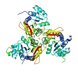 | | Crystal structure of MmtN | | 分子名称: | PHOSPHATE ION, SAM-dependent methyltransferase | | 著者 | Peng, M, Li, C.Y. | | 登録日 | 2021-11-09 | | 公開日 | 2022-04-20 | | 最終更新日 | 2024-05-29 | | 実験手法 | X-RAY DIFFRACTION (2.45 Å) | | 主引用文献 | Insights into methionine S-methylation in diverse organisms.
Nat Commun, 13, 2022
|
|
4O32
 
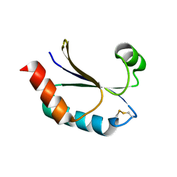 | | Structure of a malarial protein | | 分子名称: | CHLORIDE ION, Thioredoxin | | 著者 | Egea, P.F, Koehl, A, Peng, M, Cascio, D. | | 登録日 | 2013-12-17 | | 公開日 | 2014-12-24 | | 最終更新日 | 2024-03-13 | | 実験手法 | X-RAY DIFFRACTION (2.196 Å) | | 主引用文献 | Crystal structure and solution characterization of the thioredoxin-2 from Plasmodium falciparum, a constituent of an essential parasitic protein export complex.
Biochem.Biophys.Res.Commun., 456, 2015
|
|
8HLE
 
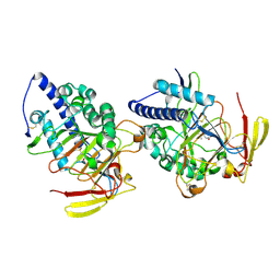 | | Structure of DddY-DMSOP complex | | 分子名称: | 3-[dimethyl(oxidanyl)-$l^{4}-sulfanyl]propanoic acid, DMSP lyase DddY, ZINC ION | | 著者 | Peng, M, Li, C.Y, Zhang, Y.Z. | | 登録日 | 2022-11-30 | | 公開日 | 2023-10-04 | | 最終更新日 | 2023-12-20 | | 実験手法 | X-RAY DIFFRACTION (1.91 Å) | | 主引用文献 | DMSOP-cleaving enzymes are diverse and widely distributed in marine microorganisms.
Nat Microbiol, 8, 2023
|
|
8HLF
 
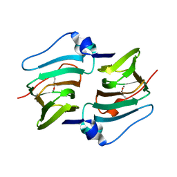 | | Crystal structure of DddK-DMSOP complex | | 分子名称: | 3-[dimethyl(oxidanyl)-$l^{4}-sulfanyl]propanoic acid, MANGANESE (II) ION, Novel protein with potential Cupin domain | | 著者 | Peng, M, Li, C.Y, Zhang, Y.Z. | | 登録日 | 2022-11-30 | | 公開日 | 2023-10-04 | | 最終更新日 | 2023-12-20 | | 実験手法 | X-RAY DIFFRACTION (1.62 Å) | | 主引用文献 | DMSOP-cleaving enzymes are diverse and widely distributed in marine microorganisms.
Nat Microbiol, 8, 2023
|
|
7VVW
 
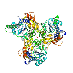 | | MmtN-SAM complex | | 分子名称: | GLYCEROL, PHOSPHATE ION, S-ADENOSYLMETHIONINE, ... | | 著者 | Zhang, Y.Z, Peng, M, Li, C.Y. | | 登録日 | 2021-11-09 | | 公開日 | 2022-04-20 | | 最終更新日 | 2024-05-29 | | 実験手法 | X-RAY DIFFRACTION (2.11 Å) | | 主引用文献 | Insights into methionine S-methylation in diverse organisms.
Nat Commun, 13, 2022
|
|
7VVX
 
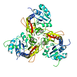 | | MmtN-SAH-Met complex | | 分子名称: | METHIONINE, PHOSPHATE ION, S-ADENOSYL-L-HOMOCYSTEINE, ... | | 著者 | Zhang, Y.Z, Peng, M, Li, C.Y. | | 登録日 | 2021-11-09 | | 公開日 | 2022-04-20 | | 最終更新日 | 2024-05-29 | | 実験手法 | X-RAY DIFFRACTION (2.51 Å) | | 主引用文献 | Insights into methionine S-methylation in diverse organisms.
Nat Commun, 13, 2022
|
|
6AK1
 
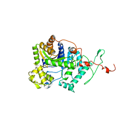 | | Crystal structure of DmoA from Hyphomicrobium sulfonivorans | | 分子名称: | Dimethyl-sulfide monooxygenase | | 著者 | Cao, H.Y, Wang, P, Peng, M, Li, C.Y. | | 登録日 | 2018-08-28 | | 公開日 | 2018-12-12 | | 最終更新日 | 2023-11-22 | | 実験手法 | X-RAY DIFFRACTION (2.284 Å) | | 主引用文献 | Crystal structure of the dimethylsulfide monooxygenase DmoA from Hyphomicrobium sulfonivorans.
Acta Crystallogr.,Sect.F, 74, 2018
|
|
7VLZ
 
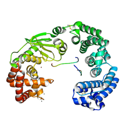 | | Crystal structure of the collagenase unit of a Vibrio collagenase from Vibrio harveyi VHJR7 | | 分子名称: | CALCIUM ION, Peptide P1, Peptide P2, ... | | 著者 | Cao, H.Y, Wang, Y, Peng, M, Zhang, Y.Z. | | 登録日 | 2021-10-05 | | 公開日 | 2022-10-19 | | 最終更新日 | 2024-10-16 | | 実験手法 | X-RAY DIFFRACTION (1.6 Å) | | 主引用文献 | Crystal structure of the collagenase unit of a Vibrio collagenase from Vibrio harveyi VHJR7
To Be Published
|
|
7ESI
 
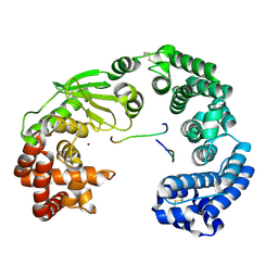 | | Crystal structure of the collagenase unit of a Vibrio collagenase from Vibrio harveyi VHJR7 at 1. 8 angstrom resolution. | | 分子名称: | CALCIUM ION, Collagenase unit (CU), Peptide P1, ... | | 著者 | Cao, H.Y, Wang, Y, Peng, M, Zhang, Y.Z. | | 登録日 | 2021-05-11 | | 公開日 | 2022-02-09 | | 最終更新日 | 2024-10-30 | | 実験手法 | X-RAY DIFFRACTION (1.8 Å) | | 主引用文献 | Structure of Vibrio collagenase VhaC provides insight into the mechanism of bacterial collagenolysis.
Nat Commun, 13, 2022
|
|
6IHK
 
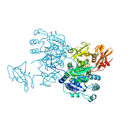 | | Structure of MMPA CoA ligase in complex with ADP | | 分子名称: | ADENOSINE-5'-DIPHOSPHATE, AMP-binding domain protein | | 著者 | Shao, X, Cao, H.Y, Wang, P, Li, C.Y, Zhao, F, Peng, M, Chen, X.L, Zhang, Y.Z. | | 登録日 | 2018-09-30 | | 公開日 | 2019-07-03 | | 最終更新日 | 2024-03-27 | | 実験手法 | X-RAY DIFFRACTION (2.23 Å) | | 主引用文献 | Mechanistic insight into 3-methylmercaptopropionate metabolism and kinetical regulation of demethylation pathway in marine dimethylsulfoniopropionate-catabolizing bacteria.
Mol.Microbiol., 111, 2019
|
|
6IJB
 
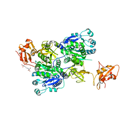 | | Structure of 3-methylmercaptopropionate CoA ligase mutant K523A in complex with AMP and MMPA | | 分子名称: | 2-[3-(2-HYDROXY-1,1-DIHYDROXYMETHYL-ETHYLAMINO)-PROPYLAMINO]-2-HYDROXYMETHYL-PROPANE-1,3-DIOL, 3-(methylsulfanyl)propanoic acid, ADENOSINE MONOPHOSPHATE, ... | | 著者 | Shao, X, Cao, H.Y, Wang, P, Li, C.Y, Zhao, F, Peng, M, Chen, X.L, Zhang, Y.Z. | | 登録日 | 2018-10-09 | | 公開日 | 2019-07-03 | | 最終更新日 | 2023-11-22 | | 実験手法 | X-RAY DIFFRACTION (2.111 Å) | | 主引用文献 | Mechanistic insight into 3-methylmercaptopropionate metabolism and kinetical regulation of demethylation pathway in marine dimethylsulfoniopropionate-catabolizing bacteria.
Mol.Microbiol., 111, 2019
|
|
6A55
 
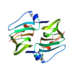 | | Crystal structure of DddK mutant Y122A | | 分子名称: | 3-(dimethyl-lambda~4~-sulfanyl)propanoic acid, MANGANESE (II) ION, Novel protein with potential Cupin domain | | 著者 | Zhang, Y.Z, Li, C.Y. | | 登録日 | 2018-06-21 | | 公開日 | 2019-02-20 | | 最終更新日 | 2023-11-22 | | 実験手法 | X-RAY DIFFRACTION (1.6 Å) | | 主引用文献 | Structure-Function Analysis Indicates that an Active-Site Water Molecule Participates in Dimethylsulfoniopropionate Cleavage by DddK.
Appl. Environ. Microbiol., 85, 2019
|
|
6A53
 
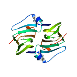 | | Crystal structure of DddK | | 分子名称: | MANGANESE (II) ION, Novel protein with potential Cupin domain | | 著者 | Zhang, Y.Z, Li, C.Y. | | 登録日 | 2018-06-21 | | 公開日 | 2019-02-20 | | 最終更新日 | 2024-03-27 | | 実験手法 | X-RAY DIFFRACTION (2 Å) | | 主引用文献 | Structure-Function Analysis Indicates that an Active-Site Water Molecule Participates in Dimethylsulfoniopropionate Cleavage by DddK.
Appl. Environ. Microbiol., 85, 2019
|
|
6A54
 
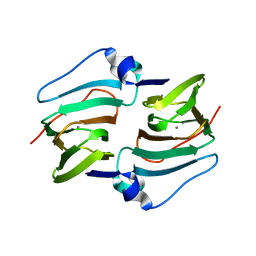 | | Crystal structure of DddK mutant Y64A | | 分子名称: | MANGANESE (II) ION, Novel protein with potential Cupin domain | | 著者 | Zhang, Y.Z, Li, C.Y. | | 登録日 | 2018-06-21 | | 公開日 | 2019-02-20 | | 最終更新日 | 2023-11-22 | | 実験手法 | X-RAY DIFFRACTION (2.3 Å) | | 主引用文献 | Structure-Function Analysis Indicates that an Active-Site Water Molecule Participates in Dimethylsulfoniopropionate Cleavage by DddK.
Appl. Environ. Microbiol., 85, 2019
|
|
2QYK
 
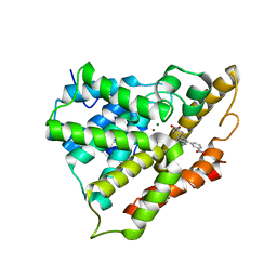 | | Crystal structure of PDE4A10 in complex with inhibitor NPV | | 分子名称: | 4-[8-(3-nitrophenyl)-1,7-naphthyridin-6-yl]benzoic acid, Cyclic AMP-specific phosphodiesterase HSPDE4A10, MAGNESIUM ION, ... | | 著者 | Wang, H, Peng, M, Chen, Y, Geng, J, Robinson, H, Houslay, M. | | 登録日 | 2007-08-15 | | 公開日 | 2008-04-08 | | 最終更新日 | 2024-04-03 | | 実験手法 | X-RAY DIFFRACTION (2.1 Å) | | 主引用文献 | Structures of the four subfamilies of phosphodiesterase-4 provide insight into the selectivity of their inhibitors.
Biochem.J., 408, 2007
|
|
5GSN
 
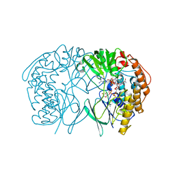 | | Tmm in complex with methimazole | | 分子名称: | 1-METHYL-1,3-DIHYDRO-2H-IMIDAZOLE-2-THIONE, FLAVIN-ADENINE DINUCLEOTIDE, Flavin-containing monooxygenase, ... | | 著者 | Zhang, Y.Z, Li, C.Y. | | 登録日 | 2016-08-16 | | 公開日 | 2017-01-18 | | 最終更新日 | 2023-11-08 | | 実験手法 | X-RAY DIFFRACTION (2.203 Å) | | 主引用文献 | Structural mechanism for bacterial oxidation of oceanic trimethylamine into trimethylamine N-oxide
Mol. Microbiol., 103, 2017
|
|
5IPY
 
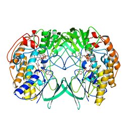 | | Crystal structure of WT RnTmm | | 分子名称: | FLAVIN-ADENINE DINUCLEOTIDE, Flavin-containing monooxygenase, NADP NICOTINAMIDE-ADENINE-DINUCLEOTIDE PHOSPHATE | | 著者 | Li, C.Y, Zhang, Y.Z. | | 登録日 | 2016-03-10 | | 公開日 | 2017-01-18 | | 最終更新日 | 2023-11-08 | | 実験手法 | X-RAY DIFFRACTION (1.5 Å) | | 主引用文献 | Structural mechanism for bacterial oxidation of oceanic trimethylamine into trimethylamine N-oxide
Mol. Microbiol., 103, 2017
|
|
5IQ4
 
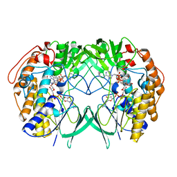 | | Crystal structure of RnTmm mutant Y207S soaking | | 分子名称: | FLAVIN-ADENINE DINUCLEOTIDE, Flavin-containing monooxygenase, NADP NICOTINAMIDE-ADENINE-DINUCLEOTIDE PHOSPHATE | | 著者 | Zhang, Y.Z, Li, C.Y. | | 登録日 | 2016-03-10 | | 公開日 | 2017-01-18 | | 最終更新日 | 2023-11-08 | | 実験手法 | X-RAY DIFFRACTION (1.5 Å) | | 主引用文献 | Structural mechanism for bacterial oxidation of oceanic trimethylamine into trimethylamine N-oxide
Mol. Microbiol., 103, 2017
|
|
5IQ1
 
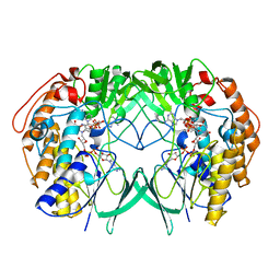 | | Crystal structure of RnTmm mutant Y207S | | 分子名称: | FLAVIN-ADENINE DINUCLEOTIDE, Flavin-containing monooxygenase, NADP NICOTINAMIDE-ADENINE-DINUCLEOTIDE PHOSPHATE | | 著者 | Li, C.Y, Zhang, Y.Z. | | 登録日 | 2016-03-10 | | 公開日 | 2017-01-18 | | 最終更新日 | 2023-11-08 | | 実験手法 | X-RAY DIFFRACTION (1.75 Å) | | 主引用文献 | Structural mechanism for bacterial oxidation of oceanic trimethylamine into trimethylamine N-oxide
Mol. Microbiol., 103, 2017
|
|
5YPO
 
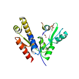 | | Crystal structure of PSD-95 GK domain in complex with phospho-SAPAP peptide | | 分子名称: | Disks large homolog 4, GLYCEROL, SAPAP | | 著者 | Zhu, J, Zhou, Q, Shang, Y, Weng, Z, Zhang, R, Zhang, M. | | 登録日 | 2017-11-02 | | 公開日 | 2018-03-14 | | 最終更新日 | 2023-11-22 | | 実験手法 | X-RAY DIFFRACTION (2.29 Å) | | 主引用文献 | Synaptic Targeting and Function of SAPAPs Mediated by Phosphorylation-Dependent Binding to PSD-95 MAGUKs.
Cell Rep, 21, 2017
|
|
5YPR
 
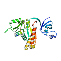 | | Crystal Structure of PSD-95 SH3-GK domain in complex with a synthesized inhibitor | | 分子名称: | Disks large homolog 4, Synthesized GK inhibitor | | 著者 | Zhu, J, Zhou, Q, Shang, Y, Weng, Z, Zhu, R, Zhang, M. | | 登録日 | 2017-11-02 | | 公開日 | 2018-03-14 | | 最終更新日 | 2024-10-30 | | 実験手法 | X-RAY DIFFRACTION (2.349 Å) | | 主引用文献 | Synaptic Targeting and Function of SAPAPs Mediated by Phosphorylation-Dependent Binding to PSD-95 MAGUKs.
Cell Rep, 21, 2017
|
|
6JMU
 
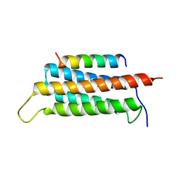 | | Crystal structure of GIT1/Paxillin complex | | 分子名称: | ARF GTPase-activating protein GIT1, Paxillin | | 著者 | Zhu, J, Lin, L, Xia, Y, Zhang, R, Zhang, M. | | 登録日 | 2019-03-13 | | 公開日 | 2020-05-20 | | 最終更新日 | 2023-11-22 | | 実験手法 | X-RAY DIFFRACTION (2 Å) | | 主引用文献 | GIT/PIX Condensates Are Modular and Ideal for Distinct Compartmentalized Cell Signaling.
Mol.Cell, 79, 2020
|
|
6JMT
 
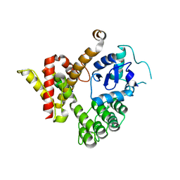 | | Crystal structure of GIT/PIX complex | | 分子名称: | ARF GTPase-activating protein GIT2, ZINC ION, beta PIX | | 著者 | Zhu, J, Lin, L, Xia, Y, Zhang, R, Zhang, M. | | 登録日 | 2019-03-13 | | 公開日 | 2020-05-20 | | 最終更新日 | 2023-11-22 | | 実験手法 | X-RAY DIFFRACTION (2.8 Å) | | 主引用文献 | GIT/PIX Condensates Are Modular and Ideal for Distinct Compartmentalized Cell Signaling.
Mol.Cell, 79, 2020
|
|
6K1M
 
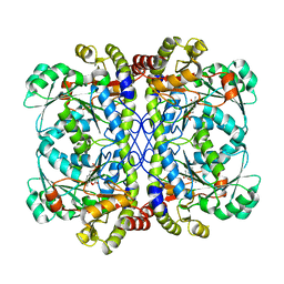 | | Engineered form of a putative cystathionine gamma-lyase | | 分子名称: | Cystathionine gamma-lyase, PYRIDOXAL-5'-PHOSPHATE, PYRUVIC ACID | | 著者 | Chen, S, Wang, Y. | | 登録日 | 2019-05-10 | | 公開日 | 2020-05-13 | | 最終更新日 | 2023-11-22 | | 実験手法 | X-RAY DIFFRACTION (2.32 Å) | | 主引用文献 | Structural characterization of cystathionine gamma-lyase smCSE enables aqueous metal quantum dot biosynthesis.
Int.J.Biol.Macromol., 174, 2021
|
|
6K1O
 
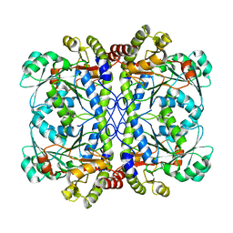 | | Apo form of a putative cystathionine gamma-lyase | | 分子名称: | Cystathionine gamma-lyase | | 著者 | Chen, S, Wang, Y. | | 登録日 | 2019-05-10 | | 公開日 | 2020-05-13 | | 最終更新日 | 2023-11-22 | | 実験手法 | X-RAY DIFFRACTION (2.033 Å) | | 主引用文献 | Structural characterization of cystathionine gamma-lyase smCSE enables aqueous metal quantum dot biosynthesis.
Int.J.Biol.Macromol., 174, 2021
|
|
