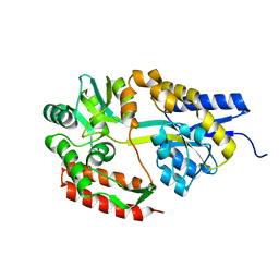
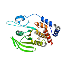
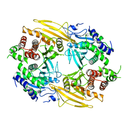
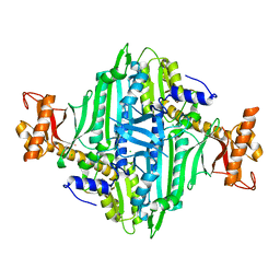
8WOP
 
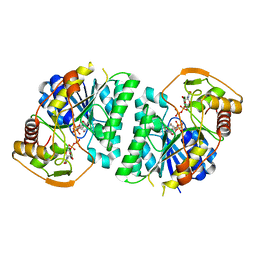 | | Crystal structure of Arabidopsis thaliana UDP-glucose 4-epimerase 2 (AtUGE2) complexed with UDP, wild-type | | 分子名称: | NICOTINAMIDE-ADENINE-DINUCLEOTIDE, UDP-glucose 4-epimerase 2, URIDINE-5'-DIPHOSPHATE | | 著者 | Matsumoto, M, Umezawa, A, Kotake, T, Fushinobu, S. | | 登録日 | 2023-10-07 | | 公開日 | 2024-05-08 | | 最終更新日 | 2024-07-10 | | 実験手法 | X-RAY DIFFRACTION (2.35 Å) | | 主引用文献 | Cytosolic UDP-L-arabinose synthesis by bifunctional UDP-glucose 4-epimerases in Arabidopsis.
Plant J., 119, 2024
|
|
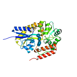
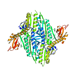
8WOV
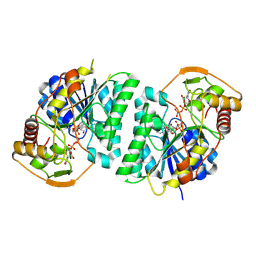 | | Crystal structure of Arabidopsis thaliana UDP-glucose 4-epimerase 2 (AtUGE2) complexed with UDP, G233A mutant | | 分子名称: | NICOTINAMIDE-ADENINE-DINUCLEOTIDE, UDP-glucose 4-epimerase 2, URIDINE-5'-DIPHOSPHATE | | 著者 | Matsumoto, M, Umezawa, A, Kotake, T, Fushinobu, S. | | 登録日 | 2023-10-07 | | 公開日 | 2024-05-15 | | 最終更新日 | 2024-07-10 | | 実験手法 | X-RAY DIFFRACTION (2.25 Å) | | 主引用文献 | Cytosolic UDP-L-arabinose synthesis by bifunctional UDP-glucose 4-epimerases in Arabidopsis.
Plant J., 119, 2024
|
|
8WOW
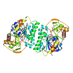 | | Crystal structure of Arabidopsis thaliana UDP-glucose 4-epimerase 2 (AtUGE2) complexed with UDP, I160L/G233A mutant | | 分子名称: | NICOTINAMIDE-ADENINE-DINUCLEOTIDE, SULFATE ION, UDP-glucose 4-epimerase 2, ... | | 著者 | Matsumoto, M, Umezawa, A, Kotake, T, Fushinobu, S. | | 登録日 | 2023-10-08 | | 公開日 | 2024-05-15 | | 最終更新日 | 2024-07-10 | | 実験手法 | X-RAY DIFFRACTION (2.6 Å) | | 主引用文献 | Cytosolic UDP-L-arabinose synthesis by bifunctional UDP-glucose 4-epimerases in Arabidopsis.
Plant J., 119, 2024
|
|
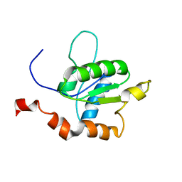
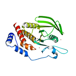
2M1W
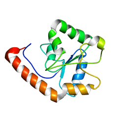 | | TICAM-2 TIR domain | | 分子名称: | TIR domain-containing adapter molecule 2 | | 著者 | Enokizono, Y, Kumeta, H, Funami, K, Horiuchi, M, Sarmiento, J, Yamashita, K, Standley, D.M, Matsumoto, M, Seya, T, Inagaki, F. | | 登録日 | 2012-12-07 | | 公開日 | 2014-01-15 | | 最終更新日 | 2024-05-15 | | 実験手法 | SOLUTION NMR | | 主引用文献 | Structures and interface mapping of the TIR domain-containing adaptor molecules involved in interferon signaling.
Proc.Natl.Acad.Sci.USA, 110, 2013
|
|
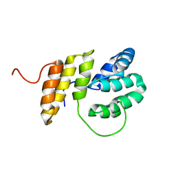
2M1X
 | | TICAM-1 TIR domain structure | | 分子名称: | TIR domain-containing adapter molecule 1 | | 著者 | Enokizono, Y, Kumeta, H, Funami, K, Horiuchi, M, Sarmiento, J, Yamashita, K, Standley, D.M, Matsumoto, M, Seya, T, Inagaki, F. | | 登録日 | 2012-12-07 | | 公開日 | 2014-01-15 | | 最終更新日 | 2024-05-15 | | 実験手法 | SOLUTION NMR | | 主引用文献 | Structures and interface mapping of the TIR domain-containing adaptor molecules involved in interferon signaling.
Proc.Natl.Acad.Sci.USA, 110, 2013
|
|
2M63
 | | The protease-resistant N-terminal domain of TIR-domain containing adaptor molecule-1, TICAM-1 | | 分子名称: | TIR domain-containing adapter molecule 1 | | 著者 | Kumeta, H, Enokizono, Y, Sakakibara, H, Ogura, K, Matsumoto, M, Seya, T, Inagaki, F. | | 登録日 | 2013-03-20 | | 公開日 | 2014-03-05 | | 最終更新日 | 2024-05-15 | | 実験手法 | SOLUTION NMR | | 主引用文献 | The N-terminal domain of TIR domain-containing adaptor molecule-1, TICAM-1.
J.Biomol.Nmr, 58, 2014
|
|
2ZYO
 | | Crystal structure of cyclo/maltodextrin-binding protein complexed with maltotetraose | | 分子名称: | alpha-D-glucopyranose-(1-4)-alpha-D-glucopyranose, solute-binding protein | | 著者 | Matsumoto, M, Yamada, M, Kurakata, Y, Yoshida, H, Kamitori, S, Nishikawa, A, Tonozuka, T. | | 登録日 | 2009-01-27 | | 公開日 | 2009-03-31 | | 最終更新日 | 2023-11-01 | | 実験手法 | X-RAY DIFFRACTION (1.55 Å) | | 主引用文献 | Crystal structures of open and closed forms of cyclo/maltodextrin-binding protein
Febs J., 276, 2009
|
|
2ZYM
 | | Crystal structure of cyclo/maltodextrin-binding protein complexed with alpha-cyclodextrin | | 分子名称: | Cyclohexakis-(1-4)-(alpha-D-glucopyranose), Solute-binding protein | | 著者 | Matsumoto, M, Yamada, M, Kurakata, Y, Yoshida, H, Kamitori, S, Nishikawa, A, Tonozuka, T. | | 登録日 | 2009-01-27 | | 公開日 | 2009-03-31 | | 最終更新日 | 2023-11-01 | | 実験手法 | X-RAY DIFFRACTION (1.8 Å) | | 主引用文献 | Crystal structures of open and closed forms of cyclo/maltodextrin-binding protein
Febs J., 276, 2009
|
|
2ZYN
 | | Crystal structure of cyclo/maltodextrin-binding protein complexed with beta-cyclodextrin | | 分子名称: | Cycloheptakis-(1-4)-(alpha-D-glucopyranose), Solute-binding protein | | 著者 | Matsumoto, M, Yamada, M, Kurakata, Y, Yoshida, H, Kamitori, S, Nishikawa, A, Tonozuka, T. | | 登録日 | 2009-01-27 | | 公開日 | 2009-03-31 | | 最終更新日 | 2023-11-01 | | 実験手法 | X-RAY DIFFRACTION (1.7 Å) | | 主引用文献 | Crystal structures of open and closed forms of cyclo/maltodextrin-binding protein
Febs J., 276, 2009
|
|
2ZYK
 | | Crystal structure of cyclo/maltodextrin-binding protein complexed with gamma-cyclodextrin | | 分子名称: | Cyclooctakis-(1-4)-(alpha-D-glucopyranose), Solute-binding protein | | 著者 | Tonozuka, T, Sogawa, A, Yamada, M, Matsumoto, N, Yoshida, H, Kamitori, S, Ichikawa, K, Mizuno, M, Nishikawa, A, Sakano, Y. | | 登録日 | 2009-01-26 | | 公開日 | 2009-02-10 | | 最終更新日 | 2024-04-03 | | 実験手法 | X-RAY DIFFRACTION (2.5 Å) | | 主引用文献 | Structural basis for cyclodextrin recognition by Thermoactinomyces vulgaris cyclo/maltodextrin-binding protein
Febs J., 274, 2007
|
|
5H08
 | | Human PTPRZ D1 domain complexed with NAZ2329 | | 分子名称: | 3-{[2-Ethoxy-5-(trifluoromethyl)benzyl]sulfanyl}-N-(phenylsulfonyl)thiophene-2-carboxamide, Receptor-type tyrosine-protein phosphatase zeta | | 著者 | Sugawara, H. | | 登録日 | 2016-10-04 | | 公開日 | 2017-07-26 | | 最終更新日 | 2023-11-08 | | 実験手法 | X-RAY DIFFRACTION (2.53 Å) | | 主引用文献 | Targeting PTPRZ inhibits stem cell-like properties and tumorigenicity in glioblastoma cells
Sci Rep, 7, 2017
|
|
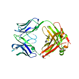
8U31
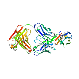 | | Crystal structure of PD-1 in complex with a Fab | | 分子名称: | Fab heavy chain, Fab light chain, GLYCEROL, ... | | 著者 | Sun, D, Masureel, M. | | 登録日 | 2023-09-07 | | 公開日 | 2024-06-19 | | 実験手法 | X-RAY DIFFRACTION (2.73 Å) | | 主引用文献 | Structure- and machine learning-guided engineering demonstrate that a non-canonical disulfide in an anti-PD-1 rabbit antibody does not impede antibody developability.
Mabs, 16, 2024
|
|
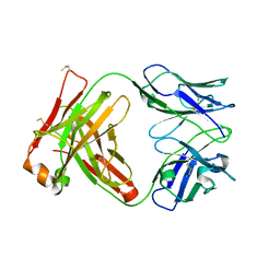
8U32
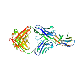 | | Crystal structure of PD-1 in complex with a Fab | | 分子名称: | 2-acetamido-2-deoxy-beta-D-glucopyranose, Fab heavy chain, Fab light chain, ... | | 著者 | Sun, D, Masureel, M. | | 登録日 | 2023-09-07 | | 公開日 | 2024-06-19 | | 実験手法 | X-RAY DIFFRACTION (2.51 Å) | | 主引用文献 | Structure- and machine learning-guided engineering demonstrate that a non-canonical disulfide in an anti-PD-1 rabbit antibody does not impede antibody developability.
Mabs, 16, 2024
|
|
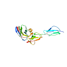
3VGP
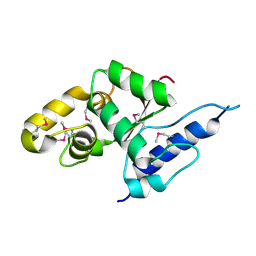 | | Crystal structure of the C-terminal globular domain of oligosaccharyltransferase (AF_0329) from Archaeoglobus fulgidus | | 分子名称: | Transmembrane oligosaccharyl transferase, putative | | 著者 | Matsumoto, S, Igura, M, Nyirenda, J, Yuzawa, S, Noda, N.N, Inagaki, F, Kohda, D. | | 登録日 | 2011-08-18 | | 公開日 | 2012-07-04 | | 実験手法 | X-RAY DIFFRACTION (1.75 Å) | | 主引用文献 | Crystal Structure of the C-Terminal Globular Domain of Oligosaccharyltransferase from Archaeoglobus fulgidus at 1.75 A Resolution
Biochemistry, 51, 2012
|
|
6J6U
 | | Rat PTPRZ D1-D2 domain | | 分子名称: | Receptor-type tyrosine-protein phosphatase zeta | | 著者 | Sugawara, H. | | 登録日 | 2019-01-15 | | 公開日 | 2019-08-28 | | 最終更新日 | 2023-11-22 | | 実験手法 | X-RAY DIFFRACTION (3.32 Å) | | 主引用文献 | A head-to-toe dimerization has physiological relevance for ligand-induced inactivation of protein tyrosine receptor type Z.
J.Biol.Chem., 294, 2019
|
|
7T97
 | |
7T99
 | |
7T98
 | |
7DZY
 | | Spike protein from SARS-CoV2 with Fab fragment of enhancing antibody 2490 | | 分子名称: | Fab Heavy chain of enhancing antibody 2490, Fab light chain of enhancing antibody 2490, Spike glycoprotein | | 著者 | Liu, Y, Soh, W.T, Li, S, Kishikawa, J, Hirose, M, Kato, T, Standley, D, Okada, M, Arase, H. | | 登録日 | 2021-01-26 | | 公開日 | 2021-06-02 | | 最終更新日 | 2024-06-05 | | 実験手法 | ELECTRON MICROSCOPY (3.6 Å) | | 主引用文献 | An infectivity-enhancing site on the SARS-CoV-2 spike protein targeted by antibodies.
Cell, 184, 2021
|
|
5AWX
 | | Crystal structure of Human PTPRZ D1 domain | | 分子名称: | BROMIDE ION, Receptor-type tyrosine-protein phosphatase zeta | | 著者 | Sugawara, H. | | 登録日 | 2015-07-10 | | 公開日 | 2016-02-17 | | 最終更新日 | 2023-11-08 | | 実験手法 | X-RAY DIFFRACTION (1.86 Å) | | 主引用文献 | Small-molecule inhibition of PTPRZ reduces tumor growth in a rat model of glioblastoma
Sci Rep, 6, 2016
|
|
7DZX
 | | Spike protein from SARS-CoV2 with Fab fragment of enhancing antibody 8D2 | | 分子名称: | Fab Heavy chain of enhancing antibody, Fab light chain of enhancing antibody, Spike glycoprotein | | 著者 | Liu, Y, Soh, W.T, Li, S, Kishikawa, J, Hirose, M, Kato, T, Standley, D, Okada, M, Arase, H. | | 登録日 | 2021-01-26 | | 公開日 | 2021-06-02 | | 最終更新日 | 2024-03-27 | | 実験手法 | ELECTRON MICROSCOPY (3.53 Å) | | 主引用文献 | An infectivity-enhancing site on the SARS-CoV-2 spike protein targeted by antibodies.
Cell, 184, 2021
|
|
7DZW
 | | Apo spike protein from SARS-CoV2 | | 分子名称: | Spike glycoprotein | | 著者 | Liu, Y, Soh, W.T, Li, S, Kishikawa, J, Hirose, M, Kato, T, Standley, D, Okada, M, Arase, H. | | 登録日 | 2021-01-26 | | 公開日 | 2021-06-02 | | 最終更新日 | 2024-03-27 | | 実験手法 | ELECTRON MICROSCOPY (3.45 Å) | | 主引用文献 | An infectivity-enhancing site on the SARS-CoV-2 spike protein targeted by antibodies.
Cell, 184, 2021
|
|
7WKI
 | |
5XIF
 | | Crystal Structure of Prolyl-tRNA Synthetase (PRS) from Toxoplasma gondii | | 分子名称: | CHLORIDE ION, Prolyl-tRNA synthetase (ProRS) | | 著者 | Jain, V, Manickam, Y, Sharma, A. | | 登録日 | 2017-04-26 | | 公開日 | 2018-03-07 | | 最終更新日 | 2023-11-22 | | 実験手法 | X-RAY DIFFRACTION (2.48 Å) | | 主引用文献 | Targeting Prolyl-tRNA Synthetase to Accelerate Drug Discovery against Malaria, Leishmaniasis, Toxoplasmosis, Cryptosporidiosis, and Coccidiosis
Structure, 25, 2017
|
|
5XIQ
 | | Crystal Structure of Toxoplasma gondii Prolyl-tRNA Synthetase (TgPRS) in complex with Halofuginone | | 分子名称: | 7-bromo-6-chloro-3-{3-[(2R,3S)-3-hydroxypiperidin-2-yl]-2-oxopropyl}quinazolin-4(3H)-one, MAGNESIUM ION, PHOSPHOAMINOPHOSPHONIC ACID-ADENYLATE ESTER, ... | | 著者 | Jain, V, Manickam, Y, Sharma, A. | | 登録日 | 2017-04-26 | | 公開日 | 2018-03-07 | | 最終更新日 | 2023-11-22 | | 実験手法 | X-RAY DIFFRACTION (2.19 Å) | | 主引用文献 | Targeting Prolyl-tRNA Synthetase to Accelerate Drug Discovery against Malaria, Leishmaniasis, Toxoplasmosis, Cryptosporidiosis, and Coccidiosis
Structure, 25, 2017
|
|


