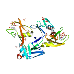
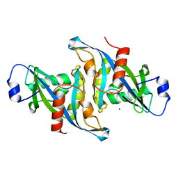
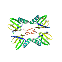
6MEB
 
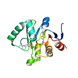 | | Crystal structure of Tylonycteris bat coronavirus HKU4 macrodomain in complex with nicotinamide adenine dinucleotide (NAD+) | | 分子名称: | NICOTINAMIDE-ADENINE-DINUCLEOTIDE, Replicase polyprotein 1ab | | 著者 | Hammond, R.G, Schormann, N, McPherson, R.L, Leung, A.K.L, Deivanayagam, C.C.S, Johnson, M.A. | | 登録日 | 2018-09-06 | | 公開日 | 2019-09-11 | | 最終更新日 | 2023-10-11 | | 実験手法 | X-RAY DIFFRACTION (1.8 Å) | | 主引用文献 | ADP-Ribose and Analogues bound to the DeMARylating Macrodomain from the Bat Coronavirus HKU4
Proc.Natl.Acad.Sci.USA, 2021
|
|
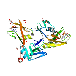
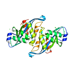
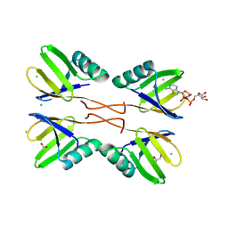
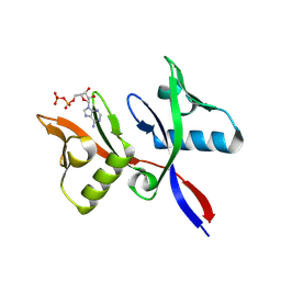
6MEA
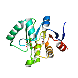 | | Crystal structure of a Tylonycteris bat coronavirus HKU4 macrodomain in complex with adenosine diphosphate ribose (ADP-ribose) | | 分子名称: | ADENOSINE-5-DIPHOSPHORIBOSE, Replicase polyprotein 1ab | | 著者 | Hammond, R.G, Schormann, N, McPherson, R.L, Leung, A.K.L, Deivanayagam, C.C.S, Johnson, M.A. | | 登録日 | 2018-09-06 | | 公開日 | 2019-09-11 | | 最終更新日 | 2023-10-11 | | 実験手法 | X-RAY DIFFRACTION (1.35 Å) | | 主引用文献 | ADP-Ribose and Analogues bound to the DeMARylating Macrodomain from the Bat Coronavirus HKU4
Proc.Natl.Acad.Sci.USA, 2021
|
|
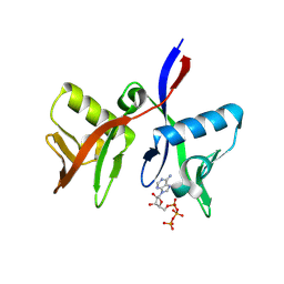
6MEN
 | | Crystal structure of a Tylonycteris bat coronavirus HKU4 macrodomain in complex with adenosine diphosphate glucose (ADP-glucose) | | 分子名称: | ADENOSINE-5'-DIPHOSPHATE-GLUCOSE, Replicase polyprotein 1ab | | 著者 | Hammond, R.G, Schormann, N, McPherson, R.L, Leung, A.K.L, Deivanayagam, C.C.S, Johnson, M.A. | | 登録日 | 2018-09-06 | | 公開日 | 2019-09-11 | | 最終更新日 | 2023-10-11 | | 実験手法 | X-RAY DIFFRACTION (1.5 Å) | | 主引用文献 | ADP-Ribose and Analogues bound to the DeMARylating Macrodomain from the Bat Coronavirus HKU4
Proc.Natl.Acad.Sci.USA, 2021
|
|
6B09
 | |
7SZ2
 | |
7SZ3
 | |
5W6Z
 | |
5W6X
 | |
5VY2
 | |
5WJI
 | |
8R6A
 | |
8R7O
 | |
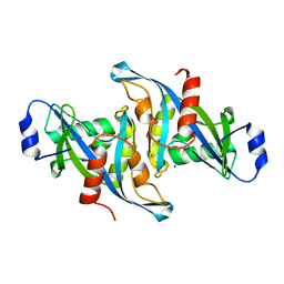
8RD1
 | | HUWE1 WWE domain in complex with compound 4 | | 分子名称: | 2-(2-hydroxy-2-oxoethyl)-1,3-bis(oxidanylidene)isoindole-5-carboxylic acid, CHLORIDE ION, E3 ubiquitin-protein ligase HUWE1 | | 著者 | Muenzker, L, Zak, K.M, Boettcher, J. | | 登録日 | 2023-12-07 | | 公開日 | 2024-07-31 | | 最終更新日 | 2024-08-07 | | 実験手法 | X-RAY DIFFRACTION (1.896 Å) | | 主引用文献 | A ligand discovery toolbox for the WWE domain family of human E3 ligases.
Commun Biol, 7, 2024
|
|
8RD7
 | | HUWE1 WWE domain in complex with ADP-ribose | | 分子名称: | ACETATE ION, CHLORIDE ION, E3 ubiquitin-protein ligase HUWE1, ... | | 著者 | Muenzker, L, Zak, K.M, Boettcher, J. | | 登録日 | 2023-12-07 | | 公開日 | 2024-07-31 | | 最終更新日 | 2024-08-07 | | 実験手法 | X-RAY DIFFRACTION (1.31 Å) | | 主引用文献 | A ligand discovery toolbox for the WWE domain family of human E3 ligases.
Commun Biol, 7, 2024
|
|
8R5N
 | |
8RD0
 | | HUWE1 WWE domain in complex with compound 3 | | 分子名称: | (1,3-dioxo-1,3-dihydro-2H-isoindol-2-yl)acetic acid, CHLORIDE ION, E3 ubiquitin-protein ligase HUWE1 | | 著者 | Muenzker, L, Boettcher, J. | | 登録日 | 2023-12-07 | | 公開日 | 2024-07-31 | | 最終更新日 | 2024-08-07 | | 実験手法 | X-RAY DIFFRACTION (1.765 Å) | | 主引用文献 | A ligand discovery toolbox for the WWE domain family of human E3 ligases.
Commun Biol, 7, 2024
|
|
8R6B
 | |


