2VRC
 
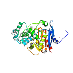
 | | Crystal structure of the Citrobacter sp. triphenylmethane reductase complexed with NADP(H) | | 分子名称: | TRIPHENYLMETHANE REDUCTASE | | 著者 | Kim, Y, Park, H.J, Kwak, S.N, Lee, J.S, Oh, T.K, Kim, M.H. | | 登録日 | 2008-03-31 | | 公開日 | 2008-09-23 | | 最終更新日 | 2011-07-13 | | 実験手法 | X-RAY DIFFRACTION (2.5 Å) | | 主引用文献 | Structural Insight Into Bioremediation of Triphenylmethane Dyes by Citrobacter Sp. Triphenylmethane Reductase.
J.Biol.Chem., 283, 2008
|
|
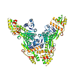
2VRB
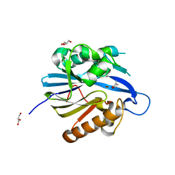
 | | Crystal structure of the Citrobacter sp. triphenylmethane reductase complexed with NADP(H) | | 分子名称: | NADP NICOTINAMIDE-ADENINE-DINUCLEOTIDE PHOSPHATE, TRIPHENYLMETHANE REDUCTASE | | 著者 | Kim, Y, Park, H.J, Kwak, S.N, Lee, J.S, Oh, T.K, Kim, M.H. | | 登録日 | 2008-03-31 | | 公開日 | 2008-09-23 | | 最終更新日 | 2024-05-08 | | 実験手法 | X-RAY DIFFRACTION (2 Å) | | 主引用文献 | Structural Insight Into Bioremediation of Triphenylmethane Dyes by Citrobacter Sp. Triphenylmethane Reductase.
J.Biol.Chem., 283, 2008
|
|
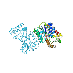
2BTN
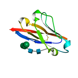
 | | Crystal Structure and Catalytic Mechanism of the Quorum-Quenching N- Acyl Homoserine Lactone Hydrolase | | 分子名称: | AIIA-LIKE PROTEIN, GLYCEROL, ZINC ION | | 著者 | Kim, M.H, Choi, W.C, Kang, H.O, Lee, J.S, Kang, B.S, Kim, K.J, Derewenda, Z.S, Oh, T.K, Lee, C.H, Lee, J.K. | | 登録日 | 2005-06-03 | | 公開日 | 2005-12-07 | | 最終更新日 | 2024-05-08 | | 実験手法 | X-RAY DIFFRACTION (2 Å) | | 主引用文献 | The Molecular Structure and Catalytic Mechanism of a Quorum-Quenching N-Acyl-L-Homoserine Lactone Hydrolase.
Proc.Natl.Acad.Sci.USA, 102, 2005
|
|
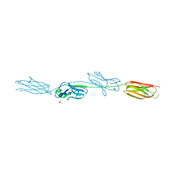
2JEL
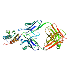 | | JEL42 FAB/HPR COMPLEX | | 分子名称: | HISTIDINE-CONTAINING PROTEIN, JEL42 FAB FRAGMENT, SULFATE ION | | 著者 | Prasad, L, Waygood, E.B, Lee, J.S, Delbaere, L.T.J. | | 登録日 | 1998-02-24 | | 公開日 | 1998-05-27 | | 最終更新日 | 2024-04-03 | | 実験手法 | X-RAY DIFFRACTION (2.5 Å) | | 主引用文献 | The 2.5 A resolution structure of the jel42 Fab fragment/HPr complex
J.Mol.Biol., 280, 1998
|
|
1P25
 | |
1P26
 | |
1P24
 | |
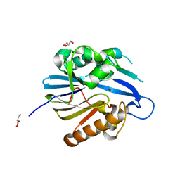
8JJW
 | | Crystal structure of QG-hNTAQ1 C28S | | 分子名称: | MAGNESIUM ION, Protein N-terminal glutamine amidohydrolase | | 著者 | Kang, J.M, Han, B.W. | | 登録日 | 2023-05-31 | | 公開日 | 2024-06-26 | | 実験手法 | X-RAY DIFFRACTION (1.4 Å) | | 主引用文献 | Structural study for substrate recognition of human N-terminal glutamine amidohydrolase 1 in the arginine N-degron pathway.
Protein Sci., 33, 2024
|
|
8JJY
 | | Crystal structure of QN-hNTAQ1 C28S | | 分子名称: | Protein N-terminal glutamine amidohydrolase | | 著者 | Kang, J.M, Han, B.W. | | 登録日 | 2023-05-31 | | 公開日 | 2024-06-26 | | 実験手法 | X-RAY DIFFRACTION (1.69 Å) | | 主引用文献 | Structural study for substrate recognition of human N-terminal glutamine amidohydrolase 1 in the arginine N-degron pathway.
Protein Sci., 33, 2024
|
|
8JK2
 | | Crystal structure of QF-hNTAQ1 C28S | | 分子名称: | Protein N-terminal glutamine amidohydrolase | | 著者 | Kang, J.M, Han, B.W. | | 登録日 | 2023-05-31 | | 公開日 | 2024-06-26 | | 実験手法 | X-RAY DIFFRACTION (1.742 Å) | | 主引用文献 | Structural study for substrate recognition of human N-terminal glutamine amidohydrolase 1 in the arginine N-degron pathway.
Protein Sci., 33, 2024
|
|
8JJG
 | | Crystal structure of QW-hNTAQ1 C28S | | 分子名称: | Protein N-terminal glutamine amidohydrolase | | 著者 | Kang, J.M, Han, B.W. | | 登録日 | 2023-05-30 | | 公開日 | 2024-06-26 | | 実験手法 | X-RAY DIFFRACTION (1.45 Å) | | 主引用文献 | Structural study for substrate recognition of human N-terminal glutamine amidohydrolase 1 in the arginine N-degron pathway.
Protein Sci., 33, 2024
|
|
8JJI
 | | Crystal structure of QR-hNTAQ1 C28S | | 分子名称: | Protein N-terminal glutamine amidohydrolase | | 著者 | Kang, J.M, Han, B.W. | | 登録日 | 2023-05-30 | | 公開日 | 2024-06-26 | | 実験手法 | X-RAY DIFFRACTION (2.206 Å) | | 主引用文献 | Structural study for substrate recognition of human N-terminal glutamine amidohydrolase 1 in the arginine N-degron pathway.
Protein Sci., 33, 2024
|
|
8JK0
 | | Crystal structure of QL-hNTAQ1 C28S | | 分子名称: | Protein N-terminal glutamine amidohydrolase | | 著者 | Kang, J.M, Han, B.W. | | 登録日 | 2023-05-31 | | 公開日 | 2024-06-26 | | 実験手法 | X-RAY DIFFRACTION (1.45 Å) | | 主引用文献 | Structural study for substrate recognition of human N-terminal glutamine amidohydrolase 1 in the arginine N-degron pathway.
Protein Sci., 33, 2024
|
|
8JJH
 | | Crystal structure of QH-hNTAQ1 C28S | | 分子名称: | Protein N-terminal glutamine amidohydrolase | | 著者 | Kang, J.M, Han, B.W. | | 登録日 | 2023-05-30 | | 公開日 | 2024-06-26 | | 実験手法 | X-RAY DIFFRACTION (1.61 Å) | | 主引用文献 | Structural study for substrate recognition of human N-terminal glutamine amidohydrolase 1 in the arginine N-degron pathway.
Protein Sci., 33, 2024
|
|
8JJX
 | | Crystal structure of QS-hNTAQ1 C28S | | 分子名称: | Protein N-terminal glutamine amidohydrolase | | 著者 | Kang, J.M, Han, B.W. | | 登録日 | 2023-05-31 | | 公開日 | 2024-06-26 | | 実験手法 | X-RAY DIFFRACTION (1.7 Å) | | 主引用文献 | Structural study for substrate recognition of human N-terminal glutamine amidohydrolase 1 in the arginine N-degron pathway.
Protein Sci., 33, 2024
|
|
8JJF
 | | Crystal structure of QE-hNTAQ1 C28S | | 分子名称: | Protein N-terminal glutamine amidohydrolase | | 著者 | Kang, J.M, Han, B.W. | | 登録日 | 2023-05-30 | | 公開日 | 2024-06-26 | | 実験手法 | X-RAY DIFFRACTION (1.51 Å) | | 主引用文献 | Structural study for substrate recognition of human N-terminal glutamine amidohydrolase 1 in the arginine N-degron pathway.
Protein Sci., 33, 2024
|
|
8JJZ
 | | Crystal structure of QQ-hNTAQ1 C28S | | 分子名称: | Protein N-terminal glutamine amidohydrolase | | 著者 | Kang, J.M, Han, B.W. | | 登録日 | 2023-05-31 | | 公開日 | 2024-06-26 | | 実験手法 | X-RAY DIFFRACTION (2.03 Å) | | 主引用文献 | Structural study for substrate recognition of human N-terminal glutamine amidohydrolase 1 in the arginine N-degron pathway.
Protein Sci., 33, 2024
|
|
8JJU
 | | Crystal structure of QD-hNTAQ1 C28S | | 分子名称: | Protein N-terminal glutamine amidohydrolase | | 著者 | Kang, J.M, Han, B.W. | | 登録日 | 2023-05-31 | | 公開日 | 2024-06-26 | | 実験手法 | X-RAY DIFFRACTION (1.46 Å) | | 主引用文献 | Structural study for substrate recognition of human N-terminal glutamine amidohydrolase 1 in the arginine N-degron pathway.
Protein Sci., 33, 2024
|
|
8JK1
 | | Crystal structure of QA-hNTAQ1 C28S | | 分子名称: | Protein N-terminal glutamine amidohydrolase | | 著者 | Kang, J.M, Han, B.W. | | 登録日 | 2023-05-31 | | 公開日 | 2024-06-26 | | 実験手法 | X-RAY DIFFRACTION (2.067 Å) | | 主引用文献 | Structural study for substrate recognition of human N-terminal glutamine amidohydrolase 1 in the arginine N-degron pathway.
Protein Sci., 33, 2024
|
|
3QMX
 | |
5XLN
 | |
2BR6
 | | Crystal Structure of Quorum-Quenching N-Acyl Homoserine Lactone Lactonase | | 分子名称: | AIIA-LIKE PROTEIN, GLYCEROL, HOMOSERINE LACTONE, ... | | 著者 | Kim, M.H, Choi, W.C, Kang, H.O, Kang, B.S, Kim, K.J, Derewenda, Z.S, Lee, J.K, Oh, T.K, Lee, C.H. | | 登録日 | 2005-05-03 | | 公開日 | 2005-12-07 | | 最終更新日 | 2024-05-08 | | 実験手法 | X-RAY DIFFRACTION (1.7 Å) | | 主引用文献 | The Molecular Structure and Catalytic Mechanism of a Quorum-Quenching N-Acyl-L-Homoserine Lactone Hydrolase.
Proc.Natl.Acad.Sci.USA, 102, 2005
|
|
4I0K
 | | Crystal structure of murine B7-H3 extracellular domain | | 分子名称: | 2-acetamido-2-deoxy-beta-D-glucopyranose, CD276 antigen, SULFATE ION, ... | | 著者 | Vigdorovich, V, Ramagopal, U, Bonanno, J.B, Toro, R, Nathenson, S.G, Almo, S.C, New York Structural Genomics Research Consortium (NYSGRC), Atoms-to-Animals: The Immune Function Network (IFN) | | 登録日 | 2012-11-16 | | 公開日 | 2012-12-05 | | 最終更新日 | 2023-09-20 | | 実験手法 | X-RAY DIFFRACTION (2.97 Å) | | 主引用文献 | Structure and T cell inhibition properties of B7 family member, B7-H3.
Structure, 21, 2013
|
|
4GOS
 | | Crystal structure of human B7-H4 IgV-like domain | | 分子名称: | V-set domain-containing T-cell activation inhibitor 1, alpha-D-mannopyranose-(1-3)-[alpha-D-mannopyranose-(1-6)]beta-D-mannopyranose-(1-4)-2-acetamido-2-deoxy-beta-D-glucopyranose-(1-4)-2-acetamido-2-deoxy-beta-D-glucopyranose | | 著者 | Vigdorovich, V, Ramagopal, U, Bhosle, R, Toro, R, Nathenson, S.G, Almo, S.C, New York Structural Genomics Research Consortium (NYSGRC), Atoms-to-Animals: The Immune Function Network (IFN) | | 登録日 | 2012-08-20 | | 公開日 | 2012-09-12 | | 最終更新日 | 2020-07-29 | | 実験手法 | X-RAY DIFFRACTION (1.59 Å) | | 主引用文献 | Structure and cancer immunotherapy of the B7 family member B7x.
Cell Rep, 9, 2014
|
|
5F1F
 | | Crystal structure of CMY-10 adenylylated by acetyl-AMP | | 分子名称: | ADENOSINE MONOPHOSPHATE, Beta-lactamase, CADMIUM ION | | 著者 | An, Y.J, Kim, M.K, Na, J.H, Cha, S.S. | | 登録日 | 2015-11-30 | | 公開日 | 2016-12-07 | | 最終更新日 | 2023-11-08 | | 実験手法 | X-RAY DIFFRACTION (1.548 Å) | | 主引用文献 | Structural and mechanistic insights into the inhibition of class C beta-lactamases through the adenylylation of the nucleophilic serine.
J.Antimicrob.Chemother., 72, 2017
|
|


